This consent is given by me personally and voluntarily. This consent is valid until revoked, provided that there is no other legal basis for the processing of personal data by the operator, and I can revoke it at any time in writing by notifying the operator at the above address.

- X-ray of the foot
- The X-ray examinations in the SM Clinic include:
- Advantages of X-ray diagnostics at SM Clinic
- Indications for an X-ray examination of the ankle
- How are the limbs scanned?
- innovations
- contraindications
- X-ray examination of the ankle at Yauza Klinikkel Hospital
- How is the investigation carried out?
- X-ray in the Kutuzovsky Medical Center
- What the doctor sees
- Preparation and execution of an X-ray examination
- Interpretation of the results
- Symptoms of Flat Feet
- contraindications
- How often can a foot x-ray be performed?
- Indications for diagnosis
- Contraindications for surgery
- What diseases the X-ray shows
- Indications for the examination.
- contraindications
- How is an ankle x-ray performed?
- How often can I have an X-ray of the joint?
- How long does the investigation take?
X-ray of the foot
An X-ray examination of the foot is a basic diagnostic method designed to determine the cause of pain at rest and when walking, to determine the nature and extent of injuries, to detect age-related and degenerative changes in bone tissue. The examination takes about 10 minutes and is not stressful for the patient. In most cases, the orthopedist recommends this examination at the first visit to make the diagnosis.
If you need an inexpensive foot x-ray, contact the SM-Clinic in Moscow. The examination is carried out on a modern X-ray machine and the results are interpreted by qualified radiologists with many years of experience.
The X-ray examinations in the SM Clinic include:
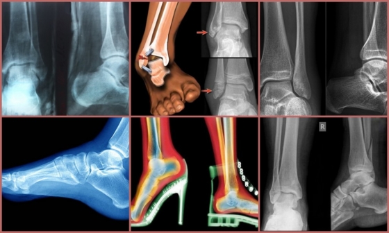
X-rays of the foot allow the doctor to examine the metatarsal, tarsal and toe bones in detail and to assess the condition of the joints formed by these bony structures. Since the X-rays pass unhindered through the soft tissue and are retained in the dense bone tissue, the contours of the structures of interest are made visible in the image.
X-rays of the foot can help diagnose conditions such as: B.
- Bone injuries - fractures, dislocations;
- Chronic joint diseases – arthritis, arthrosis;
- Gout;
- acquired and congenital foot deformities – flat feet, club feet, valgus deformity of the first toe;
- Benign and malignant tumors;
- Pyoinflammatory processes in the foot (deep acetabulum, phlegmon).
A foot x-ray is most commonly requested by a trauma surgeon who needs to determine the nature of a bone injury or identify signs of a chronic musculoskeletal disorder that is causing discomfort to the patient. Sometimes the patient is referred by doctors from other disciplines for an X-ray examination of the foot.
Advantages of X-ray diagnostics at SM Clinic
X-ray examination of the ankle joint makes it possible to detect various types of pathological processes. X-ray examination helps in the initial diagnosis of the following diseases
- Disorders of the integrity of the joint-forming bones – talus, distal part of the fibula and tibia (fractures, breaks are diagnosed);
- Disorders of joint congruence due to dislocations and subluxations;
- Primary tumors and secondary tumors involving bone tissue;
- calcium salt deposits in the soft tissue components of the joint;
- Necrotic processes in bony structures;
- flat feet (longitudinal and lateral), clubfoot;
- degenerative-dystrophic joint damage (arthrosis or osteoarthritis);
- decreased bone mineral density characterized by osteoporosis;
- Arthritis - an inflammatory lesion in a joint;
- Arthrosis – a purulent inflammatory lesion of the bone marrow and adjacent bone tissue.
Indications for an X-ray examination of the ankle
An X-ray examination of the ankle joint is recommended if appropriate. These are situations in which a visual assessment of the musculoskeletal structures is required to accurately describe the clinical situation. Indications are:
- trauma (it is recommended that an X-ray be taken as soon as possible after the traumatic event);
- swelling of the foot and/or ankle;
- pain and accompanying claudication;
- Restriction of motor activity in the ankle joint;
- Pallor of the skin in the ankle and foot area, especially in connection with low local temperatures;
- irregular wearing of footwear;
- visible deformations of the foot.
In addition to the initial diagnosis, x-rays of the ankle joint are taken to dynamically assess the previously identified abnormalities. The diagnostic procedure is usually repeated some time after the patient has undergone treatment or surgery. Modern digital scanners produce the lowest dose of radiation required, allowing repeat examinations when appropriate.
How are the limbs scanned?
The examination of the legs takes an average of 10-15 minutes. Depending on the area to be examined, the patient must lie, stand or sit during the examination. The specialist will also advise if the limb needs to be flexed or straightened. The laboratory technician will then carefully immobilize the limb. The patient is required to remain stationary for a few seconds and is then sent back to await the scan and description of the examination.
If necessary, an X-ray examination of the lower limbs with weight-bearing foot can be performed. This helps to assess the shape of the foot, which is characteristic when walking. Such x-rays show the foot in relation to pressure, making flat feet more visible.
innovations
The modern technologies used in our hospital allow us to do this.
- X-rays of the entire length of the limbs or local (bones, joints, spine),
- Functional examinations while standing – X-rays of the entire length of the lower limbs under weight.
These examinations are often indispensable in the planning of orthopedic operations and in the assessment of axial curvature in traumatology, orthopedics and neurology. - The images are sent directly to a server where they are available to the attending physicians. At the patient's request, the results can be recorded digitally, printed on film and made available to physicians around the world.
- If necessary, a second opinion can be obtained from a specialist in the observed pathology.
The information on this website should not be used for self-medication and self-diagnosis. Diagnostic examinations in case of an exacerbation should only be carried out by a doctor! For a correct diagnosis and treatment, please consult your doctor.
contraindications
X-ray examination of the ankle is not indicated in patients in severe condition.
- CT examination serves to obtain very accurate and high-quality images, to detect abnormalities in the bone structures of the joint and pathological accumulations of fluid in the joint cavity (pus, blood). It is associated with a specific radiation exposure.
- MRI – does not contain ionizing radiation, allows you to assess the condition of the cartilage and soft tissues that form and surround the ankle (muscles, tendons, ligaments, cartilage), as well as blood flow in them.
- Ultrasound – detects abnormalities in the soft tissues adjacent to the joint, a completely safe and inexpensive examination method.
X-ray examination of the ankle at Yauza Klinikkel Hospital
The X-ray examination of the ankle is performed in our hospital by experienced radiologists using a digital diagnostic radiography system (Philips, The Netherlands), which offers the following advantages
- High-quality images at maximum examination speed thanks to digital signal transmission;
- Convenient procedure (flat panel detectors allow you to capture images in the desired projection without having to move the patient);
- Immediate availability of images for treating doctors (all images are sent directly to the hospital server via Wi-Fi).
The results of our radiological diagnostics comply with all strict international protocols and are accepted by all medical institutions in the world.
Prices for Services You can take a look at the price list or call the phone number on the website for details.
The information on this website should not be used for self-medication or self-diagnosis. In the case of an acute illness, diagnostic tests should only be carried out by a doctor! In order to make a correct diagnosis and prescribe treatment, you should consult your doctor.
How is the investigation carried out?
No preparation is required for an ankle x-ray. X-rays of the ankle joint are usually taken in two projections: straight and lateral. The examination in the straight projection is carried out with or without rotation of the foot. The shinbone, ankle joint, talus bone and ankle joint can be seen. In the lateral view, the patient lies on his side, the healthy leg is bent at the knee and drawn towards his stomach. The pictures show the shinbone, the ankle joint, the ankle bone, the heel bone and the ankle joint. In some cases, the doctor recommends taking the x-ray under stress - power flexion - to assess the condition of the ligaments. If necessary, the examination is carried out under local anesthesia.
The evaluation is carried out by comparing the recording with a normal reference. Height, shape, bone structure, height and arch angle, and other criteria are determined.
The conclusions regarding the norm or the detected abnormalities are recorded in a report, which serves as the basis for a definitive diagnosis.
X-ray in the Kutuzovsky Medical Center
The medical center is located in the immediate vicinity of the Slavyansky Bulvar metro station at 5 Davydkovsky Street and offers a full range of medical services. The center has developed into a trusted clinic characterized by international best practice, state-of-the-art equipment and high standards of service. It is important to us that patients feel cared for, that the staff is friendly, that they understand the doctor's instructions, that they receive an accurate diagnosis and appropriate treatment. Our prices are affordable – the price of an ankle x-ray is 2250 rubles. Choose a convenient time and make an appointment at Kutuzovsky LDC.
You can also receive a complete diagnosis of musculoskeletal problems of the lower limbs at our center and have an MRI of the calf muscle and an X-ray of the foot.
What the doctor sees
An x-ray of the ankle shows that a person without specialist training will not be able to qualitatively decipher the findings. However, the doctor will easily be able to recognize the following pathological changes:
- anomalies in the shape and correct mutual alignment of the hock bones;
- consequences of trauma;
- Increase in bone dimensions due to increased load;
- abnormalities of the upper layer of the bone and joint;
- detachment of the periosteum from the bone;
- formation of necrotic areas;
- formation of fracture lines in the bone;
- Deformation;
- proliferation of connective tissue;
- Change in the size of the joint space.

The doctor determines the area to be examined based on the clinical signs. Only a highly qualified doctor is able to detect all of these abnormalities. In the early stages of pathology they may be barely noticeable.
Important!!!
If the results of the x-ray are not enough to make a correct diagnosis, the doctor may recommend additional testing.
Preparation and execution of an X-ray examination
No special preparation is required to prepare for an X-ray examination. It is important to remove all metal objects in order not to distort the diagnostic results.
The procedure is performed under the supervision of a radiologist. The patient is placed on a special table or couch. The ankle area must be within reach of the device.
The patient must lie still during the x-ray. It's even better not to breathe. The slightest movement distorts the results of the examination.
After the first x-ray, the patient changes position to image the leg in a different projection. The doctor then shares the results, which are forwarded to the treating physician for prescription.
Interpretation of the results
The first person to understand what the X-ray of the foot shows is the radiologist. He or she does not make a preliminary diagnosis, but simply describes the anatomical features of the bones in the foot. The radiologist is responsible for detecting fractures, tumors, inflammation and other abnormalities. Based on the description of the recording, the diagnosis is made by the attending physician - surgeon, orthopedist, oncologist or other.
A correct x-ray of the foot of a healthy person should show bones with a homogeneous structure and intact. The outlines of the bones should be uniform and clear, without darkening.
Symptoms of Flat Feet

To diagnose a flat foot, a stress X-ray of the foot is usually taken in lateral projection. To do this, the foot is placed on a surface with the inside facing the X-ray cassette. The other foot is pushed to the side. With this positioning, three lines are marked on the images:
- I – runs from the first toe to the heel bone;
- II – runs from the place where the calcaneus merges into the first line to the wedge joint of the calcaneus;
- III – runs from the same joint to the first metatarsal bone.
Match these lines to each other and measure the height and angle at which they intersect. The height is a straight line descending from the intersection of the second and third lines to the first horizontal line. The normal height should be more than 35mm and the angle should be between 125° and 130°.
A deviation from these norms indicates the presence of a flat foot. To determine the degree of pathology, it is necessary to assess how much the actual foot parameters deviate from the norm:
- Grade I. The height of the arch of the foot is 25-35 mm, the angle is 131-140°. With this pathology, the patient suffers from leg fatigue during physical activity.
- Degree II. The height of the arch is 17-24 mm and the angle is 141-155°. In this case, the patient experiences pain when walking for hours and during physical activity.
- Grade III. The height is less than 17mm and the angle is greater than 155°. With this pathology, the patient suffers from constant pain in the lower legs, lower back and feet.
contraindications
During an X-ray examination of the foot, the patient is exposed to minimal radiation. Absolute contraindications for X-rays of the feet does not exist, but in some cases the prescription must be done with caution. This includes pregnant women and children under 15 years old.
How often can a foot x-ray be performed?
To determine how often a foot x-ray can be taken in two projections, you need to know how much radiation the x-ray machine emits in a single exposure. You can then refer to SanPiN, which tells you the radiation dose a patient can receive per year. By comparing these two parameters, the frequency of the diagnostic procedure can be concluded.
X-rays are usually ordered first when there is evidence of pathology. After a certain period of time (e.g. after a month) a second referral will be made to assess the effectiveness of the treatment. This frequency of x-rays is acceptable.
Make an appointment for a consultation or diagnosis today!

Indications for diagnosis
An x-ray of the foot clearly shows the metatarsal, tarsal and toe bones, the joints and the gaps between them. The black and white photograph allows the specialist to assess the anatomical features of the foot and the structure of the hard tissue, detect fractures, cracks, dislocations and tumors, and diagnose flat feet. As a rule, an orthopedist/traumatologist refers the patient for an X-ray examination, but a surgeon, oncologist, rheumatologist or endocrinologist (important for diabetics) may also request the diagnosis.
An X-ray examination of the foot is recommended in these cases:
- Complaints of pain in the foot when walking and at rest
- Lower limb injuries
- Congenital or acquired diseases – clubfoot, fallen arches, flat feet
- Overlapping big toe on right or left foot
- heel spur
- Symptoms suggestive of osteoarthritis – swelling, redness of the skin.
For bone fractures, an x-ray can help assess the type of injury, the number and location of fragments, and the progress of bone healing. An X-ray may be necessary if a purulent process in the bones of the foot is suspected, such as: B. cellulitis or a deep hip joint injury.
An x-ray is one of the mandatory examinations before an operation on the metatarsal or tarsal bones, as well as for later assessment of the effectiveness of the treatment.
Contraindications for surgery
There are no absolute contraindications to conducting an X-ray examination, but there are some restrictions for women during pregnancy and breastfeeding. In this case, specialists weigh the possible risks and prescribe diagnostics only if there are strict indications.
Relative contraindications include:
- Restriction of movement if the person is unable to assume the posture required for the examination
- high doses of X-rays within the last year
- Large-scale wounds and injuries in the foot area.
What diseases the X-ray shows
The radiologist will describe the x-ray image in detail and prepare a written report. Based on the results, the treating doctor will either make an accurate diagnosis or refute it if no pathological changes are found. Fractures, breaks, dislocations, subluxations, inflammation and bone abnormalities are clearly visible on the x-ray.
What diseases can be diagnosed during the examination?
- Osteoporosis, rickets
- flat feet
- Valgus deformity of the big toe
- Osteoarthritis and arthritis
- Salt deposits and osteophytes
- Congenital osteosclerosis
- Age-related degenerative changes in the foot.
An x-ray of the foot of a healthy person should not show any abnormalities in tissue structure and integrity. When deciphering the results, the diagnostician pays attention to the contours of the joints and bones - they should be clear, without darkening or deformation.
Indications for the examination.
- Trauma is the most common reason for referral for an ankle x-ray
- Pain in the joint - during movement or at rest - associated with traumatic and non-traumatic damage to the joint
- limited mobility, limited range of motion, symptoms indicating a pathological process in the joint
- Swelling and deformation of the joint.
X-rays can be used to detect various diseases of the ankle joint and its surroundings:
- Fractures and ruptures, sprains, tendon tears
- Congenital and acquired anomalies – flat feet, club feet.
- Inflammatory diseases – arthritis, osteoarthritis, synovitis, bursitis, synovial bursitis
- Degenerative joint diseases – osteoarthritis, osteoporosis.
Because healthy and damaged tissue reflects X-rays differently, each disease has a specific image on the X-ray. This allows the radiologist to make a statement about the condition of the organ in the image and the treating doctor to make a diagnosis and recommend treatment.
contraindications
X-ray examination is contraindicated in pregnant women, since exposure to radiation is dangerous for the unborn child, and in patients in serious condition requiring resuscitation.
How is an ankle x-ray performed?
The ankle joint has a complex, block-like shape. In order to better see the structures in this area, multiple projections are used in the X-ray examination.
There are two direct projection options - with and without foot rotation. In the first case, the patient lies on his back, the heel is pressed against the cassette and the foot is turned inward by 15-20 degrees together with the shin. In a non-rotational ankle exam, the patient lies supine, heel resting on the cassette, and toes pointing up. The direct projection images of the ankle show the lower parts of the tibia, the outer and inner malleolus, and the talus. An inward x-ray shows the outer part of the ankle, and a joint space status x-ray shows dislocations and subluxations.
The x-ray is taken in lateral projection with the patient lying on their side. The ankle is placed with its outer surface on the cassette and the heel pressed firmly against the table, allowing the foot to rotate 15-20 degrees inward. The patient bends the healthy leg and pulls it towards the stomach so that it does not obstruct the view. This x-ray shows the talus and calcaneus, the distal parts of the tibia, and what is known as the posterior ankle, which is the edge of the articular surface of the tibia that often detaches during trauma.
If there is no fracture, stress x-rays are taken to assess the ligamentous system. This examination can be performed either sideways or directly. Since the manipulation can be painful, local anesthesia is used.
X-rays of the ankle require no special preparation. Before the examination, it is better to wear comfortable shoes and clothes that allow quick access to the area to be examined.
How often can I have an X-ray of the joint?
X-rays of the hip and all other joints can be taken as often as the doctor recommends. You are not allowed to decide for yourself whether you want to undergo this examination. In any case, there should be an interval of at least three weeks between examinations. For children, x-rays may only be taken once every six months.
X-rays are electromagnetic waves that lie between gamma rays and ultraviolet radiation. The level of radiation exposure depends not only on how often the test is performed, but also on the power of the equipment used and the duration of the procedure. In the event of an overdose, the RNA and DNA strands are severely damaged. The result can be genetic changes, mutations and cancer. The modern digital devices are much safer than the old film based devices. In addition, different areas of the body have different X-ray conductivity.
To reduce radiation exposure during the procedure, special aprons (shields) are used. Lead is used to make these medical accessories. The apron is used to cover important organs. The X-ray tube used during the examination is also fitted with a special protective shield to protect the patient from unnecessary radiation.
It should be clear that X-rays are very helpful in diagnosis and that the harmfulness of X-rays is relatively small. If they are not treated in time, this can lead to much greater health damage. In addition, the treatment of neglected pathologies requires more money. It is also important to know that each person absorbs an average of around 2.4 million S per year from the environment, which is many times more than regular medical examinations.
After the X-ray examination, it is important to help the body recover faster. This can be done by eating cottage cheese, whole wheat bread, tomatoes, beets, walnuts, bananas, olives, plums, and green tea. If there are no contraindications, you can drink a good glass of red wine.
How long does the investigation take?
Patients' feedback confirms that the X-ray examination does not take long. It is carried out quickly and smoothly. On average, the procedure takes about 15 minutes.
After an X-ray examination of the shoulder joints and all other bones, the doctor determines the integrity of all components. If there is a fracture, its size and location are described. The medical records always indicate which radiation dose was administered.
Now you know where to get an x-ray of your knee or other joint, how an ankle x-ray is done, and the ins and outs of this procedure. Read patient reviews to learn more!
Read more:- Anatomy of the heel bone x-ray.
- Anatomy of the foot x-ray.
- Structure of the human ankle.
- dislocation of the ankle.
- Sesame legs of the hock.
- Treated subluxation of the ankle.
- Damaged ligaments of the ankle photo.
- fracture of the ankle.
