Parents often ask the orthopedist what to do if their child is already 1 year old and has a clubfoot? At this stage, Ponseti therapy is the most productive method:
- Congenital clubfoot: causes, symptoms, treatment
- Causes of congenital clubfoot
- Classification: Types of clubfoot in children.
- Symptoms of clubfoot in children
- Diagnosis and Symptoms of Clubfoot
- Treatment of clubfoot
- Symptoms of Clubfoot
- diagnosis
- Treatment of anomalies
- Methods of treatment of congenital clubfoot
- prevention
- Causes of Clubfoot
- Clubfoot classification and clinical picture
- ICD-10
- classification
- What is clubfoot?
- Severity of clubfoot in children
- A little about the disease
- Causes and symptoms of clubfoot in children
- Causes of clubfoot in children
- Who can correct clubfoot?
Congenital clubfoot: causes, symptoms, treatment
Clubfoot is a malformation of the bones and muscles of the foot that causes the foot to deviate inward. Most club feet are congenital, so it is especially important to begin treatment as soon as possible after birth. Congenital clubfoot can be diagnosed as early as the third month of pregnancy through an ultrasound scan.
Many parents wonder why their child has congenital clubfoot? How can this be determined? Let's try to understand this.
Causes of congenital clubfoot
There has been a lot of research into this condition, but so far the exact causes are unknown. There are currently three theories that this defect could have arisen:
- Mechanically. Proponents of this theory believe that it is caused by excessive pressure of the uterine walls on the foot (caused by the narrowness of the uterus, lack of water, tumor, breech position of the baby).
- Neuromuscular. In this case, the cause lies in abnormal development of the fetus, which is due to the influence of external negative factors: drug abuse, alcohol, smoking during pregnancy, infectious diseases, constant stress, medications, electromagnetic radiation.
- Genetically. Proponents of this theory believe that clubfoot is inherited.
Classification: Types of clubfoot in children.
Unilateral clubfoot causes one leg to be shorter than the other. There are the following levels of severity:
- Easy – ankle movement is fully possible;
- Moderate – joint mobility is somewhat limited, difficulty and give are felt when trying to adjust the position;
- Severe – the bones and ankle joint are deformed and readjustment of the foot using manual correction methods is impossible.
A severe form is associated with dysfunction of the muscles and nerves of the foot, and other developmental disorders may also be present.
Depending on the changes in the bones and joints of the foot and the presence of other accompanying problems, the following types of clubfoot are distinguished:
- Idiopathic: associated with a reduction in foot waist;
- Positional (postural): There is a subluxation of the joint between the talus and the heel bone, but the bones themselves are normally developed;
- Syndromic: in combination with other developmental disorders, e.g. B. congenital kidney defects;
- Secondary clubfoot: caused by neuropathy and myopathy.
Symptoms of clubfoot in children
Clubfoot usually appears at birth: the foot is tilted inwards and upwards and the mobility of the foot is limited. If the child can already walk, he or she supports only the outer edge of the foot. The child steps on the foot he is standing on. The gait is wobbly and uneven.
Without treatment, the bony misalignment worsens and subluxations occur in the small joints of the foot. The outer edge of the foot skin becomes thicker. The muscles of the lower leg that are not involved in walking gradually weaken and atrophy. This is followed by dysfunction of the knee and pelvic joints. The later treatment begins, the more serious and difficult it is to correct.
The main symptoms of clubfoot in children include:
- The sole of the foot is bent at the ankle joint;
- the sole is turned inwards with the outer edge hanging downwards;
- the forefoot is turned inward;
- The muscles of the lower limbs are underdeveloped and disappear in older children;
- A staggered gait is noticeable;
- If the disease is unilateral, one leg is several centimeters shorter than the other.
The children tire quickly when walking or standing, rub their skin and complain of pulling pain in their feet.
Diagnosis and Symptoms of Clubfoot
Thanks to modern diagnostic methods, doctors can determine the presence of clubfoot long before the child is born. It is not too difficult to detect clubfoot during an ultrasound examination of the pregnant woman by an experienced specialist. As soon as a child is born with this pathology, clubfoot is diagnosed almost immediately. The following symptoms are characteristic of this condition:
Their severity varies. They depend on the severity of the disease, which is divided into three groups:
- Lightweight - there is no restriction of movement in the ankle joint and the alignment of the foot is corrected by applying light pressure with the hand;
- Moderate – ankle movement is significantly restricted and there is a springy stiffness associated with some resistance when attempting to correct foot position;
- Severe – there is a severe deformity of the foot and ankle that cannot be corrected by manual manipulation.
There is also a definition of clubfoot depending on the suspected etiology (cause of occurrence). There are three forms of congenital clubfoot:
- Typical. The exact cause of this type of pathology cannot yet be determined. It is characterized by dysplasia (underdevelopment, underdevelopment) of the ankle joint and a violation of the anatomical structure and location of the muscles and ligaments. With this form, a one-step correction is not possible even in the first days after birth - a long, step-by-step treatment of clubfoot in a newborn is necessary;
- Positional clubfoot – caused by shortening of muscles and ligaments without damage to bones and joints. It is caused by abnormalities in the fetal development period. The treatment of this form is less complicated and allows a complete recovery without lasting consequences;
- Secondary – results from various congenital anomalies of the neuromuscular system. The clinical picture and treatment options as well as the results are directly related to the underlying disease;
Treatment of clubfoot
Parents are often horrified by the diagnosis. They believe that complete correction of the foot anomaly is impossible. However, this view is wrong. There are several treatment options for congenital clubfoot with a very high success rate. In most cases, they therefore have a good chance of curing their child.
In the initial phase of treatment, conservative techniques are used that produce good results for mild to moderate club feet:
- The first is a tight bandage on the foot, which is previously applied by hand in an anatomically correct position. This technique is started immediately after discharge from the hospital and the umbilical wound has completely healed. With a slight deformity, the feet can be returned to their normal position 2-3 months after birth;
- The most common method for correcting moderate clubfoot in children is the Ponseti method, named after the American doctor who first used it in the middle of the last century. This method involves applying a gradual cast over a period of 4-6 weeks, changing every 7 days up to the upper third of the thigh. Subsequently, the child must wear a special orthosis that keeps the foot in the correct position until the age of 3-4 years;
- When treating the severe form, which also includes deformity of the joints, it is sometimes necessary to perform surgery on the ligaments and muscles, sometimes on the joints of the foot. A plaster cast will also be applied after the operation.
Important!!!
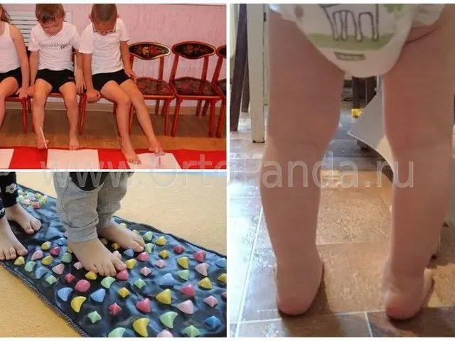
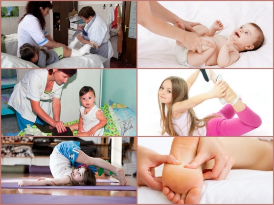
These treatment methods are complemented by physiotherapy (application of paraffin, ozokerite and mud), massage and therapeutic exercises, as well as the obligatory wearing of orthopedic shoes for up to 5-6 years.
A comprehensive approach to the treatment of foot pathologies guarantees radical correction of the anomaly in 90 % of cases. Even if a complete cure is not achieved, after all these procedures the child's condition improves to such an extent that he is able to lead an active life with minimal restrictions during puberty.
Symptoms of Clubfoot
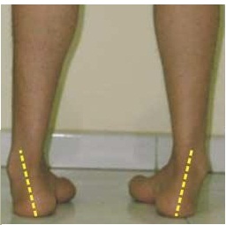
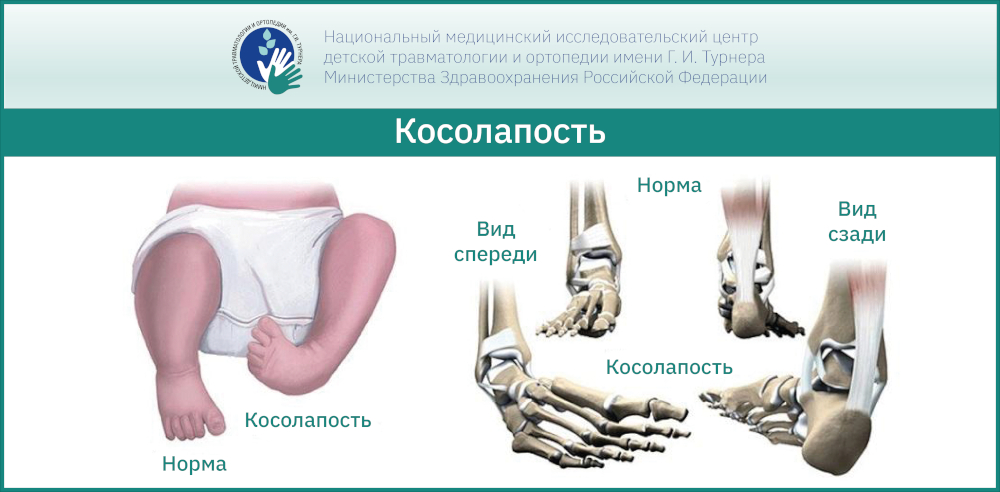
Equinus, varus (the foot is flexed and the toes are turned inward) and supination (the sole is turned up and inward). The mobility of the ankle joint is limited. Due to the changed foot position, a child with clubfoot does not walk on the sole of the foot, but rather on the outer edge. A specific gait pattern develops in which the affected person steps on the supporting leg with every step.
Over time, this condition worsens. The bones of the foot become more deformed and the ankle joints become subluxated. The skin on the outer surface of the foot becomes rough. The muscles of the lower leg that are not involved in walking atrophy and the knee joints are disrupted. The later treatment of congenital clubfoot begins, the more difficult it will be to correct the problem and restore the shape of the foot.
diagnosis
It is important to determine whether it is a true clubfoot (caused by a malformation of the foot bones) or a deformity. With clubfoot, the patient's foot is more mobile and is actively or passively brought into a normal position. The equinus is only weakly pronounced. There are transverse folds on the back of the foot, which indicate sufficient mobility. A positional clubfoot usually resolves spontaneously within the first few weeks of life, but if this form of clubfoot is diagnosed, conservative treatment is always indicated.
X-rays are not meaningful in children under 3 months of age because the bones at this age are usually still cartilaginous and are not visible on X-rays. For children older than 3 months, radiographs are taken in two projections: anteroposterior and lateral. The x-rays are taken with the sole and dorsiflexion of the foot at its maximum. An ultrasound scan is carried out to examine children under 3 months old. This method is completely harmless, but less informative because only one of two levels (side or top view) can be viewed.
Treatment of anomalies
With early diagnosis and detection of foot pathology at an early age, it is possible to correct the alignment of the arch of the foot using conservative methods. In severe cases, disease progression requires the patient to be hospitalized.
Warning.
The classification of clubfoot, the diagnosis of the degree of damage and the selection of treatment methods are carried out only by a qualified doctor - a podiatrist, under whose supervision the duration of therapy is adjusted.
Methods of treatment of congenital clubfoot
The diagnosis of the disease is made in the delivery room, and after 7-10 days comprehensive treatment begins on an outpatient basis under the supervision of the doctor. The child's foot is immobilized with a plaster cast, which is changed weekly until the desired therapeutic effect - maximum correction of the deformity - is achieved.
After the cast is removed, a custom-made splint is made for the young patient, which is placed on him in a relaxed position while sleeping. Maintaining the effect and preventing relapse.
In most cases, the treatment of congenital clubfoot in children is carried out using the method of Dr. Vilensky. A number of therapeutic measures are carried out by parents and specialists:
- plastering;
- Strengthening of joints with special devices – orthopedic braces;
- Applications with paraffin, mud;
- Therapeutic exercises depending on the specified load on the lower limbs;
- Gymnastics – complex exercises for the whole body;
- foot massage;
- Warm baths with pine extracts.
After the main treatment, it is recommended that children wear orthopedic shoes to consolidate the result. Botox injections are another popular method for correcting foot defects.
prevention
To prevent clubfoot, it is important to be active during pregnancy, eat a healthy diet, maintain a healthy daily routine, and give up unhealthy habits.
Prevention of clubfoot includes walking barefoot on uneven surfaces, regular massages of the lower limbs (to improve blood circulation and muscle mobility), relaxing foot baths, water treatments and exercise.
Don't forget a balanced and healthy diet, hardening your body, wearing good shoes, moderate physical activity, walking outdoors and swimming.
Warning!!!
At the first signs of a foot deformity, you should consult your doctor immediately to avoid serious complications.
Bad feet are not a punishment! With comprehensive measures to improve the child, the pathology can be eliminated effectively and without possible consequences.
Causes of Clubfoot
Congenital clubfoot can also be caused by a genetic pathology, but more often by an external factor acting on the fetus. According to research, the most common cause of congenital clubfoot in a child is:
- Fusion of the amniotic membrane (the watery membrane that surrounds the fetus in the womb) with the lower extremity;
- Mechanical compression of the amniotic membrane on the fetal foot or traction on the foot by the umbilical cord;
- Abnormal formation and development of the lower limbs in the first trimester of pregnancy. This can occur as a result of infectious viral diseases and severe stresses on the mother during this period (long-term observations show that the number of newborns with this pathology increases sharply in the war and post-war years);
- pressure of the wall of the uterus on the foot due to insufficient amniotic fluid (dehydration);
- Mechanical impact of a uterine tumor on the baby's foot;
- toxoplasmosis acquired during pregnancy;
- The child has a spinal nerve disease.
Important!!!
Modern diagnostic equipment for examining pregnant women (ultrasound examination) allows doctors to detect clubfoot before the birth of the child. In this case, adults are ready to take urgent measures to correct clubfoot. The success of treatment depends on the timeliness of these measures.
Clubfoot in a child can be caused by a variety of injuries: severe burns, mechanical damage to the bones and musculoskeletal system of the foot. Fractures of the foot and lower leg that have not healed properly, frequent ankle sprains, sprains and torn ligaments lead to clubfoot when the child walks. Rickets and poliomyelitis can also cause acquired clubfoot.
Clubfoot classification and clinical picture
Why does a child have a clubfoot? Doctors differentiate between four main forms of equinovarus foot deformity in children (the so-called clubfoot). This classification focuses on etiopathogenesis - the causes and mechanisms of the malformation. Clubfoot can be:
- Typical. The exact cause of this form of pathology has not yet been clarified. It is characterized by dysplasia (underdevelopment, underdevelopment) of the ankle joint and a violation of the anatomical structure and location of the muscles and ligaments. With this form, one-stage correction is not possible even in the first days after birth - long, gradual treatment is required;
- Postural defects – caused by shortening of muscles and ligaments without damage to bones and joints. It is caused by abnormalities during fetal development;
- Secondary - caused by various congenital anomalies of the neuromuscular system. The clinical picture is directly related to the underlying disease;
- Arthrogryposis caused by a severe joint pathology.
In the typical and positional form, both limbs are often affected, while in the secondary and arthrogryposis only one limb may be affected - the child then stands on one leg. Depending on the extent of the affected limb, the degree of abnormality varies:
- Mild - there is no restriction of movement in the ankle joint, the alignment of the foot is corrected with light pressure from the hand. Sometimes the child walks normally in summer sandals, but with clubfoot in winter shoes - voluminous and soft;
- Moderate – ankle joint movement is significantly restricted and there is a springy stiffness associated with some resistance when attempting to correct the alignment of the foot;
- Severe – severe deformity of the foot and ankle that cannot be corrected with manual treatment.
ICD-10
Foot deformities are a group of pathological conditions in which the appearance of the foot is altered. This group includes a variety of disorders resulting from trauma, malformations, paresis, paralysis and a number of diseases. The severity of foot deformities and disabilities can vary greatly, ranging from almost complete loss of function to severe disability. However, even small foot deformities negatively affect the upper body, causing pain, fatigue when walking, poor posture, premature fatigue of the back and lower leg muscles, and ultimately increase the likelihood of osteochondrosis and arthrosis of the small and large joints of the lower limbs.
Foot deformities are treated by orthopedists and traumatologists. Depending on the cause of the disease, neurologists, neurosurgeons, rheumatologists and other specialists may also be consulted.
classification
There are the following basic types of foot deformities:
- horseshoe– Accompanied by a sustained plantar flexion (plantar flexion). Active dorsiflexion of 90 degrees or less is not possible or only possible with difficulty. In severe cases, even passive flexion cannot return the foot to its normal position.
- heel foot – Is characterized by persistent dorsiflexion. In severe deformities, the dorsal surface of the foot contacts the anterior surface of the tibia.
- Hollow foot (stiff, supinating) – This is accompanied by an increase in the curvature of the longitudinal arch. In severe cases, the patient relies only on the heads of the metatarsal bones and the tuberosity of the heel bone, with the metatarsal bones not touching the surface.
- Flat feet (soft, pronating) – characterized by a flattening of the transverse or longitudinal vault. With longitudinal flat feet, the outer edge of the foot does not rest on the surface, as is the norm, but rather on the sole of the foot. Transverse flatfoot is accompanied by a widening of the forefoot and an increase in the distance between the heads of the metatarsal bones.
In practice, a combination of several types of foot deformities often occurs. In addition to the condition of the bones, joints, tendons and ligaments, the size and type of misalignment can also be influenced by pathological changes in the upper extremities, especially the ankle joint.
What is clubfoot?
Congenital clubfoot is a complex malformation of the foot in which the external shape of the foot is influenced by changes in the skeletal, articular, soft tissue, nervous and vascular systems of the lower limbs.
Clubfoot can be detected by ultrasound as early as 3 months of pregnancy, but there are cases where parents only get a 'surprise' after birth. The pediatrician can make the first diagnosis at birth. In any case, it is only after birth that the pediatric orthopedist can determine the degree of deformity, the need for treatment and the treatment plan. In larger cities, children are examined by pediatric orthopedists in the maternity ward in the first few days after birth.
Severity of clubfoot in children
There are 4 degrees of congenital clubfoot:
- Easy – complete elimination of all elements of deformity is possible without effort;
- Moderate – primary correction of moderate foot position is possible;
- Severe – elements of the deformity cannot be corrected all at once;
- Atypical – deep longitudinal furrow, originally small feet, short toes.

A little about the disease
In a young patient, the feet cannot stand upright because the feet and joints are deformed. The crooked position of the feet causes pain when walking and standing. Motor functions are also impaired. Ultimately, this leads to curvature of the spine and dysfunction of the internal organs. In addition to health problems, a child's character can also change drastically if he or she begins to think about aesthetics during puberty. Doctors warn parents that treatment should begin as soon as possible, that is, as soon as the diagnosis is made.
Clubfoot in children can be caused by birth trauma or abnormal skeletal maturation during the fetal period.
Experts also point out that clubfoot often occurs later in life and is referred to as 'acquired'.
Depending on the nature of the manifestation of the pathology, it is divided into typical and atypical, into unilateral or bilateral (if one or both feet are diagnosed).
In addition, four levels of severity are distinguished:
- Light, in which the bones are not deformed and the ankle is mobile.
- Moderate, when ligaments, joints and muscle tissue are deformed.
- In the severe stage, both feet are deformed.
- In a very severe stage, there is a risk that the child will become disabled and require surgery.
Causes and symptoms of clubfoot in children
The disease is very common worldwide, with doctors detecting it in one in 1,000 children. Unfortunately, orthopedic surgeons do not immediately recognize them at a young age. Only from the age of 2 can an accurate diagnosis be made and the cause of the disease known.
In most cases it is caused by external factors:
- There have been cases of bone fractures followed by abnormal bone fusion.
- Other injuries are due to bruises, burns, torn ligaments and muscles.
- Consequences of life-threatening diseases (polio, rickets, hip dysplasia, valgus).
- Scoliosis (curvature of the spine).
- Increased muscle tension in the lower back.
- Swelling in the foot area.
- Parents' incorrectly chosen shoes (too tight).
- Genetic disorders, when close relatives suffer from the same disorder;
- abnormalities during pregnancy (smoking, taking prohibited drugs, alcohol, working in a dangerous position);
- multiple pregnancy;
- umbilical cord or uterine wall obstructing the baby's feet;
- Excessive toxemia, avitaminosis, low water pressure or uterine hypertonicity;
- Improper positioning of the baby before birth.

Causes of clubfoot in children
With clubfoot, the child develops an ugly, clumsy gait, and the shoes quickly become a one-sided fall. Congenital clubfoot occurs in three out of every 100 children, with boys twice as likely as girls to be born with the condition.
In addition to congenital clubfoot, there is also acquired clubfoot, which is caused by a sedentary lifestyle and lack of foot movement.
Congenital clubfoot is caused by a genetic predisposition or mechanical damage. It can be detected through an ultrasound scan soon after birth or during pregnancy.
Acquired clubfoot is diagnosed at the age of 3 years. It is caused by a malfunction in the body. The danger with this form is that it is not always noticed by parents at a young age, and some mothers only raise the alarm after some time.
Who can correct clubfoot?
Clubfoot is treated by an orthopedist. If you suspect an illness, you should take your child to a doctor as soon as possible. Clubfoot can be easily treated, especially if the problem is identified at a young age.
Clubfoot treatment can be conservative or surgical.
Surgical intervention is rarely indicated - usually when the disease is detected late and conservative treatments can no longer help. After the operation, the patient is again prescribed physiotherapy and massage.
Read more:- What is clubfoot?.
- Congenital clubfoot.
- 1 year old child with clubfoot.
- clubfoot.
- Clubfoot in 7-year-old children.
- How to remove a clubfoot.
- Clubfoot Treatment.
- What to do if your child has clubfoot?.