Until recently, the mainstay of surgical treatment was the traditional approach, using either cardiothoracic techniques:

- Arthritis in the fingers.
- Causes of the inflammatory process in the finger joints
- What is hand arthritis?
- What to do when you're young: simple advice
- Stages of Heberden nodule formation on the fingers
- Treatment of Heberden and Bouchard nodules
- Physiotherapeutic treatment of Geberden nodules
- How is it diagnosed?
- How do you treat a painful thickening of the big toe?
- A few words about prevention
- Causes of thickened fingers
- This article has been reviewed by
- Types of thickenings on the toes
- Why does the bone of the wrist hurt?
- What are the symptoms of bone pain?
- risk factors
- signs
- Treatment of Dupuytren's contracture
- Forecast. prevention
Arthritis in the fingers.
The inflammation of the small joints usually does not develop spontaneously, but rather as part of a general illness. Arthritis in the fingers is a characteristic symptom of rheumatoid arthritis, but can also be a manifestation of another disease. In order to avoid serious complications and disabilities, it is important to consult a doctor in a timely manner, determine the cause of the disease and initiate treatment. In this article you will find all the information you need about this disease.
Finger arthritis is an inflammation of the metatarsophalangeal and interfinger joints. The disease occurs in all age groups. Statistically speaking, women aged 40 and over are more commonly affected. The reason for this is that women put more strain on their hands and fingers than men. ICD-10 code M13 for finger arthritis of unspecified origin.
Depending on the underlying cause, the disease may only occur in the small joints of the fingers or be combined with damage to the large joints. The onset can be acute, subacute or chronic, but the course is usually long-lasting or chronic. An exception is post-traumatic arthritis of the fingers, which, with appropriate treatment, leads to complete healing. However, if it is not treated in time, it also becomes chronic.
Causes of the inflammatory process in the finger joints
The causes of the disease can be different. The small finger joints are most commonly affected by rheumatoid arthritis, an autoimmune disease (allergy to one's own tissue) that is based on a hereditary predisposition. The outbreak of the disease is usually triggered by an infection. Finger arthritis can develop over time in genetically predisposed individuals. As a result, other larger joints may also develop.
Psoriatic arthritis is another common cause of inflammation of the small joints of the hands. It is also an autoimmune hereditary disease that primarily affects the finger joints (distal joints) with characteristic nail damage. It usually occurs against the background of pre-existing skin manifestations of psoriasis, but sometimes the symptoms of finger arthritis appear first.
Finger joint arthritis often develops together with gout. The disease has a metabolic origin (metabolic) - the metabolism of uric acid salts is disturbed, which are deposited in the joint and periarticular tissue and trigger an inflammatory process.
Post-traumatic arthritis of the fingers is sometimes work-related. It develops with constant minor trauma to the hands of jewelers, seamstresses, hairdressers, etc. The inflammatory process is constantly maintained by additional injuries and takes a chronic course. Acute arthritis can follow an acute injury (sports, home) which then resolves completely.
Other types of arthritis rarely affect small joints. Hormonal changes (teenage years, pregnancy, menopause), stress, frequent colds and allergies, bad habits, professional activity are some of the triggers for the development of arthrosis of the small joints of the hand.
What is hand arthritis?
The joint contains cartilage tissue that changes for one reason or another. Damaged areas form. They become thin - osteophytes form. The fingers begin to thicken unsightly, making it difficult to perform normal activities that require fine motor skills.
Neither hand arthrosis nor gonarthrosis are hereditary: only a predisposition to it is inherited. Metabolic characteristics, the nature of the tissue, and the elasticity and density of the articular cartilage can be inherited. If your close relatives suffer from osteoarthritis, remember: you are at risk!
What to do when you're young: simple advice
Certain professions - pianists, massage therapists, office workers and people who constantly use keyboards - are particularly prone to osteoarthritis in the hands. If you anticipate the possibility of developing this disease, you should try to choose another profession in your youth.
- Avoid injuries to the joints of the fingers and wrists. These most often provoke the development of dystrophic changes.
- Be especially careful when icing, as you intuitively put your hands underneath you if you fall.
- Don't overwhelm yourself with exercises with dumbbells.
Stages of Heberden nodule formation on the fingers
It is difficult to diagnose the stage of Bouchard's node formation 'by eye' - only the size of the osteophyte can be an indication. In the early stages, itching and burning may also be felt in the affected joints.
The best way to accurately determine the stage of the lump is an X-ray examination:
- at . stage 1 Small osteophytes become visible on the x-ray;
- under 2 stage the osteophytes begin to partially close the joint spaces, causing narrowing and 'lines' of subchondral sclerosis appear;
- on page stage 3 large osteophytes and significant narrowing of the joint space are visible;
- large osteophytes almost completely occlude the joint spaces and deform the bone heads so that they become flat.
There are no changes in the blood or urine at any stage.
Treatment of Heberden and Bouchard nodules
Treatment of Heberden and Bouchard nodules is mainly conservative. If medications, physical therapy, or exercise are ineffective and the disease progresses, topical corticosteroid injections are indicated. Surgical treatment of Heberden nodules of the fingers is very rare when the joint is almost completely immobilized or there is permanent soft tissue trauma.
Despite the swelling and redness of the skin in Bouchard nodules, the problem is more of an aesthetic nature. If the patient has no complaints, there is no treatment for Geberdin nodules. Doctors usually prescribe chondroprotectors and supportive exercises.
The treatment of Heberden nodules is no different from the treatment of Bouchard nodules.
The therapy is usually non-specific and is carried out in conjunction with the treatment of the underlying disease – deforming osteoarthritis. Therefore, the affected joints must be protected and tightening (fixing) orthoses must be worn at night or during working hours.
Physiotherapeutic treatment of Geberden nodules
If the symptoms are mild, physiotherapy is carried out prophylactically. It aims to slow the progression of osteoarthritis. Physiotherapy techniques can reduce inflammation, prevent muscle loss, reduce pressure on joints and improve metabolism in tissues.
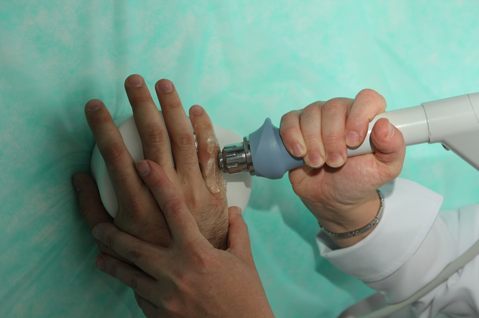
Physiotherapy is an effective treatment for Bouchard nodules
If Heberden's nodules on the fingers cause pain and discomfort to the patient, physiotherapy is used:
- paraffin therapy or ozokerite (if there is no inflammation of the synovial membrane);
- medicinal phonophoresis (if there are signs of inflammation);
- electrophoresis;
- laser therapy
- Magnetic therapy
- cold therapy;
- electromyostimulation;
- shock wave therapy
- Massage;
- balneotherapy and mud treatment;
- Remedial gymnastics.
How is it diagnosed?
The diagnosis of joint pathology is complex and represents an important step in treatment. The differential diagnosis includes rheumatoid arthritis and other joint diseases.
The diagnosis is made after an examination that includes:
- Complaints and anamnesis
- physical examination
- general blood test – no change
- biochemical blood test and CRP (C-reactive protein) – no abnormal values
- Rheumatism test – negative
- X-ray of the hand – joint space narrowing, marginal osteophytes
- MRI -.
MRI is the most effective and informative method for studying joint pathologies.
How do you treat a painful thickening of the big toe?
Unfortunately, this problem is mostly caused by chronic diseases and cannot be completely cured. Only the hirgroma can be removed once and for all. All other growths persist throughout life, but exacerbations can be avoided by achieving lasting remission.
Rheumatoid arthritis is treated with corticosteroids or nonsteroidal anti-inflammatory drugs. The same nonsteroidal anti-inflammatory drugs are also effective against gout, as is colchicine.
Osteoarthritis is treated with chondroprotectors, finger exercises and physiotherapy treatments: multi-channel electrical stimulation and laser therapy. With multi-channel electrical stimulation, the muscles are stimulated with short electrical pulses to relieve tension and improve blood circulation. Laser therapy involves directing a laser beam at the joint prominence to reduce inflammation and increase blood flow to the tissue.
Osteoarthritis is also treated with physiotherapy: ultrasound (reduces tissue volume), shock wave therapy (stimulates blood circulation, eliminates swelling and pain using sound pulses) and laser therapy.
Surgical intervention may be necessary if the cartilage and joint surfaces are severely damaged in advanced osteoarthritis or osteoarthritis with swelling, stiffness and pain in the fingers. In particularly severe cases, an endoprosthesis is prescribed, in which the worn joint is replaced by an artificial joint. Surgery may be performed if a malignant tumor is present. The oncologist develops an individual treatment program for each patient.
Conventional medicine is powerless against the appearance of a lump and pain on the finger joint. No homeopathic remedy contains ingredients that will make the lump disappear. Therefore, relying on chance is highly undesirable with such an illness: any hope for a miracle can ultimately lead to serious consequences.
A few words about prevention
The best prevention of growths is a healthy lifestyle: eat a balanced diet, eat fresh fruits and vegetables regularly, avoid bad habits, sweets and fried foods. Wear protective gloves when coming into contact with chemicals. Avoid stress and lack of sleep.
If you are a student who has a bump on your thumb and it hurts, you are probably writing too much and holding your pen incorrectly. In this case, the bump is just a blister that will disappear over time. Do not put a band-aid on them or try to remove them by any other means. Try holding the pen differently and not squeezing it too tightly with your fingers.
Causes of thickened fingers
- lung cancer;
- Chronic obstructive pulmonary disease;
- bronchiectasis;
- Tuberculosis;
- lung tumor;
- Changes in the normal anatomical structure of the lungs;
- cirrhosis
- congenital heart defects;
- infectious damage to the heart valves;
- granulomatous intestinal inflammation;
- intestinal infantilism;
- Ulcerative colitis.
Thickening of the fingers is observed against the background of joint diseases such as arthrosis, infectious arthritis, metabolic disorders, skin diseases and nail fungus. Thickening of the toe joints can be caused by excessive and prolonged pressure on the skin from tight, uncomfortable footwear, arch injury, flat feet, and limping. More rarely, joint deformities are the result of vibration sickness, HIV infection and prolonged stays at high altitudes.
This article has been reviewed by
Types of thickenings on the toes
Depending on which part of the limb's skeleton the lesions occurred, a distinction is made between thickenings on the toe joints and thickenings on the ankle joints. Visually, the shape of the finger or toe bones changes and they look like a 'drum' - the nail plate is characterized by a clockwork deformity with bulging thickening of the end phalanges of the fingers.
A typical sign of the defect is Chamroth syndrome - an atrophy of the cleft when the nails of opposite hands are brought together. In a patient with tympanic toe, the angle between the nail base and the nail plate is smoothed, and instead a deep and wide distal angle is created, which exceeds 180 degrees when viewed from the side. The transformation of the tissue between the nail and the nail plate into a spongy substance creates a false sense of nail plate mobility when the nail base is compressed. In some cases, thickening of the finger bones is accompanied by pain and limited mobility.
Other symptoms depend on the underlying disease. So, deforming arthrosis of small joints progresses with the formation of Geberden and Bouchard nodes - thickenings on the dorsal and / or lateral surface of the distal interphalangeal joints of the fingers. These are characterized by pain in the fingers, a burning sensation, swelling, swelling and stiffness in movement.
Why does the bone of the wrist hurt?
If the bone in your wrist hurts, it can have many causes. The condition is often caused by an injury (bruise, dislocation, sprain or fracture). This bone pain is usually accompanied by swelling of the surrounding soft tissue, which turns blue.
Arthritis or osteoarthritis can also cause bone problems. The former is an inflammation of the joint caused by an infection, an allergic reaction or an autoimmune disease, while the latter is accompanied by degenerative changes, usually associated with age.
Osteoarthritis is often caused by occupational diseases. For example, if you sit at the computer for a long time, you can develop tunnel syndrome. This is caused by compression of the nerve in the wrist. An excessive load, e.g. B. in athletes, can cause tendinitis, an inflammation of the wrist tendon. Kinbeck disease (or avascular necrosis) is an occupational disease of locksmiths, carpenters, crane operators and carpenters. Blood flow to the wrist is reduced and the bone begins to break down. In right-handed people the right wrist bone hurts, in left-handed people the left wrist bone hurts.
Carpal thygroma (also known as a 'nodule') is a benign tumor. The patient often describes the condition as 'the bone has popped out of the wrist and it hurts'. A cyst forms on the bone of a joint (sometimes on several bones) - a thick capsule filled with fibrin threads, serous fluid and mucus, which as it grows compresses the neurovascular bundles, sometimes causing severe pain. Cysts often form in certain professions: pianists, computer technicians, spinners, painters, teachers, professional tennis, golf and badminton players. In addition, genetic predisposition, trauma, inflammatory processes and operations on the wrist play a role.
What are the symptoms of bone pain?
Depending on the cause of the pathology, the pain may be accompanied by other symptoms. In the event of a serious injury, not only does the pea bone (or other bone) in the wrist hurt, but there is also swelling of the injured area and restriction of movement of the wrist. If there is a sprain or fracture, the pain is particularly severe and the joint becomes deformed.
Tunnel syndrome is accompanied by numbness and weakness of the hand. There may be a burning, tingling sensation, especially at night and in the morning, and swelling is likely after sleeping.
With tendonitis, a characteristic crunching sound can be heard in the joint. Lifting objects with the affected hand may be difficult.
In carpal tunnel syndrome, pregnant women may experience not only bone pain but also itching, burning, tingling and trembling in the fingers. These symptoms mainly occur at night.
In acute arthritis, the pain syndrome is pronounced, there is swelling and redness in the affected area, and the body temperature often rises. In chronic arthritis, the symptoms are muted and the patient may not seek medical attention for a long time, leading to destruction of the joint. Osteoarthritis is characterized by gradually increasing pain in the bones, limited mobility and painful tenderness. A grinding or clicking noise can be heard in the affected joint. If left untreated, the pain increases and the hand gradually loses mobility.
In Kinnbeck disease, the pain is felt around the clock and increases during manual activities or when pressing on the affected bone.
With a hirgroma, the affected person initially does not feel any pain, but only notices that the bone on the outside or inside of the wrist is growing (it appears as if the bone is enlarging). The skin in this area gradually darkens and thickens, and the bone begins to hurt when the wrist is moved. As the hirgroma enlarges, it becomes painful and the patient can no longer move the wrist normally.
risk factors
Despite its precise location (joints only), osteoarthritis of the big toe is a multifactorial disease. A number of causes precede the appearance of symptoms. This includes:
- inherited predisposition;
- Injuries at home and at work (arthritis of the big toe is common in footballers and ballet dancers);
- tight and uncomfortable footwear;
- Obesity with regular overload of the feet;
- severe hypothermia;
- Congenital and acquired foot deformities (flat feet, developmental anomalies);
- Problems with the endocrine system, hormonal disorders and changes (thyroid disease, diabetes, menopause, taking steroids, etc.).
- Long-term pressure on the foot area (low physical activity);
- age-related changes.
If you have two or more risk factors for osteoarthritis of the big toe, you should consider having a check-up. We have extensive experience in diagnosing osteoarthritis in its early stages and treating it at all stages.
signs
- Rapid fatigue of the legs;
- pain syndrome (the pain can be aching or stabbing);
- 'cheerful' sensitivity;
- poor blood circulation in the feet (toes and feet feel cold);
- joint stiffness, occasional 'crunching', especially with sudden movements;
- swelling and inflammation of the tissue around the bone of the big toe;
- deformation of the joint area;
- Nodules (Gegerden) on the joint surface.
This list includes the symptoms of the different stages of the disease. Depending on the patient, these can occur sooner or later, so a specialist should be consulted at the slightest signs.
Be careful!!! The widespread nature of the disease has led to the use of many folk methods to treat osteoarthritis, resulting in patients only seeing a doctor at a late stage.
Treatment of Dupuytren's contracture
The most effective treatment for Dupuytren's contracture is surgery. This treatment can restore the function of the hand and lengthen the fingers, but it is not yet possible to stop the disease itself, that is, the progression of the contracture. Effective conservative treatments have not yet been developed.
There are four main types of surgical treatment:
Needle fasciotomy and collagen fasciotomy are minimally invasive techniques. Recovery takes about a week and complications are rare. After surgery, the contracture often develops again. Recurrence can occur after 3-4 months or after several years, depending on the person.
Needle fasciotomy Typically performed in a single session. After treating the hand with an antiseptic solution, the surgical area is anesthetized with a local anesthetic. The nodules and ligaments of the modified palmar aponeurosis are then severed at different levels using needles of different diameters through a percutaneous incision, ie without cutting into the skin.
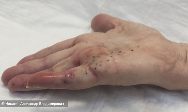
This results in the integrity of the plexuses being broken and the finger becoming erect. The severed fibers of the aponeurosis itself remain under the skin, which is the reason for frequent recurrences. Despite its apparent simplicity, fasciotomy should only be performed by a doctor specializing in hand surgery due to the risk of injuring important structures: tendons, vessels and nerves. Needle fasciotomy is contraindicated in cases of intolerance to the procedure under local anesthesia, in cases of thinning of the skin and scarring due to open hand surgery, in cases of open wounds or infections of the hand.
Fasciotomy with collagenase bacteria Clostridium histolyticum is carried out in two sessions. During the first appointment, collagenase is injected into the node or fascia. Within a day, the nodes and aponeuroses soften and become soft. At the second appointment, after the anesthesia, the doctor forcibly stretches the fingers and pulls the trains apart. The contraindications for this procedure are the same as for needle fasciotomy, but there is also an allergy to collagenase. In addition, the effects on breastfeeding and pregnant women have not been studied, so they are not treated in this way. The operation is also not performed on patients under 18 years of age [14].
Forecast. prevention
Regardless of the surgical technique, the development of a contracture cannot be completely stopped. Even after a dermofacicectomy, recurrences can occur around the transplanted flap, and there are cases where patients have undergone more than 30 surgeries. For this reason, the doctor cannot guarantee that the contracture will not recur after treatment. The risk of recurrence is higher in younger patients.
In 2012, tamoxifen was proposed to reduce the risk of recurrence after fasciectomy in high-risk patients [4] . However, the effectiveness of this method has not yet been confirmed.
There is no specific prophylaxis for this disease. All that can be done is to reduce the risk factors and seek medical help early.
In the future, new methods for treating and preventing Dupuytren's contracture could be developed thanks to the involvement of experts in molecular biology, genomics, histology and radiology.
Read more:- Orthopedic Clinic.
- Orthopedic reports.
- Foot.
- Bone anatomy of the leg.
- paraparesis.
- Equino valgus.
- How muscles and bones are related.
- At what age do girls' legs grow?.