Over time, the pain becomes a habit and the person's gait changes. The sufferer begins to plant the foot away from the affected area, relying primarily on the toes and lateral surface of the foot. This often leads to the development of transverse flatfoot, which further aggravates the condition.
- Anatomy of the Female Genitalia
- External genital organs
- pubic bone
- clitoris
- Small labia
- Structure of the human knee joint
- Human elbow joint structure
- causes
- symptoms
- Types of leg fractures
- hip fractures
- Ideal foot shape
- How to correct the curvature of the legs due to their shape
- Field report about the lipofilling of the lower legs
- What is the risk of injury?
- Features of lower limb injuries
- Definition of a muscle
- Structure of the human muscle
- Skeleton of upper and lower limbs
- Lesson Summary 'Skeleton of the Upper and Lower Limbs'
- Treatment in the Health Energy Clinic
- Benefits of the clinic.
Anatomy of the Female Genitalia
EVERY WOMAN SHOULD KNOW THE STRUCTURE OF HER SEX ORGANS!
The female genital system (genitals) is an amazing mechanism that enables woman to create new life and experience the joy of motherhood. Knowing how it works gives insight into the drives of parents and doctors.
anatomy – The science of structure. The reproductive organs are only part of the reproductive system and their structure is discussed below. To understand why certain processes occur in these organs, one must understand the structure of the reproductive system as a whole. Many of you have heard the expression, 'All diseases come from the nerves. '
How true this statement is, we can see from the fact that some neurological and mental disorders and diseases are accompanied by menstrual disorders. The work of all organs is controlled by the nervous system. It establishes our connection to the environment and allows the body to adapt (or not) to changes in it.
But nerves alone won't get you very far. On the beach you can tell with a single glance whether a person is male or female. Why is that? This is due to a unique substance in our body – the sex hormones.
Hormones play a major role in both the development and functioning of the sex organs. The sex glands or ovaries are part of the body's endocrine system, and the sex hormones are not only responsible for the development of sex characteristics. They affect all types of metabolism in the body and the functioning of other organs and systems.
The genital system is both part of the endocrine system and connected to the nervous system. Such an orchestra must have a conductor. This is the neuroendocrine gland - the pituitary gland. Located in the brain, it communicates between the nervous and endocrine systems.
Nerve impulses trigger the production of hormones in the pituitary gland. The hormones of the pituitary gland reach the sex glands (ovaries) via the bloodstream and influence the production of the ovarian hormones (progesterone and estradiol) there. By altering metabolism in tissues, hormones produced by the endocrine glands affect the functioning of organs and systems, including the nervous system. The figure shows schematically how the genital organs, nervous system and endocrine system of the human body are interconnected.
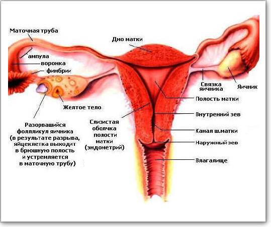
External genital organs

1 – pubic mound; 2 – clitoral hood; 3 – clitoral glans; 4 – labia minora; 5 - external urethral opening; 6 – hymen (is the boundary between the external and internal genitalia); 7 - barium; 8 - anus; 9 - vaginal opening; 10 - labia majora.

pubic bone
The pubic bone (pubis) is an erection located in front of and slightly above the pubic conjunctiva and is covered with hair with the upper limit of hair growth running horizontally (in males, hair growth runs up the midline).
clitoris
The clitoris is a small (up to 1-1.5 cm) but very delicate and important organ, mainly composed of the cavernous bodies. The male penis has a similar structure. The cavernous bodies have cavities that are filled with circulating blood. During sexual arousal, these cavities fill with blood and the clitoris becomes larger and thicker - an erection. The cavernous body is not able to contract like blood vessels, which is why injuring the clitoris is dangerous because it bleeds profusely.
Small labia
The labia minora (labia minora) are two folds of skin that lie between the labia majora and the vaginal opening. At the front they are connected to each other and form the clitoral foreskin. Normally, the labia majora extend slightly beyond the limits of the labia majora and are pale pink to dark brown in color at the rear. The labia majora has a large number of vessels and nerve endings and is an erogenous zone that enlarges due to the influx of blood during sexual stimulation.
The labia majora vary in shape and size, and are often asymmetrical. If the shape and size of the labia are physically or psychologically uncomfortable, surgical correction of the shape and size of the labia is performed.
Structure of the human knee joint
The knee is a complex and large joint in the human musculoskeletal system. It allows movement of the lower limbs in relation to the hips and supports body weight. Movement of the knee joint is essential for everyday activities such as walking, running, sitting and standing.
The knee is a modified joint, a type of synovial joint, made up of three functional parts: the patellar (patellofemoral) joint, which includes the kneecap (patella) and is located at the front of the thigh, and the medial and lateral tibial joints, which connect the femur connect to the shin. The knee also has a cartilage pad, the meniscus, which acts as a shock absorber inside the knee. In addition to the joint capsule and ligaments that support the knee, there are also some important structures that surround the knee and help soften the joint and protect it from friction. These are small 'sacs' of synovial fluid called bursae.
Human elbow joint structure
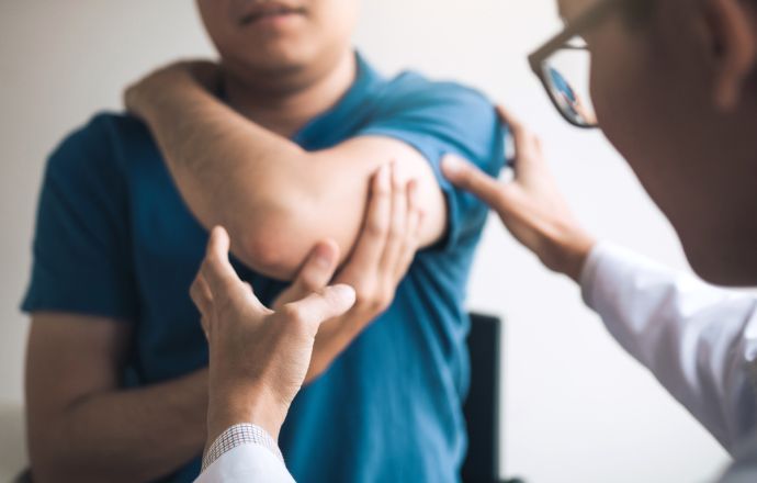
The elbow joint is a joint formed between the distal end of the humerus and the proximal end of the forearm. Like all other joints, the structure of the elbow consists of a smooth layer of cartilage. There is also a joint capsule that surrounds the joint and provides strength and smooth sliding.
The fluid formed by the synovial membrane of the joint capsule fills the empty space between the bones and lubricates the joint, reducing friction and wear. The extensive network of ligaments surrounding the joint capsule help keep the elbow stable and withstand mechanical stress. There is also a ring-shaped band that runs from the elbow bone around the head and holds the bones of the forearm together. These ligaments are responsible for the movement and extension of the elbow and protect it from sprains and strains.
causes
The causes of X-shaped limb deformities are varied. The condition can be caused by both congenital and acquired factors. The most important include:
- Early start of walking. Most often, valgus deformity of the lower limbs develops as a result of excessive loading on the child's immature legs. In such cases, the muscles and ligaments are not yet strong enough to keep the child's legs in the correct position, and poor coordination makes the problem worse because the child will spread their legs too far when walking to avoid falling.
- Constitutive Features. The development of an X-shaped leg deformity can be caused by the child being overweight. However, if you are not overweight, you are more likely to be asthenic than hypersthenic, which may be due to a congenital weakness of the musculoskeletal system, which is more common in people with an asthenic constitution. Girls are more likely than boys to suffer from valgus leg curvature due to the gendered structure of the pelvis, and the wider the pelvis and the shorter the femur, the more pronounced the X-shaped leg curvature can be.
- Impaired calcium metabolism. Certain kidney diseases and other conditions negatively affect skeletal formation and lead to impaired calcium metabolism and consequently reduced bone strength. Rickets used to be considered one of the main causes of valgus, but this problem is no longer relevant. Nevertheless, rickets sometimes occurs, so it should be excluded in the differential diagnosis.
- inheritance. X-shaped legs are inherited in a number of cases. This disorder is caused by abnormal ossification of the outer condyle of the femur. Unlike acquired valgus curvature, genetic pathology is detected immediately after birth, since newborns should usually have a physiological O-shaped deformity. A congenital X-shaped deformity is always associated with flat feet and a valgus deformity of the femoral neck.
- trauma and tumors. Unilateral deformities can be caused by intra-articular fractures of the tibial and femoral condyles, compression fractures of the metaphysis, and diaphyseal fractures of the femur and tibia with unresolved angular misalignment. Curvature of the limbs is possible with malignant and benign tumors of cartilage and bone tissue.
- Congenital diseases. Sometimes a unilateral X-shaped deformity develops with congenital anomalies of the lower limbs (hip dysplasia and congenital hip dislocation, tibial hypoplasia, knee joint anomalies, etc.).
symptoms
Children with X-shaped legs tire easily and often complain of pain in their lower limbs. Your gait becomes clumsy and unsteady. Sometimes muscle cramps also occur. To accurately determine the severity of the anomaly, the child is asked to stand with his legs together, and then the distance between the feet is measured. If this distance exceeds 4-5 cm in a child under 4 years old, then further examinations, conservative treatment and dynamic observation are required.
Long-term persistence of a valgus deformity leads to anatomical changes in the knee joints, the feet and in some cases the spine. The inner collateral ligaments of the knee joints are overstretched, the joints become unstable and laterally overstretched. The patient's X-shaped feet become flat, contributing to a flatfoot pattern that makes walking difficult and causes pain and fatigue. If one leg is more bent than the other, the child's trunk deviates from the vertical axis when standing, which can lead to postural disorders and scoliosis.
Types of leg fractures
hip fractures
A hip fracture is a serious injury associated with severe pain and bleeding from the fracture. The severity of the injury and the need for immobilisation of the fragments with skeletal traction or a solid cast results in a severe limitation of mobility which, particularly in the presence of other injuries or comorbidities, can cause dangerous complications such as pressure sores and congestion pneumonia. Fat embolism can occur in the first three days after injury.
femoral neck fractures are intra-articular and are more common in elderly patients with osteoporosis. A leg fracture is caused by a fall at home or on the street; with a significant loss of bone strength, the integrity of the bone can be compromised even by an awkward turn in bed. The patient complains of slight pain in the joint, which increases with movement. The leg is externally rotated and the patient cannot lift the heel off the bed when lying supine. Shortening of the limbs can be observed with displaced fractures. The swelling at the injury site is usually mild.
The diagnosis is confirmed by a radiological examination of the hip. Due to insufficient blood supply, the femoral neck does not heal properly, adequate bone marrow usually does not form, and the fractures are glued together by connective tissue, leading to a high rate of disability. The preferred treatment for these leg fractures is therefore surgery – osteosynthesis with a triangular nail, endoprosthesis or bone graft.
If the general condition does not allow surgery, skeletal traction is used. Elderly patients are put in a plaster splint with a crossbar that prevents limb rotation. This allows the fibular callus to form while the patient remains sufficiently physically active.
Ideal foot shape
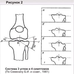
There are many opinions about the correct shape of the feet, but they are all theoretical. When determining the curvature, doctors are guided by the classification of Artemiev AA, proposed in 2001.
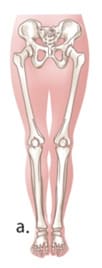
This doctor was the first to talk about the parameters of the correct form in practical terms. Beautiful legs have three spindle-shaped slits between the insides of the legs. These slots are bounded by the perineum, closed knees, upper shin muscles, and ankles (Fig. 1).
If you walk up to a tall mirror and put your feet close together, you can see three windows. If you observe such a view, rejoice - your lower limbs are perfect. If not, it might be crooked legs.
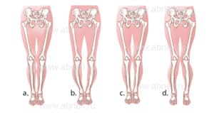
Fig.2. a - correct shape, b - true curvature O, c - false distortion, d - true curvature X.
1. Curvature O. With this anatomical feature, the knees cannot be brought closer together without effort. The result is an O-shaped gap bounded by the ankles and groin. 2. X-shaped. In this case, it is not possible to bring the ankles together with the hips closed. It is a relaxed posture in which the patient makes little effort to position the legs correctly.
3. wrong. The first two are due to bone deformity, while in this case the bones are straight. The problem is due to an abnormal distribution of the soft tissues. The legs are not joined at the top of the shin but meet at the ankles, knees and hips. Outwardly, the legs look crooked, although in reality they are not. Artemyev considers that the true curvature of the legs is due to curved shins. In some cases, the curvature is caused by changes in the knee joint that are due to the anatomy of the condyles. (Fig.3).
How to correct the curvature of the legs due to their shape
If you don't want to hide your deformities, you need to see a surgeon. True curvatures are corrected by orthopedic traumatologists as part of osteoplastic surgery. It is less about correcting a cosmetic defect and more about maintaining your health. Even a slight curvature of the bone changes the load on the joint, which can lead to serious diseases. If there is a misalignment, the plastic surgeon corrects the shape of the tibia by injecting autologous fat or inserting an implant. The inside of the legs becomes fuller, the hole between the knees and ankles disappears.
Field report about the lipofilling of the lower legs
'The operation went quickly and I have fond memories of my time there. The doctor and all the staff were friendly and attentive to me. And most importantly, in less than two weeks I've bought myself a lot of beautiful clothes that I could only lick my eyes at before. Tight trousers, mini skirts, slits, playful ruffles, tapered and loose: everything suited me. It was amazing!'
Yulia, 34. Because of its simplicity, Artemyev's classification is used with pleasure by doctors and patients. It is more tailored to the needs of orthopedists and traumatologists. For plastic surgeons, the proposed framework is too narrow. Only one curvature type is suggested for the correct bone shape. However, this does not correspond to the practical observations. Otherwise, a correction method would have to be applied to all patients with an incorrect curvature, which is not the case in practice. Cases are individual and require a thorough assessment of the condition to find a solution. This is the only way to correct imperfections and bring the shape of the legs closer to the ideal. Analyzing patient photos, we came to the conclusion that there are several variants of false leg curves. Therefore, we have created a contour classification for lower limb deformities that meets the needs of plastic surgeons. Using these guidelines, it is easy to choose a correction method depending on the type of soft tissue distribution.
What is the risk of injury?
It is obvious that the ability to walk, run and stand securely on one's feet is very valuable and important for every human being. When a person loses their mobility, they are unable to lead a full life, perform routine household chores, or work in jobs that require some physical activity.
Leg and pelvic injuries are one of the causes that can contribute to a person becoming disabled and unable to walk, move, or run. Injuries to the lower limbs are caused by mechanical impact when the integrity of muscles, ligaments, joints, bones and skin is compromised and when parts of the joint system can change position and not work together normally.
Lower limb injuries are generally divided into two groups: closed and open injuries. The former pose the greatest risk to individuals because they cannot be seen and assessed visually - the damage is beneath the intact skin. It can involve tears in tissue, fractures, dislocations and, less commonly, bruised muscles and ligaments.
Open wounds and lacerations mean that the skin at the site of the injury is also torn, cut or bruised and internal injuries may be visible. This principle of discernment is particularly important for those who need to provide first aid in the event of an accident, natural disaster or accident, because even the absence of visible external signs of injury is no guarantee that the person is well.

What are lower limb injuries? They are all differentiated by the type of disorders they cause in the body.
Features of lower limb injuries
A contusion, accompanied by swelling, bruising, and pain, is considered the most minor of all lower-limb injuries. It can occur after a fall or a violent impact. The integrity of the blood vessels in the skin is compromised, resulting in a bruise that discolors over time. In addition, soft tissue contusions can be associated with more serious injuries such as fractures and lacerations.
A dislocated joint is a pathological condition in which parts of the joint, most commonly the condyle, move out of their usual 'place', e.g. B. moves the condyle out of the acetabulum.
These injuries can be caused by sudden and abnormal movements that are outside the joint's functional range of motion, or by impact and heavy weights.
A bone fracture is the occurrence of a crack or break in the bone, which can result in the spread of bone fragments in a wound. Fractures can be closed or open - in the first case, the skin remains intact, in the second case, the skin is crushed or cut by protruding bone splinters or by mechanical impact. The affected person feels severe pain, the fracture site is swollen and blood circulation is impaired. Painful sensations make it very difficult to move and kick the injured leg.
Tears and dislocations of tissues, ligaments and muscles are caused by sudden, forced movements that exceed the joint's capacity. These injuries are common in athletes, but even ordinary people are not immune to painful and dangerous injuries. People who overexert themselves and work out in the gym without following training and safety codes are particularly vulnerable to these injuries.
Tissue crushing occurs as a result of a strong impact – this disrupts the normal structure and vitality of tissue, blood circulation and metabolic processes. Such wounds are dangerous because they are prone to infection and inflammation and take a long time to heal.
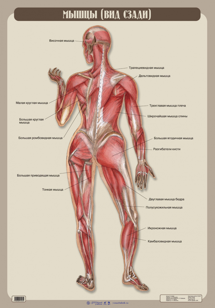
Definition of a muscle
muscle (Latin. muscles) is an organ of the animal or human body composed of muscle tissue. Muscle tissue is a complex structure. The cells of the myocytes and the endomysium, the covering, form separate muscle bundles that combine to form a muscle that is covered by a protective layer of connective tissue. fascia.
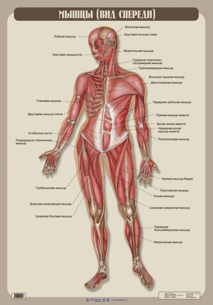
The muscles of the human body can be divided into:
As the name suggests, skeletal type muscles are attached to the bones of the skeleton. The other name is cross striped.This group includes the muscles of the head, limbs and trunk. Their movements are free, meaning humans can control them. This human muscle group This muscle group enables movement in space and can be developed or 'pumped up' through exercise.
Smooth muscle is part of the internal organs such as the intestines, bladder, blood vessel walls and heart. Thanks to their contraction, blood pressure rises during stress or when a chunk of food moves in the intestine.
The heart muscle is characteristic only of the heart and ensures the continuous circulation of blood in the body.
It is interesting that the first contraction of the muscle already takes place in the fourth week of life of the embryo - this is the first heartbeat. From then until death, the heart does not stop for a minute. The only reason the heart stops beating during life is open heart surgery, but then an artificial heart beating machine is in action for this important organ.

Structure of the human muscle
The structural unit of muscle tissue is the muscle fiber. Even a single muscle fiber is capable of contraction, suggesting that a muscle fiber is not just a single cell, but an efficient physiological entity capable of performing a specific action.
A single muscle cell is from one sarcolemma - A tough, elastic membrane made up of proteins collagen i elastin. The elasticity of the sarcolemma allows the muscle fibers to stretch and, in some of them, perform miracles of flexibility - splits and other tricks.
Like the branches of a broomstick, the sarcolemma is densely packed with fibers myofibrilscomposed of individual sarcomeres. Thick strands of myosin and thin strands of actin form a multinucleated cell, and the diameter of a muscle fiber is not a strictly fixed value and can vary in a fairly wide range from 10 to 100 µm. Actin, a component of the myocyte, is an integral part of the cytoskeletal structure and has the ability to contract. Actin consists of 375 amino acid residues that make up approximately 15 % of the myocyte. The remaining 65 %s of the muscle protein are myosin. Two polypeptide chains of 2,000 amino acids make up the myosin molecule. When actin and myosin work together, an actomyosin-protein complex is formed.
Skeleton of upper and lower limbs

In this lesson, students will continue with an introduction to the human skeleton. You will look at the skeletal structure of the upper and lower limbs. The skeleton of the upper limb margin and the skeleton of the free upper limb, the skeleton of the lower limb margin and the skeleton of the free lower limb are examined in detail. The video lesson contains informative material that broadens the students' general perspective and stimulates their interest in the topic
Lesson Summary 'Skeleton of the Upper and Lower Limbs'
The upper limbs are represented by the hands. Hands are characterized by a high degree of mobility, with their help man performs various labor activities and manipulates objects.
The Lower Limbs are the legs. They carry a heavy load and completely take over the function of movement. They are massive and have large and stable joints.
It follows, Main functions of the limbs – Support, moving the body in space. i Support for work-related activities.
The upper and lower limbs are attached to the spine by the bones of the limb girdle: upper part of the shoulder girdle i edge of the lower limbs.
Skeletal structure of the upper limbs. It is represented by the shoulder girdle and the free thigh.
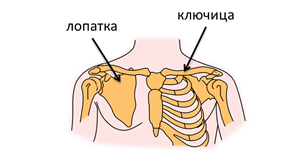
The skeleton of the upper extremity consists two shoulder blades i two collarbones. shoulder blade – is a flat, paired bone with a triangular shape. The scapulae lie loosely between the back muscles. They form the connection between the humerus and the collarbones. If necessary, they participate in the movement of the arm together with the collarbones.
collarbone – The clavicle is a small, paired bone with a sigmoidal shape. It connects the shoulder blade (scapula) to the breastbone (sternum).
It connects the shoulder to the body. The clavicle lengthens the shoulder joint at some distance from the rib cage and gives freedom of movement to the arm. The long collarbones, the position of the shoulder blades, the flat and wide chest and the large number of muscles make the arm very flexible. It is characterized by high precision of movement, which allows, for example, a circus performer to juggle several objects at the same time and a watchmaker to assemble miniature clocks from parts that are difficult to see with the naked eye.
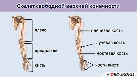
The upper limb consists of three parts: the arm, the forearm i the hand. The skeleton of the free upper limb consists of the upper arm bone (humerus), two forearm bones - radius and ulna (on the side of the little finger) - and the hand bones.
Treatment in the Health Energy Clinic
If you experience heel pain or discomfort, come to the Health Energy Clinic for an appointment. An experienced podiatrist will listen to you, examine you and perform the necessary tests. After the diagnosis, we will suggest modern treatment methods:
- Blockages that allow for quick elimination of pain;
- all kinds of physical therapy;
- professional therapeutic massage;
- Movement therapy training and supervised exercise hours.
In addition, we prescribe medication according to the latest standards and monitor the effect of the treatment.
Benefits of the clinic.
'Health Energy' is a modern and multidisciplinary clinic that offers a comprehensive range of therapeutic and preventive services. Here are the benefits of coming to us:
- overview and advanced early detection programs;
- state-of-the-art diagnostic equipment;
- Qualified nursing staff who undergo regular further training;
- A comprehensive approach to diagnosis and treatment.
We do everything to make every patient feel as comfortable as possible from the first moment in the clinic. Reasonable prices make quality medicine affordable.
A heel spur is just a small bony protuberance, but one that can completely change a person's life. Don't delay seeing a doctor and make an appointment with an orthopedist at Energy of Health.
Read more:- Long legs, small body.
- Which legs are considered long in women?.
- The parts of the human leg are named.
- X-shaped legs photo.
- parts of the human leg.
- How much does crooked leg surgery cost?.
- Can orthopedic shoes be returned?.
- What to do if your teen has crooked legs.