Patients who need an X-ray of the joints often ask about the harm of the procedure. Radiation exposure in this type of diagnosis is minimal thanks to modern equipment. The ViTerra Clinic uses the latest radiological equipment with minimal radiation exposure. X-rays are taken quickly, which reduces the stress on the body. An X-ray of the knee joint, for example, results in a radiation dose of 0.001 mSv - almost as much as a person absorbs in their daily activities. Therefore, after 2-4 weeks, the X-ray can be repeated to assess the effectiveness of the prescribed treatment or the dynamics of tissue healing.

trauma x-ray
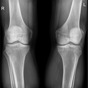
X-ray examination of joints and bones – is one of the leading methods for diagnosing bone and joint diseases. It is often used in orthopedics and traumatology to diagnose anomalies in these structures and is one of the fastest and most meaningful examination methods. The examination is performed by a radiologist. He or she analyzes the image and creates a written report that is sent to the attending physician.
X-rays are used when degenerative or inflammatory diseases are suspected and when evaluating injuries. X-ray images help the trauma surgeon to make an accurate diagnosis and differential diagnosis, identify complications or secondary manifestations of diseases of other organs and systems, decide on surgical or conservative treatment tactics, and then monitor the effectiveness of the prescribed treatment. X-ray examination of the hip and bones is more accessible than more complex examinations (CT, MRI, etc.) and allows to obtain accurate information about the condition of the tissues in a very short time.
Indications and contraindications
The specialist may suspect bone and joint disease based on symptoms such as pain, redness and swelling, restriction of joint mobility and fever. Trauma is also a reason for an X-ray examination - it is important to assess the condition of the bone tissue, to exclude serious damage and to take timely measures if it is detected.
Pregnancy is a relative contraindication for an X-ray examination.However, there are cases when such diagnostics are justified.
In order to get reliable information about the structures under study, it is necessary to keep calm. Your doctor will inform you when it is necessary to 'freeze' yourself. Therefore, another contraindication for an X-ray can be a mental illness, in which the patient is unable to keep still.
Symptoms and Signs of Injury
It is almost impossible not to notice disruptions in tissue integrity. A symptom common to all injuries is pain. However, the nature of the pain is different:
- with bruises - moderate, constant, sometimes throbbing, depending on which nerve is closer;
- in sprains, it is sharp when the fibers are severed, and then quieter but more intense when moving;
- in dislocations - may be absent or, conversely, be stronger than the severity of the injury;
- With fractures – sharp at break, painful afterwards, with increasing longitudinal load.
With bruises (when no vital organs were affected), there are no other symptoms except pain, bruising, and sometimes swelling. Strains and sprains are accompanied by restricted movement and swelling. A dislocation is accompanied by a faint cracking sound, a fracture by a distinct crunching sound. In the latter case, the swelling is more pronounced.
How to avoid fractures and injuries
Occupational injuries, like sports injuries, require special preventive measures. We will focus on domestic situations that anyone can get into.
Injuries in winter are quite common. The easiest way to avoid them is to avoid falls. While it's impossible to completely eliminate these risks, following a few simple rules can help reduce them.
Don't ignore the outside factors. The home environment should be safe - not where all family members slip and fall, trip or bump into visible objects.
When injuries are unavoidable, efforts should be made to reduce the severity of the injuries. The prerequisite for a fracture is a force that exceeds the strength limit of the bone. The lower the force of the impact, the more likely it is a bruise.
For bruises
Place a cold compress on the bruised area and apply a bandage to hold it in place.
The only way to tell if it is a fracture is to take an X-ray. As long as you have even a suspicion of a rupture, you have to be careful. Immediately (but at the same time carefully) apply a splint to the injured limb, using improvised means and elastic or ordinary bandages. Limit the movement of the bone at the site of injury, trying to fix not only the bone, but also the joints above and below. For open fractures, first place a sterile bandage on the wound before applying a splint.
If a spinal fracture is suspected, the casualty should be placed on a firm, level surface such as a board or boards. In the case of fractures and dislocations in the cervical spine, the bones are immobilized with a wire splint that is applied from behind.
For wounds.
If the wound is deep, rinse well with running water (about 5 liters). Do not remove deeply buried foreign objects from the wound. Stabilize the foreign body with a volumetric bandage and, if necessary, fix it with splints.
If the bleeding is profuse, try to stop it: put pressure on the injured vessel over the wound, apply a tight bandage, or apply a tourniquet. Remember that a tourniquet can last 1.5 hours in warm weather and 1 hour in cold weather. After this time, loosen the tourniquet for 5 minutes by pressing your finger on the affected vessel over the wound, and then tighten again.
Bandage the edges of the wound with a sterile gauze bandage or a cotton ball soaked in hydrogen peroxide or alcohol (vodka, cologne). Thoroughly wipe the skin around the wound. Then drizzle the edges with iodine, without touching the wound itself. Apply a dry, clean bandage.
Minor abrasions and scratches can be rubbed with a solution of hydrogen peroxide or alcohol and iodine, then apply a bandage.
'And the poor grasshopper has a dislocated shoulder...'.
However, if there is a fracture and a long immobilization in a plaster cast, do not forget about treatment methods that can significantly speed up recovery.
The newly developed ALMAG+ from ELAMED is not only suitable for adults with musculoskeletal disorders. It can also be used in children as young as one month old to recover from various childhood injuries (including sports injuries), such as bruises and fractures.
The main principle of the pulsed magnetic field in the heart of the ALMAG+ device is blood flow regulation.
- relieving pain and inflammation;
- enhancing the effectiveness of drugs;
- improve blood circulation;
- strengthening the growing walls of vessels, veins and arteries;
- nourishing the cartilage;
- Speeding up recovery and shortening healing time.
Recovery time after an injury can also be accelerated with physical therapy at home.
Another advantage is that the device does not cause the child any discomfort or pain. This means the child is more likely to be quiet during treatment, which also affects recovery.
ALMAG+ helps reduce the negative effects of broken bones, torn ligaments and bruises.
Leaflet 'How to protect yourself from frostbite and hypothermia'.
frostbite Frostbite is an injury (up to and including necrosis) of any part of the body caused by exposure to cold. Frostbite is most common in winter, when the temperature is below 10-20 oC.
The affected area of skin is pale and reddens when heated, sometimes purpling; swelling occurs. The first symptoms of frostbite are a burning sensation, tingling, and then numbness in the affected area.
Initially, there is pallor, coldness, and loss of sensation, but these manifestations occur with all frostbite. Therefore, the most characteristic symptom is the formation of blisters, which are filled with clear contents in the first days after the injury.
In the initial phase, there is pallor, coldness and sensory disturbances, but these phenomena occur with all degrees of frostbite. Therefore, the most characteristic symptom is the formation of blisters with clear contents in the first days after the injury.
These occur with prolonged exposure to cold, a drop in tissue temperature when it is at its highest. All layers of the soft tissues become necrotic, and bone and joint damage often occurs.
First aid for frostbite
The first measure of frostbite symptoms is to take the victim to the nearest warm room, remove frozen shoes, socks and gloves. For first-degree frostbite, warm the hypothermic area with warm hands until red, massage lightly, rub with a woolen cloth, breathe, and then apply a gauze bandage. For second-fourth degree frostbite, do not use rapid warming, massage or rub the affected area with warm hands, massage lightly, rub with a woolen cloth, breathe, and then apply a cotton gauze bandage.
Cover the affected area with an insulating bandage (a layer of gauze, a thick layer of cotton wool, another layer of gauze, and a piece of oilcloth or rubber). The affected limbs can be immobilized with improvised means (board, plywood, thick cardboard) placed over the bandage and tied. The victim may be given hot drinks, hot food, one tablet each of aspirin, analgesic, two tablets of No-shpa, and papaverine. It is not advisable to rub snow on the affected person as the blood vessels in the hands and feet are very delicate and can therefore become damaged and the microfluidics that form on the skin can introduce infections. Rapid heating of frozen limbs by fire or the uncontrolled use of heaters and similar heat sources should be avoided, as this aggravates the course of frostbite. For mild generalized hypothermia, the most effective method is to warm the casualty in a warm bath with the water initially at 24oC, which is then raised to normal body temperature. In the case of moderate to severe general hypothermia with respiratory and circulatory problems, the casualty should be taken to the hospital as soon as possible.
Ice

Everyone has dealt with ice injuries at some point in their life. In winter, our streets turn into an ice rink. How do you get through the time until spring without sprains and broken bones? Is it possible to avoid winter injuries and what to do if a fall occurs?
Slipping and falling and waking up with a cast is no big deal. It's much harder to stay on your feet. Doctors stress that you need to know how to fall. However, their advice is difficult to implement because a fall on ice lasts only a short time. But they can very well help to ensure that you do not seriously injure yourself.
Doctors therefore advise paying attention to winter shoes - to fall less often. Shoes should be suitable: without heels and with ribbed non-slip soles. Strips of tape or tape can be glued to the soles.
Do not wear long trousers as they can snag on the floorboards and be trampled on by people walking behind you.
Don't carry bags with long handles - they shift the center of gravity and reduce maneuverability.
Do not put your hands in your pockets, but stretch them out slightly. This will help you keep your balance.
Walk on the side of the pavement - the edges are less slippery.
The safest way to walk on black ice is to 'Cow on the Ice'. – Walk with small steps, as if you were walking on a slide, like on a small ski. It is advisable to walk as slowly as possible. The faster the step, the greater the risk of falling.
If you feel like you're falling, crouch down - this will reduce the fall height. Don't stretch your hands out in front of you. It's a common misconception that you should relax when you fall. In a relaxed state, the risk of fracture increases because the force of the impact is on the bone. In the event of a fall, you should brace yourself against it.
Pull your head onto your shoulders, push your elbows to your sides and slightly bend your legs. It's better to fall sideways; never land on your outstretched arms. Many people do this automatically, resulting in a broken forearm, a common ice injury.
signs
If you experience sudden, severe, or increasing wrist pain, it's important to see a doctor right away. You should also see a doctor if your wrist injury is accompanied by the following symptoms:
A wrist injury can be diagnosed in a number of ways. The first step is usually a medical history and an orthopedic examination. This may be followed by further investigations, including:
- X-rays of the wrist
- Ultrasound of the wrist
- Computed tomography of the wrist
- Magnetic resonance imaging of the wrist
- blood test
- Taking a sample of fluid or tissue from the joint.
Treatment
Treatment of a broken wrist bone involves the use of a cast and immobilisation of the wrist, and in some cases even surgery. Treating Pain and Other Symptoms:
If the wrist instability is due to a structural change in the bone, the bone defect must be corrected by osteotomy, bone wedge grafting or internal fixation. Carpal instability can lead to severe joint wear. This condition usually requires more radical treatment. Treatment of bone necrosis includes immobilization with a cast or surgery.
Read more:- Anatomy of the heel bone x-ray.
- How do you tell if it's an ankle fracture or a dislocation?.
- Fracture of the calcaneus of the foot.
- How to distinguish a fracture from an ankle sprain.
- Bone structure of the navicular foot.
- Photograph of the periosteal foot.
- Fracture of the lateral condyle.
- Fracture of the articular process of the heel bone.