Many people have probably heard the advice: smile and apply blush to the apples of your cheeks. Always remember that your makeup technique depends on your face shape.
- How to choose blush by texture?
- Dry blush
- Baked blush
- Flaky blushes (flaky blushes)
- Blush in balloons
- Blush for fair and 'cool' skin
- Subluxation of the elbow extensor tendon
- Stenotic synovitis
- Treatment
- rehabilitation
- Wrist movement tests
- Crush and hold test
- Key retention test
- Full Grip Test
- Screening test for radial nerve palsy
- Tests for instability
- Watson test (scaphoid displacement test)
- Scapholunate ball test
- What are deep reflexes?
- Types of tendon reflexes
- diagnosis
- Treatment of radial nerve neuropathy
- therapeutic exercises
- prevention
- Welcome!' – a network of clinics for spine and joint diseases
- Symptomatic signs
- The nature of radial syndrome and its causes
- classification
- Basics
- How you choose
How to choose blush by texture?
There are now various blush textures on the decorative cosmetics market: dry, baked, loose, ball, cream and liquid.
Dry blush
The classic type of blush. It is applied to the cheeks with a special brush. It can have a matte, satin or pearl finish.
Compact blush is most often made in a flat, round or oval container called a refill. These are supplemented with flat brushes or powder.
In cosmetic stores you can find palettes consisting of several shades of the same color. Typically from light to intense. Or from matte (under the cheekbone) to pearly (on the upper cheekbone).
Baked blush
It contains shimmer or light-reflecting particles that create a shimmering effect. This blush gives the skin an additional glow and can therefore replace highlighters (glow products).
Baked blushes do not contain any chemical additives that make them compact. They do not look like crumbly chalk and have a higher concentration of pigment.
Flaky blushes (flaky blushes)
Rarity. As a rule, their consistency is similar to mineral powders.
Applying it to the face requires skill. The advantage of these cosmetics is the thin, natural coverage and the effect of 'highlighted' skin.
Loose blush is ideal for girls and women with problematic or reactive skin.
Blush in balloons
They are also called 'meteorites' because of the similar powder name from the Guerlain brand. A few years ago it was very popular among girls and women because it was easy to apply and gave a beautiful shine.
Blush has the same properties. They refresh and make the skin shine. The secret lies in the pearls of different shades that mix and complement each other. They create a unique, multidimensional color that melts into the skin.
Blush for fair and 'cool' skin
The lightest shade of pink Baby pink – Ideal for people with porcelain or 'cool' skin. Other light blushes have a slightly reddish hue.
Cosmetic products with peach is a universal color. They look particularly good on skin with yellow and orange undertones.
Blush with shimmer – Ideal for velvety white skin. Looks radiant and natural, without make-up effect.
To compensate for the gray haze caused by a lack of sun and vitamins in winter, use a classic blush Blush.
Source: piqsels.com
Subluxation of the elbow extensor tendon
The ulnar extensor muscle runs in the sixth canal of the posterior surface of the forearm, where it is supported by a fibrous sheath in the ulnar groove. When the tendon is subluxated, this covering ruptures. The mechanism of injury usually involves a sudden upward movement of the hand, flexion, or lateral movement of the hand.
Patients with this injury complain of pain in the dorsal wrist, clicking noises when rotating the hand (supination and pronation), and swelling or swelling. Routine x-rays usually do not provide any information, but MRI scans may show damage to the tendon sheath and displacement of the tendon from its position.
Treatment of acute subluxation should involve mandatory immobilization of the wrist with splints or elastic materials. Rehabilitation can begin after 6 weeks when the tendon sheath rupture has healed.
Stenotic synovitis

In stenotic elbow extensor synovitis, pain occurs on the posterior side of the wrist, while flexor synovitis occurs on the palmar side. This is not very common but should be considered in the differential diagnosis of wrist pain.
In stenotic synovitis, patients complain of diffuse pain throughout the wrist and swelling on the posterior surface of the wrist. Many patients are athletes whose activities involve frequent, repetitive wrist movements. Symptoms can be reproduced by asking the patient to resist pressure on the back of the hand. An MRI scan is helpful in making the diagnosis.
Conservative treatment of synovitis includes avoiding wrist strain, bandages or splints (depending on severity), cold compresses, injections of anti-inflammatory drugs into the wrist area, and NSAIDs. Osteopathy can be a good addition to shorten the rehabilitation time.
Treatment
Cold, nonsteroidal anti-inflammatory drugs may provide temporary relief.
Change in load: change of workplace, change of grip, new tool. All of these can make daily activities more enjoyable.
If the hand still cannot do without movements that have become painful due to the disease, surgical correction can be considered.
The optimal operation for this pathology is resection of part of the elbow head under arthroscopic guidance (wafer procedure). 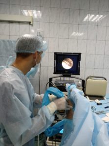
In this procedure, the surgeon inserts a camera and instruments into the wrist through small punctures in the skin (3 mm). After examining the joint, the area of damage is located and any irregular areas of the capsule, cartilage and triangular cartilage complex (TFCC) are scraped with a shaver. 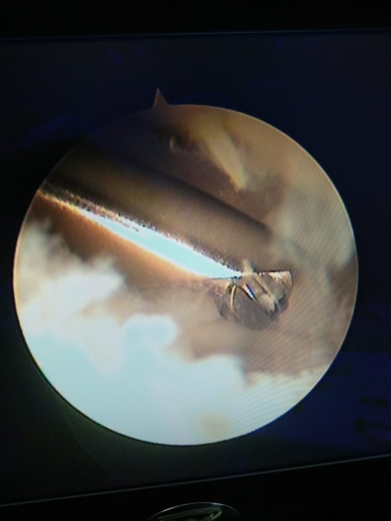
The head of the ulna bone is visible through the abraded central portion of the TFCC. Using a bone drill, the bone is resected to a depth of 5 mm and a width of approximately 1-1.5 cm.
This manipulation reduces pressure on the sickle-shaped bone and prevents further impingement.
rehabilitation
It is best to leave the hand untreated for the first week. Although there are hardly 2-3 wounds on the skin from the arthroscopic approaches, the internal work is very serious. To ensure a pleasant 'recovery' in the early postoperative period, anti-inflammatory medication should be taken regularly.
After 10 days, the stitches can be removed and the bandage becomes unnecessary. Hand strength does not return for at least six to eight weeks, and full recovery may take six months or longer.
For more effective and faster rehabilitation, exercises with a carpal tunnel therapist are necessary. Special techniques to reduce swelling and gently mobilize the joint help in the first 3-4 weeks. This is followed by strengthening and stabilization exercises.
Pain in the shoulder
Wrist pain
Pain in the toes.
Numb fingers
Wrist movement tests
Shows lack of movement and sensation due to nerve impairment.
Crush and hold test
methodology. The patient is asked to pick up a small object (e.g. a needle) from a table with the first and second fingers.
Evaluation. A satisfactory result requires normal sensitivity. The patient must repeat the test with his eyes closed. This test cannot be performed if cochlear and intercostal muscle function is impaired.

Key retention test
methodology. The patient is asked to hold the key between the first and second fingers in the usual manner.
Evaluation. If sensation on the radial surface of the index finger is impaired, as may occur with damage to the radial nerve, holding the key is not possible.

Full Grip Test
methodology. The patient is asked to hold a pencil with all fingers while the doctor tries to pull it out.
If finger flexion is restricted, the test is repeated with a larger object. Evaluation. If the median or ulnar nerve is damaged, full flexion of the fingers is not possible and grip strength is limited. In such cases the test is considered positive.

Screening test for radial nerve palsy
method. The patient is asked to extend the wrist with the arm flexed to 90° at the elbow.
Evaluation. With radial nerve palsy, the wrist extensors are impaired and the patient cannot straighten the wrist. The wrist hangs downward in a deformed position called the 'female wrist'. In the second phase of the examination, the patient is asked to withdraw the first finger. With radial nerve palsy, the patient is unable to retract the first finger because the long muscle that retracts the first finger is paralyzed.
Tests for instability
Watson test (scaphoid displacement test)
Used to assess wrist stability.
method. The test is carried out in a sitting position with the patient leaning on his elbows. The doctor fixes the scaphoid between the thumb and index finger. The doctor uses his thumb to press on the navicular cusps (distal pole of the bone), keeping the navicular bone in an upright position. The wrist is then placed in ulnar deviation, which should cause flexion of the scaphoid, but this is prevented by the pressure of the physician's thumb.
Evaluation. If the test is positive, the proximal pole of the navicular bone is displaced toward the dorsal edge of the fossa, subluxated, and impinges on the physician's index finger. A clicking sound can be heard, which is accompanied by pain and is a sign of a ligament injury in the ankle joint. However, this does not provide any information about the severity of the injury.

Scapholunate ball test
methodology. The doctor presses firmly on the crescent bone and carpal bone of the patient's wrist with the thumbs and index fingers of both hands and moves them against each other in the dorsal and palmar directions, respectively.
Evaluation. Instability occurs when the ligament complex between the scaphoid and femur offers less resistance to the shear forces. A painful displacement indicates damage to the ligament. Instability of the navicular-lunar ligament occurs as a result of a fall on the thumb with pronation of the forearm, a stretched wrist and a bent elbow, or as a result of being hit by a ball in competitive sports. This results in a tear in the ligaments between the scaphoid and the crescent bone.

What are deep reflexes?
Deep reflexes are involuntary muscle contractions that occur in response to a stimulus using muscle spindle receptors. This process occurs in the form of involuntary muscle contractions with passive stretching of the tendons.
This type of stretch is often detected by a small, jerky blow to the tendon attachments of the muscle, delivered with a special neurological hammer. When determining the response, the patient should assume a relaxed state and avoid tension and stiffness.
All muscles must be completely relaxed, otherwise it is not possible to determine the presence and extent of a reflex. If the patient feels tension in any part of the muscle, the reflex becomes inaccurate or disappears altogether.
If reactions are difficult to detect, the doctor will ask the patient to move away from the area being examined, such as For example, when examining foot reactions, clenching the teeth tightly or interlacing the fingers of both hands and stretching the arms with effort is called the Jendrassik technique.
The degree of deep reflexes is usually assessed using a point system:
- 4 points. – Maximum overreaction;
- 3 points – Reactive but with normal severity;
- 2 points. – Reactivity with normal expression is assessed;
- 1 point - small amount;
- 0 points. – complete absence.
The severity of the reaction in healthy patients can vary greatly. Typically, the reactions in the foot area are very pronounced and much easier to trigger than the reactions in the hands.
Types of tendon reflexes
One of the most revealing tendon reflexes is the Achilles tendon reflex. It is triggered when a neurological hammer hits the site of the Achilles tendon. This causes the foot to contract and flex. This reflex is triggered by various methods, namely:
- The patient must sit down.. He sits with his knees on the surface of a sofa or chair. The feet should hang freely downwards.
- The patient lies on his stomach.. The doctor should hold the patient's two feet with his left hand at a right angle to the lower leg.
- The patient should assume a 'supine position'.. The leg is bent at the large joints and rotated outwards. The foot is then flexed in dorsiflexion and planted. This procedure involves plantar flexion of the foot.
- Flexion elbow response. This reaction occurs when the technician hits the phalanx area of the thumb, which he places on the patient's ulnar flexion surface. He uses his finger to apply pressure to the tendon of the biceps muscle, which is located in the crook of the elbow. The patient's hand should be half bent at the elbow and the forearm should rest completely relaxed on the surface of the thigh. This reaction is accompanied by a motor reaction, which manifests itself as flexion of the arm at the elbow joint.
- Extensor elbow reflex. It is expressed by a hammer blow to the site of the tendon of the triceps brachii muscle (triceps brachii), which is located 1.5-2 cm above the elbow process. In this procedure, the patient's arm is grasped just above the elbow through the shoulder region and held in this position. During the detection of this reflex, the arm is extended in the area of the elbow joint.
- Knee or pelvic reflex (knee reflex). This reaction is triggered when a hammer is placed on the tendon of the quadriceps muscle, which is located below the kneecap. A contractile response then occurs, followed by extension of the tibia. This reaction is triggered in two ways: first, when the patient is in the supine position and the therapist places his hand under the bluntly bent knee; on the other hand, when the patient sits low and the legs hang down. The process of reaction dissolution is carried out using the Jendrassik technique. In this technique, the patient is instructed to squeeze the fingers together and forcefully stretch them to the side. When testing this reflex, an extension of the leg in the knee joint is observed.
- Fasciculations – is a visually noticeable, involuntary twitching of individual muscle segments that occurs in the absence of a general contraction of the entire muscle. This contraction is caused by a spontaneous contraction of the muscle group. To determine fascioculation, a thorough examination of the patient is performed, focusing on hypotrophic and paretic muscle fibers. During the examination, the patient lies on his back and is maximally relaxed. The examination should be carried out in a warm room.
- Fibrillate – is the spontaneous contraction of individual sections of muscle fibers. In contrast to the previous reflexes, fibrillations are not detected visually. They are detected using electromyography.
diagnosis
The basic method for diagnosing n. neuropathy is as follows. The neurological examination consists of a sensory examination and special functional tests to assess the function and strength of the muscles innervated by the radial nerve. During the examination, the neurologist may ask the patient to stretch his arms forward and keep his hands horizontally (a panting hand is noted on the affected side); to lower the arms along the torso and rotate the hand forward (disturbed supination is detected); to withdraw the thumb; adjust the hand and spread the fingers (the fingers bend and slide into the healthy hand on the affected side).
Functional and sensory testing can differentiate radial neuropathy from ulnar and median nerve neuropathy. In some cases, radial neuropathy is similar to CVII radial syndrome. It should be noted that the latter is also associated with impairment of wrist flexion and shoulder attachment; a characteristic radial pain that increases when sneezing and moving the head.
Electromyography, which reveals a reduction in the amplitude of muscle action potentials, and electroneurography, which detects a slowing of nerve impulse conduction along the nerve, can help clarify the issue of radial nerve damage. Of great diagnostic value are the type (compressive, post-traumatic, ischemic, toxic, etc.) and the cause of neuropathy. For this purpose, an orthopedist, traumatologist or endocrinologist can be involved, who will carry out X-rays of the bones of the arm, forearm and hand, CT scans of the joints, blood chemistry studies, blood sugar measurements and other examinations.
Treatment of radial nerve neuropathy
The main areas in the treatment of neuropathy of the radial nerve are the elimination of the etiopathogenic factors of the pathology, supportive metabolic and vascular therapy of the nerve and the restoration of function and strength of the damaged muscles. With any pathology, radial nerve neuropathy requires a comprehensive treatment approach.
If indicated, etiopathogenetic therapy can include antibiotic therapy, anti-inflammatory therapy (ketorolac, diclofenac, ibuprofen, UHF, magnetic therapy) and antiedematous therapy (hydrocortisone, dipropane), detoxification by drip infusion of sodium chloride and glucose solutions, compensation of endocrine disorders, a Reduction of dislocations, bone reduction in fractures, application of an immobilizing bandage, etc. п include. Neuropathy of traumatic origin often requires surgical treatment: neurolysis, nerve transplantation.
Metabolic (calf blood hemodialysate, vitamin B1, vitamin B6, thioctic acid) and vasoactive (pentoxifylline, nicotinic acid) drugs are used for early nerve regeneration. To rehabilitate the innervated muscles, neostigmine, massage, physiotherapy and electromyostimulation are prescribed.
therapeutic exercises
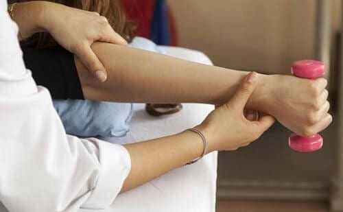
Therapeutic exercises are the primary method of rehabilitation for radial neuropathy.
The exercises are tailored to the patient's condition and the type of nerve damage. The exercises are led by a trainer and then carried out independently.
- The arm bent at the elbow should be placed on a table perpendicular to the surface. The forearm should be parallel to the table top. Slowly and gently separate the thumb and index finger by lowering the thumb and raising the index finger. Perform 10 reps.
- Do not change position. The index finger begins to descend while the middle finger gently rises. 10 standard reps.
- Bring the phalanges of the fingers of your healthy hand to work. Hold it in the palm of your hand and bend it gently. As a final movement, make a fist out of the painful hand with your good hand. Repeat the manipulation 10 times in a row.
The following exercises are considered effective and gentle
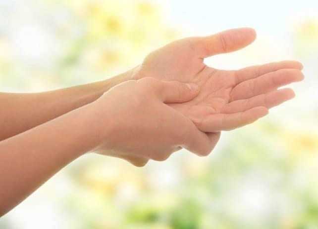
- Massage the phalanges of the affected fingers, gradually straightening them;
- Lift each finger individually, then pull back with your good hand, starting with the thumb;
- Keep the weight as light as possible at first and gradually increase it;
- Rotate each finger alternately and then all at the same time;
- place your hand with your palm on the table, press your fingers together and relax them abruptly, without helping yourself with your other hand.
The improvement in mobility will be noticeable after just a few sessions.
prevention
In order to avoid the development of radial neuralgia of the left or right hand and necrosis, one should not forget about the prevention of the disease. This condition occurs not only after trauma, but also as a result of metabolic disorders. Lack of exercise also contributes to this.
The measures to maintain health are simple and useful for general well-being:
- self-massage of the hands and forearms when fatigue occurs;
- a healthy lifestyle and physical activity, preferably through regular sports training;
- timely treatment of viral and infectious diseases;
- Sleeping in a suitable position that does not compress the limbs;
- adequate intake of vitamins and important micronutrients.
If physical activity is low, e.g. B. by sitting at the computer, you should exercise regularly. Stretch at regular intervals, at least every 25 minutes. Also pay attention to a balanced diet and enrich your diet with B vitamins.
The most important aspect of preventing radial nerve palsies (neuromas) is avoiding trauma. When you play sports, spend time outdoors or do physical work, follow the safety rules.
Welcome!' – a network of clinics for spine and joint diseases
For more than 10 years we have specialized in the non-surgical treatment of musculoskeletal disorders in various stages. ,Hello!' are clinical bases of the country's leading universities that train specialists for the most sought-after professions in medicine. The clinics' doctors are a team of experts from the RUDN faculty who, together with leading Israeli specialists, have developed unique treatment protocols in accordance with the standards of the Russian Ministry of Health.
Symptomatic signs
The distal part of the upper limb (the hand) is very mobile and therefore most susceptible to injury. Most wrist contusions occur from strong blows and compressions.
The injury is accompanied by symptomatic symptoms:
- swelling of tissue;
- Decreased sensation of the palmar surface corresponding to the 1st, 2nd and 3rd;
- Bleeding into the joint cavity and surrounding tissues;
- Radiation of pain syndrome to the fingers;
- Loss of motor function (in severe trauma);
- Subungual hematoma and throbbing sensation (when the metacarpophalangeal joint and nail phalanx are bruised);
- Swelling and discoloration of the injured nail.
Differential diagnosis and subsequent treatment prevent complications caused by bruises in the post-traumatic period.
The nature of radial syndrome and its causes
It is an inflammatory or compressive-inflammatory lesion of the anterior and posterior spinal nerve roots. It first appears at the junction of the roots (indicated by arrows in the photo below).

Most often a single nerve is the problem, but in some cases a pair of nerves or adjacent nerves on one side of the spine may be affected.
The common causes of radicular syndrome include degenerative-dystrophic processes in the spine:
- Osteochondrosis – bulging (prolapse) of the intervertebral disc, significant reduction in its height, formation of osteophytes;
- Deformed spinal osteoarthritis or spondylolisthesis;
- Habitual lateral displacement of the vertebra;
- Complication of a vertebral fracture;
- Neoplastic process.
Radiculopathy is often a complication of infectious diseases. These include influenza, tuberculosis, osteitis and syphilis.
Other causes of sciatica include rheumatism, brucellosis, tick-borne encephalitis and chronic encephalitis.
As a reminder. Sciatica is often confused with lumbago. However, the causes of this type of back pain are different and rather 'trivial'. These include lifting heavy weights, hypothermia, overloading of back muscle groups, sudden movements, muscle spasms due to trauma and poisoning.
Unlike lumbago, radicular syndrome does not appear immediately. It is preceded by a long-term pathological process.
However, it is important to know that untreated lumbago can develop into radiculopathy.
classification
The International Classification of Diseases, Revision 10 (ICD 10) codes radiculopathy as M54.1 and radicular syndrome due to spondylosis as M47.2.

The main terms radiculopathy or radiculitis can be accompanied by auxiliary adjectives or prefixes when clarifying the diagnosis in the patient's anamnesis:
- acute or chronic, depending on the 'age' of manifestation;
- depending on the location of the lesion – radiculoneuritis (inflammation of the root sheath and nerve), meningoradiculitis (inflammation of the middle part of the nerve), myelopolyradiculoneuritis (inflammation of the sheath and the middle part of the nerve);
- depending on the location of the inflamed nerve, most commonly cervical, cervicothoracic, thoracolumbar and lumbosacral.
Since one of the most common causes of sciatica is a herniated disc, there is a separate term for these radiculopathies - the discogenic radicular syndrome (r. discogena). In ICD 10, cervical sciatica is listed as M50.1 and lumbar or other sciatica is listed as M51.1.
Discogenic lumbosacral sciatica, which is associated with pain along the sciatic nerve (see image above), is also coded under M51.1.
Basics
A brush is a tool used to paint various surfaces: walls, ceilings, furniture, windows, doors, etc. It is also used to create effects: textures, gradients and other decorative elements. It has a long handle and bristles. The bristles are made from natural animal hair or synthetic fibers. The handle can be made of wood, plastic or metal. With the right brush, the quality of the paint can be significantly improved and the time required for the paint itself can be reduced. It is a very important tool in home renovations and creative art projects.
There are many types of brushes to buy. Natural brushes, made from the bristles of animals such as pigs or deer, have a great ability to absorb paint and create a smooth surface. However, they are more expensive and require more maintenance than artificial brushes made of nylon or polyester. Depending on the size and shape of the fibers, the brushes are selected for a specific application. A longer brush head is needed for large areas, while a smaller head is better for finer details and corners.
Round Brush – a round brush used for detail and contour painting. It is usually made of natural bristles.
A brush with short, dense bristles for working on large areas. They come in flat or round shapes and are made from synthetic fibers.
Precision brushes are brushes with long bristles that are suitable for working in hard-to-reach areas, such as: B. in corners or other difficult places. They are usually made from natural bristles.
How you choose
Each type has its own characteristics. The choice of brush depends on many factors, including the type of surface, application technique, material, size, shape of the fibers and the characteristics of the handle, which must fit well in the hand. It is also important that the brush must be easy to clean and durable. Some important factors to help you find the right tool for each project: Type of color and material. Natural bristles are better for oil paints and synthetic materials for water-based paints and varnishes. Natural bristles are more expensive, but offer better processing quality. While natural bristles are traditionally the first choice for many professional painters, synthetic materials are becoming increasingly popular because they are lightweight, durable and easy to care for. Size The small size is suitable for detailed work and hard-to-reach areas, while the wide size is better for large areas and quick application. The size is indicated on the packaging. shape Round pens are suitable for circular movements and curved lines. Flat pens are suitable for working on large areas. Macro and cooler are for hard to reach places. processing Brushes should have a firm grip on the bristles, be easy to handle and have a balanced weight distribution.
Even when using such a simple tool, safety precautions must be taken to avoid injury or damage. ✓ Wear safety glasses and gloves and do not raise the brush above head height to avoid paint coming into contact with skin or eyes. ✓ Work in a well-ventilated room or outdoors. Paint fumes are very dangerous! ✓ When painting high surfaces, a ladder or other special equipment is required to climb safely. ✓ Clean the brush thoroughly after use and store it in a place protected from sunlight and moisture.
Read more:- pronation and supination.
- Pronation and supination of the shoulder.
- Pronation and supination in anatomy.
- What is pronation and supination?.
- Pronation and supination of the knee joint.
- Bone structure of the navicular foot.
- Syndrome of the tibial nerve.
- shoulder brace.