The squat/extension test is used to detect a tendon tear.
- tendonitis
- says the CMRT specialist
- Causes of tendinitis
- Anatomical features of the knee joint
- Typology of quadriceps tendon rupture
- Clinical Significance
- Trigger points
- Clinical relevance
- Injuries to the groin
- mechanism of injury
- Pain in the joint
- Drive channel syndrome
- types of injuries
- Symptoms of an injury.
- Functional coordination between the broad muscles of the thigh
- Our courses are suitable for both beginners and experienced trainers and enable higher income and professional development.
- Posterior thigh muscle
- Biceps femoris (m. biceps femoris)
- The main functions of the biceps femoris are:
- Semitendinosus muscle (M. semitendinosus)
- Main functions of the semitendinosus muscle:
- Muscles on the inside of the thigh
- Thin muscle (M. gracilis)
- The main functions of the tibialis are:
- Pectoral muscle (M. pectineus)
- Main functions of the pectoralis major muscle:
- Adductor longus muscle (M. adductor longus)
- Main functions of the adductor longus muscle:
- Short adductor muscle (M. adductor brevis)
- Types of injuries: inflammation, sprains, tendon tears
- Symptoms of an injury to the triceps muscle
tendonitis
A neurologist is responsible for treating this disease.
Tendonitis of the adductor muscle of the thigh occupies a special place among the diseases that affect people with an active lifestyle. The inflammatory process, spreading to the tendon and surrounding tissues, significantly limits motor activity and leads to long-term disability. Only timely medical care and strict adherence to medical advice can quickly suppress the inflammation and prevent recurrence.
says the CMRT specialist
Date Published: June 24, 2021 Date Reviewed: January 13, 2023 All facts have been verified by a doctor.
Causes of tendinitis
- sports injuries
- Excessive physical stress
- Muscle imbalance, weakness of the muscles of the lower limbs
- Intense training without proper preparation
- metabolic disorder
- rheumatism
- immunodeficiency
- infectious diseases
- Flat feet, different leg lengths and other anatomical abnormalities
In addition, tendinitis can develop against the background of taking quinolone and fluoroquinolone antibiotics.
Anatomical features of the knee joint
The quadriceps femoris consists of four small muscles whose tendons are pulled and attached to the post in the area just above the kneecap. The quadriceps and its tendon, the kneecap and its tendon form the extensor system of the knee.
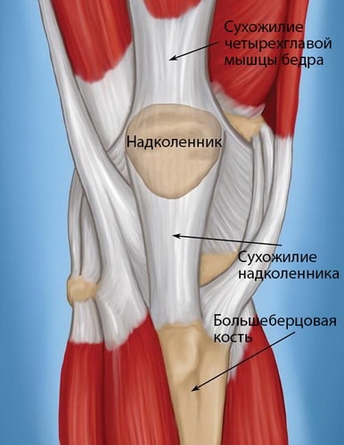
The tendons in the knee area that attach the muscles to the bony areas.
Typology of quadriceps tendon rupture
In medicine, ruptures of the quadriceps tendon are divided into the following two categories:
Partial ruptures. This involves separating some of the fibers while leaving the overall integrity of the tendon intact.
Complete crack. What is characteristic is the complete separation of the tendon into two separate fragments. It is important that in this case the patient completely loses the extension movement of the knee.
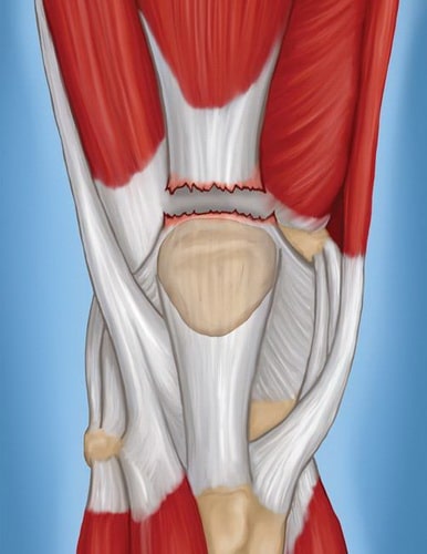
The photo shows the complete separation of the tendon.
Clinical Significance
The role of hamstring tendon activity in knee dysfunction
Cadaveric studies suggest that hamstring tendons relieve pressure on the anterior cruciate ligament (ACL). There is also evidence that people with PCC dysfunction may experience difficulty during certain activities, such as: B. when climbing, have increased activity of the hamstring. Decreased hamstring strength is also associated with poor functional performance in people after a patellarectomy.
Strengthening the hamstring can prevent lower back injuries during the following activities Lift Exercise?
Improper flexion technique when lifting has been linked to lumbar spine injuries. Flexion of the knee joints during lifting reduces the external bending moment in the lumbar spine. However, this leads to flexion moments in the hip and knee joints, which must be compensated for by the extensor muscles of these joints. The role of quadriceps femoris strength in limiting the ability to lift weights is well established. However, because the hamstring muscles are important hip extensors, they can also help a person bend and lift properly. However, the role of adductor weakness in limiting the ability to bend and lift weights is not well understood. Further research is needed to determine the relationship between Achilles tendon strength and weightlifting ability and to determine whether Achilles tendon training can improve weightlifting ability. It is also necessary to investigate how strengthening the hamstring muscles affects the occurrence of lumbar spine injuries.
Trigger points
Consequences of hamstring weakness
Tendon weakness leads to a significant loss of knee flexion strength. However, flexion of the knee joint in an upright position is often a consequence of the weight of the limbs and is controlled by the eccentric contraction of the quadriceps muscle of the thigh. Therefore, weakness in knee flexion in an upright posture leads to mild impotence. In contrast, weakness in hamstring strength can lead to greater functional impairment in the hip joint, where the hamstrings provide much of the pulling force. The weight that creates a flexion moment in the knee joint during a squat creates a flexion moment in the hip joint. This flexion moment is balanced by the extension moment, which is at least partially generated by the tendons. Consequently, hamstring weakness can cause significant difficulty in bending and lifting.
Consequences of a hamstring tendon strain
The consequences of a hamstring strain are complex because all muscles except the short head of the biceps femoris cross in both the hip and knee joints. As with the rectus femoris muscle, the two-joint structure must be taken into account when assessing hamstring tightness. Hamstring tension results in limited extension amplitude in the knee joint when the hip joint is flexed or limited flexion amplitude in the hip joint when the knee joint is not flexed. A study of more than 200 people without lower limb dysfunction found a mean hip flexion amplitude of 68.5° ±6.8° in men and 76.3° ±9.5° in women, with the knee joint not flexed. When the hip joint is flexed, the tendons relax slightly, allowing full extension of the knee joint. If the position of the hip joint has no influence on the amplitude of knee joint extension, then any limitation in the amplitude of knee joint extension is due to tensions in the unicompartmental structures such as the joint capsule and ligaments of the knee joint.
Clinical relevance
Injuries to the groin
A sprain or injury to the adductor major muscle is considered the most common form of groin strain or injury. This problem occurs mainly in track and field athletes, hockey and football players (ie mainly men). The cause of groin pain is difficult to diagnose because several muscles are involved: the hip muscles, the adductors and the glutes, as well as the vicinity of the pelvis, hip joint and sacrum.
mechanism of injury
- The mechanism of adductor strain is thought to be related to the need to decelerate the lower limbs quickly during rapid abduction and external rotation, such as occurs during ice skating or rapid changes of direction. When skating, the body needs to be stabilized more at the hips and hip joint due to the thin shoulder on which the skaters have to balance. In addition to the instability inherent in skating, these athletes use explosive movements to extend, retract, and rotate the hip and rely on eccentric contraction of the propulsion muscles to slow the leg during the stride. These repeated eccentric contractions of the adductors during fast and slow skating can lead to adductor inflammation.
- The main symptoms after an injury are pain, swelling, any activity that requires bringing the knees together is painful, and outward movement of the hip is limited by the spasm and pain.
- Complete tears are rare.
Pain in the joint
Tight adductors can cause knee pain, especially in runners. The function of the adductors is to bring the hips together, internally rotate the thigh, and stabilize the hip. The adductor major muscle has a relatively mixed ratio of muscle fiber types, although with a larger proportion of type I muscle fibers. The postural fibers (type 1) tend to shorten with chronic overload. These muscles may be torn at their origin on the pelvic bones or in their core.
Drive channel syndrome
This is a rare cause of acute arterial occlusion in young men. It is the result of compression of the artery (superficial femoral artery) by an abnormal musculotendinous pull that originates from the adductor major muscle and lies next to and above the adductor tendon. The pathogenetic mechanism of this syndrome is similar to that of a pinched popliteal fossa and can occur after exercise.
Because this syndrome occurs in young men in whom acute arterial occlusion can result in loss of limbs, it is important to recognize the presence of obvious ischemic symptoms after exercise in a healthy young man. Treatment consists of separating this abnormal traction and restoring arterial blood flow through appropriate measures. Detecting bilateral lesions can help prevent future problems, even if symptoms are unilateral.
types of injuries
These tears can occur where the tendon is attached to the bone, where the muscle meets the tendon, or in the tendon itself. When small parts of the bone and tendon are torn off, it is called a burst fracture.
The following factors can lead to a biceps injury:
- Excessive force on the arm during sports, lifting and carrying heavy objects.
- Age-related changes (tendons lose strength in people over 40 years old)
- Chronic inflammation in this area (rheumatoid arthritis, impingement, elbow bursitis, etc.) leads to tendon deterioration and therefore increases the risk of injury even after minor injuries.
- Taking certain medications (e.g. statins)
- Microtrauma
Symptoms of an injury.
- Sharp pain at the time of injury.
- Clicking, popping noise.
- Muscle cramps and spasms (sprains).
- Swelling and swelling
- Possible subcutaneous bleeding
- Relaxation of the muscles when flexing the biceps (upper third of the arm)
- Spherical thickening in the upper arm
- Decreased strength of the extremity
The diagnosis of a biceps brachii injury includes
Functional coordination between the broad muscles of the thigh
The myoelectric balance of the quadriceps muscle is essential for normal movement of the kneecap.
Proprioceptive muscle afferents help maintain correct posture. Recent studies suggest that activation of these afferents enables the contralateral quadriceps muscle to improve its coordination and thus postural balance.
The PM of the thigh can activate its fibers longitudinally. It can also activate the proximal fibers when the distal fibers do not contract. When the quadriceps remains active, the most distal fibers are activated while the most proximal fibers are not activated (likely due to a mechanism that delays the onset of fatigue).
The contribution of the hamstring muscle to the patellar tendon is small, so it is unable to generate sufficient force to medially stabilize the patella during knee extension. Rather, as it contracts, it stretches the SMP femoral aponeurosis, thereby counteracting the lateral forces on the patella generated by the femoral LSM. The LSM of the femur acts as an indirect stabilizer of the patella by concentrating its force on the medial axis of the femur.
The force generated by the LSM of the thigh increases as the knee flexion angle increases. This mechanism is due to the length of the fibers compared to the connective tissue structure of the muscle. Longer fibers have greater strength and better elasticity or resilience of connective tissue. When the knee is extended, the LSM of the hip generates a small force to maintain the position with minimal effort.
Our courses are suitable for
Suitable for both beginners and experienced trainers, enabling higher income and professional development.
The User purchasing the Services on evotren.ru, hereinafter referred to as the 'Customer', on the one hand, and Evotren LLC, hereinafter referred to as the 'Contractor', represented by the CEO FG Kapishev, acting within the framework of the Charter, on the other hand, have entered into this Agreement (hereinafter referred to as the 'Agreement') to purchase services from the Contractor as follows
1. TERMS AND DEFINITIONS USED IN THE AGREEMENT
1.1. customer – a natural person, a sole proprietorship or a legal entity, regardless of its legal form, who has placed an order with the Contractor under the terms of this Agreement by purchasing the Services of the Contractor.
1.2. contractor – is the legal entity providing services to the Client under the Agreement.
1.3. Services – Services consisting in providing access to the study of the distance learning materials indicated in the description of the information courses.
1.4. site – The contractor's information resource, located on the Internet at: edu.evotren.com
1.5. The contractor's personal account – The program interface on the website for studying the informational material and other necessary information, accessible to the customer after authorization by a login and password.
1.6. Order – An automatically generated document detailing the services requested by the customer. The order is placed by filling out the necessary forms on the contractor's website -www.evotren.ru.
1.7. Acceptance of the terms of the contract – The acceptance of the contractual conditions by the customer takes place by paying for the services in cash or by non-cash or electronic means of payment. Acceptance of the terms and conditions of the contract is deemed to have occurred when the customer has paid for the services in cash or by non-cash or electronic means of payment.
Posterior thigh muscle
These muscles are grouped together under the name biceps femoris. These muscles determine the shape of the back of the thigh, its curve. They also play a role in filling the space between the thighs.

Biceps femoris (m. biceps femoris)
A long, spindle-shaped muscle that extends across the back of the thigh. As the name suggests, it consists of two heads: a long head and a short head. The long head attaches its upper end to the ischial tuberosity of the pelvic bone and its lower end to the shinbone (tibia). The short head attaches its upper end to the back surface of the thigh bone and its lower end to the shinbone (tibia).

The main functions of the biceps femoris are:
Shin flexion (flexion of the leg at the knee)
Hip extension (pulling the hip back or straightening the torso from a flexed position)
M. The biceps femoris plays an active role in flexing the leg, in any movements that require the hip to be retracted, and in pulling the trunk from the flexed position.
Lack of mobility and strength of the biceps femoris muscle often causes back pain, poor posture and knee joint problems.
Semitendinosus muscle (M. semitendinosus)
A long, flat, downward-running muscle that lies medial (toward the middle of the body) to the biceps femoris. The upper part of the muscle attaches to the ischial tuberosity of the pelvic bone. The lower part attaches to the shinbone (tibia).
Main functions of the semitendinosus muscle:
Hip extension (pulling the hip back or pulling the body out of a flexed position)
Knee flexion (bending of the leg at the knee)
M. The semitendinosus muscle actively participates in flexing the leg, in all movements that require the hip to be retracted, and in pulling the body out of the flexed position.
Muscles on the inside of the thigh
These muscles are commonly referred to as adductors. Their main job is to bring the femur bone inward.

Thin muscle (M. gracilis)
The gracilis muscle is a long band muscle that rises above the other muscles on the inside of the thigh. Its upper part is connected to the pubic bone and its lower part to the shinbone (tibia).
The main functions of the tibialis are:
Adduction of the hip (pulls it inward)
M. The gracilis actively participates in all leg movements: running, walking, squatting, maintaining body balance.
Pectoral muscle (M. pectineus)
A flat muscle that attaches its upper end to the pubic bone and its lower end to the inside of the middle part of the thigh.
Main functions of the pectoralis major muscle:
Hip extension (pulls the hip inward)
Hip flexion (pulls the hips toward the body).
M. Pectineus actively participates in all leg movements: running, walking, squatting, maintaining body balance.
Adductor longus muscle (M. adductor longus)
The adductor longus muscle is a flat, thick muscle. It attaches its upper end to the pubic bone and its lower end to the inner, medial part of the femur.

Main functions of the adductor longus muscle:
Adduction of the hip (pulls it inward)
M. The adductor longus muscle actively participates in all leg movements: running, walking, squatting, maintaining body balance.
Short adductor muscle (M. adductor brevis)
A short adductor muscle that is flat and extends downward. Its upper end is attached to the outside of the body and to the pubic bone. The lower (wide) end is attached to the inside of the thigh.

Types of injuries: inflammation, sprains, tendon tears
A tendon tear can occur at the point where the tendon attaches to the bone, at the point where the muscle attaches to the tendon, or in the tendon itself. When small pieces of bone separate along with the tendon, it is called a tear Tendon rupture.
The main causes of triceps brachii tendon injuries include:
- Excessive physical activity
- Local inflammatory processes (deep scratches, abrasions) cause inflammation of the tendon.
- People older than 40 years
- Chronic diseases (arthritis, bunions, etc.).
- Taking corticosteroid medications
- Mechanical injuries
Symptoms of an injury to the triceps muscle
- Pain and swelling on the back of the elbow (characteristic of tendinitis)
- When stretching or flexing the joint, the patient tightens the muscle, causing the tendon to stretch and causing pain
- Characteristic clicking sound
- Muscle cramps (sprains)
- nodules under the skin
- swelling and redness of the skin,
- Painful palpation of the tendon
- Decreased limb strength.
Inflammation and sprain of the tendon of the triceps muscle is treated conservatively:
- Immobilization of the injured tendon (interruption of all pain-causing activities)
- Applying cold to the painful area.
- Use of non-steroidal anti-inflammatory drugs
- Use of orthoses and splints to relieve the tendon and promote its regeneration.
- Physiotherapy (electrophoresis, ultrasound, cold therapy, etc.).
- Exercise therapy (recommended once inflammation and pain have resolved)
If a tendon ruptures, surgical treatment is recommended. During rehabilitation, heavy physical work and sports should be avoided. The patient is recommended to wear an immobilizing bandage, do physiotherapy and carry out other therapeutic exercises under the guidance of the attending physician.
Timely and qualified treatment is the key to a speedy recovery. If left untreated, these injuries can lead to irreversible changes to the tendon tissue.
Qualified medical care can be obtained in Moscow at NCC #2 (Central Clinical Hospital of the Russian Academy of Sciences).
Read more:- The flexor muscles of the foot.
- Tibialis posterior muscle.
- lower leg flexion.
- Exercises for the triceps tibialis muscle.
- Which muscle extends the lower leg and flexes the thigh.
- Square soleus muscle.
- Pronator - what does that mean?.
- abdominal muscle.