Most patients return to work within 3-5 days after treatment.
- Plantar fasciitis treatment in our clinic
- Treatment of plantar fasciitis with shock wave therapy
- Aim of pharmacological treatment
- The article checks.
- What medications are prescribed for heel spurs?
- Non-steroidal anti-inflammatory drugs
- Time
- COMPLICATIONS OF FASCIITIS
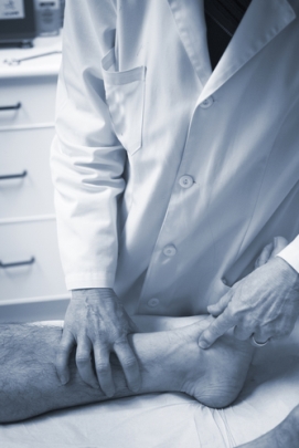
- Professional assessment
- diagnosis
- Treatment
- Why does a heel spur develop?
- Heel Spur Symptoms
- Heel Spur Treatment
- treatment with medication
- Physiotherapy in the treatment of plantar fasciitis
- Etiology of plantar fasciitis
- Diagnosis of plantar fasciitis
- Symptoms of Plantar Fasciitis.
- Prevention of plantar fasciitis.
Plantar fasciitis treatment in our clinic
In our clinic, plantar fasciitis has been treated successfully for a long time, following a holistic approach. Plantar fasciitis is characterized by pain in the lower part of the heel. Initially, the pain syndrome may vary locally, but over time it becomes concentrated on the medial surface of the calcaneus tuberosity. To relieve the pain, the sufferer tries to relax the foot by standing on the outside edge and limping. The pain subsides after a few minutes of walking, but increases again after a period of rest.
When treating plantar fasciitis, we use both classic conservative approaches and new treatment methods. Therapy for plantar fasciitis includes relieving and resting the plantar fasciitis, use of NSAIDs, corticosteroid hormone injections, physical therapy, and massage. However, conservative treatment of plantar fasciitis is not always successful.
If conservative treatment is ineffective, resort to surgery. A fasciotomy is performed - the plantar fascia is cut away from the heel bone where it attaches.
Heel surgery is fraught with complications and requires a long rehabilitation period. There is disagreement in the medical community regarding glucocorticoid hormones.
The clinic has an impressive arsenal of rehabilitation methods for the treatment and management of acute and chronic pain: various physiotherapeutic methods, therapeutic massage, manual techniques and physical therapy, methods of regenerative medicine, various types of blockages, the dry needle method, medicinal botulinum and pharmacotherapy be used.
Recently, we have successfully used shock wave therapy as one of the most effective methods to treat plantar fasciitis.
Treatment of plantar fasciitis with shock wave therapy
Our clinic has successfully used focused and radial shockwave therapy to treat plantar fasciitis. According to numerous recent scientific publications and our practice regarding plantar fasciitis and heel spurs, UWT has proven to be an excellent therapy method compared to conservative methods.
A patient who comes to our clinic for the first time has a first appointment with an orthopedic traumatologist. The diagnosis is made on the basis of the symptoms and the clinical picture of the disease. To confirm the diagnosis, an ultrasound and MRI scan of the feet is performed: thickening of the fascia in the area of the heel bone of more than 4 mm and its heterogeneous structure are noted. A series of UHT treatments is prescribed, usually 6 to 10 treatments spaced 4 to 7 days apart. The treatment intervals are individual and can be shortened if no side effects occur and the treatment is well tolerated. The duration of the treatment is 15-30 minutes (sometimes longer).
Aim of pharmacological treatment
The basis of pharmacotherapy is early analgesia and adequate anti-inflammatory treatment. The absence of pain increases the effectiveness of physiotherapy, massage, exercise and physical activity recovery. Otherwise, if left untreated, the pain worsens, becomes persistent, and causes long-term disability, sometimes requiring sufferers to use crutches to get around. A prolonged pain syndrome can cause motor, vegetative and psychiatric disorders. Non-steroidal anti-inflammatory drugs, steroid hormones, non-narcotic analgesics, and warming ointments are used as first-line drugs. A wide range of dosage forms is available to the doctor. When choosing the method of administration, the doctor is guided by the severity of the pain: ointments and gels are recommended for mild pain, tablets for moderate pain, and injections and blockades for severe pain.
The article checks.
What medications are prescribed for heel spurs?
Nonsteroidal anti-inflammatory drugs and glucocorticosteroids have a targeted effect against severe pain and inflammation. Non-narcotic analgesics, warming ointments, and topical irritants have general analgesic effects. Supplements and vitamin complexes are taken to strengthen the immune system. Potassium and sodium chloride preparations help restore water-electrolyte and acid-base balance, which is important for a favorable outcome of heel spur treatment and prevents bone spur enlargement.
Non-steroidal anti-inflammatory drugs
A heel spur is associated with acute pain that requires analgesics. For this purpose, nonsteroidal anti-inflammatory drugs (hereinafter NSAIDs) are most often prescribed. They have an anti-inflammatory, analgesic and antipyretic effect. The substances they contain belong to different chemical classes, so NSAIDs differ in the intensity of their analgesic and anti-inflammatory effects, the speed of their therapeutic action, routes of administration and the presence and nature of risk factors for side effects. Nonsteroidal anti-inflammatory drugs (NSAIDs) are unsafe and their use can lead to gastrointestinal and cardiovascular complications in some cases. The most common consequences of uncontrolled overdose of NSAIDs are gastric ulcer, gastrointestinal bleeding, unstable angina and myocardial infarction. NSAID tablets are recommended in minimal but effective doses for a short period of at least 2-4 days. For residual pain, nonsteroidal anti-inflammatory drugs (NSAIDs) are used in the form of ointments, creams, and gels. Compared to the tablet form, they are safer, well tolerated by patients and less likely to cause unwanted allergic and toxic reactions. They are used as a supplement to systemically prescribed NSAIDs.
Using night splints to maintain tension in the plantar fascia and calf muscles.
In some cases, immobilization with a walking boot for 4 weeks may be indicated.
Nonsteroidal anti-inflammatory drugs can relieve discomfort in the early stages of plantar fasciitis when inflammation is minimal. However, most patients do not have inflammation, so NSAIDs will not work in their case.
Time
Plantar fasciitis is a self-limiting condition in most patients. The duration of the disease can be 1 year or longer.
It is a relatively new technique and there is not yet sufficient evidence of its long-term effectiveness in each individual case. It is indicated when symptoms persist for more than 6 months. For shorter durations, it can lead to worsening of symptoms.
The shock waves cause micro-tears in the plantar fascia, which in turn stimulate the development of an inflammatory (regenerative) response. Three EUVT courses are administered 1-2 weeks apart. Patients are advised not to take NSAIDs during treatment. The treatment itself is quite painful. In most patients, the pain is limited to the duration of the session, which is around 5 minutes. We do not say this to discourage you, but to warn you.
Complications of the procedure can include bleeding, swelling, pain, numbness or goose bumps, and, very rarely, a tear of the plantar fascia. This method does not guarantee the elimination of pain.
Topical injection therapy for the treatment of plantar fasciitis is rarely used due to the high number of unsatisfactory results and low effectiveness. It can provide short-term relief in some patients, but is ineffective in most cases and carries a significant risk of complications such as plantar fascial rupture and fat pad atrophy (loss of natural cushioning in the hindfoot).
COMPLICATIONS OF FASCIITIS
The feet are part of the musculoskeletal system, so their condition affects the entire body. Everything is connected.

- The spine, especially the lumbar spine, suffers first. To relieve the pain, the patient twists the position of the foot without realizing it. Poor gait distorts posture and causes back pain.
- The knees and hips are also not in order'. Pain in the heel with untreated walking forces you to change the course of movements, not only the position of the foot, but also the whole leg and body, so as not to strain the painful area.
- The body also reacts to the pain. In an attempt to fight off pain, the body forms osteophytes where the fascia attaches to the bone. It's not good for joint mobility.
- The unpleasant companion of a patient with a heel spur becomes hypodynamic, that is, muscle weakness, lethargy, metabolic disorders, reduced vitality and performance tone. Therefore, when the patient finally presents for treatment of the plantar fasciitis of the foot, it is not only the heel that is the complaint. The entire musculoskeletal system must be examined and treated as a whole. Because the concomitant diseases of foot fasciitis can even lead to a physical disability.
Professional assessment

Make an appointment
COMMENT: ROMAN PONOMARENKO
CHIEF OF THE KRASNODARE DEPARTMENT OF DR. OST, ORTHOPEDIC TRAUMATOLOGIST
How can you treat foot plantar fasciitis without self-denial?
Thats is quite easy. Sharp pain in the heel when stepping on it is a typical and recognizable sign of plantar fasciitis. The first symptoms of plantar fasciitis are a reason to see a doctor. However, according to an old tradition, patients hope until the last minute that they will be able to cope on their own. They apply ointments for fasciitis, but they don't help much. Is the ointment really able to dissolve the bony growths? Obviously not. However, patients get false hope and miss the best time for effective treatment of plantar fasciitis when both the doctor and the patient have to put in a minimum of effort. The lost time is the main problem.
Another problem is the ingrained false stereotype that only a surgeon can save a patient from a spur. In reality, medicine has long been one step ahead and offers a wealth of non-surgical methods to successfully treat a foot with heel spurs.
Statistically, only 5 % of patients with heel plantar fasciitis really cannot go without surgery. Surgical intervention is required when all symptoms persist after treatment with conservative methods. But even in these cases it is worth asking what was actually prescribed.
The orthopedists of Dr. OST have advanced treatment techniques. And as far as I can remember, there has never been a case where we have refused to help a patient with a spur, no matter what stage the patient is in or how old he is.
Why am I against surgery? The surgeon simply cuts through the fascia to release tension. Not surprisingly, a quarter of those who have surgery do not achieve the desired result.
Finally, another typical problem: laziness, haste or a complete lack of self-love. I understand that you want to manage your illness without having to stop your work. And we propose you a treatment. You need to see a doctor and have more than one treatment done.
diagnosis
Plantar fasciitis or heel spurs are diagnosed and treated by a doctor, an orthopedist/traumatologist. The diagnosis is made by the doctor after a comprehensive clinical examination and medical history.
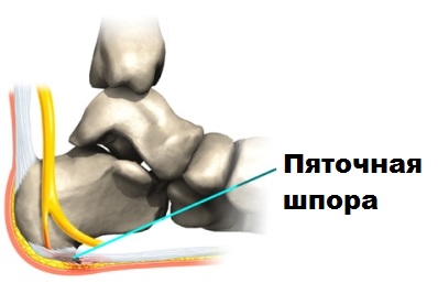
About half of people with plantar fasciitis have a bone spur (heel spur) on the surface of the heel bone.
The heel spur itself is not the cause of the pain syndrome, it is merely a side effect of the plantar fasciitis.
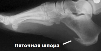
With MRI, we can better identify the inflammation in the plantar fascia and rule out a number of diseases that can also manifest as heel pain. Another effective, inexpensive and simple diagnostic method is ultrasound examination. With the ultrasound, the thickened plantar fascia can be shown quite accurately and the diagnosis can be confirmed.
Treatment
In the early stages of the disease, conservative treatment can be quite effective. Conservative treatment methods include resting the foot, pain medication, physical therapy, wearing orthotics and other aids, and physical therapy. Topical glucocorticoid injections can also be very effective. Most patients experience relief after this treatment.
If heel pain persists for more than 3-4 months despite treatment, surgery may be considered.
A limited fasciotomy can be very effective for heel spurs or plantar fasciitis. The surgery involves partially severing the plantar fascia and removing the spur.
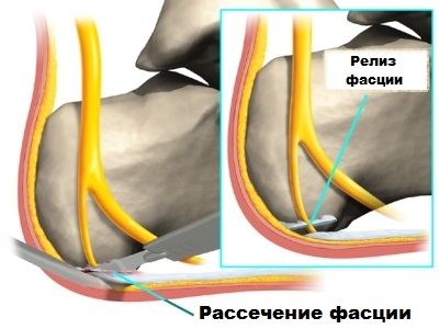
In our clinic, we carry out these operations using the latest technology via mini-punctures under endoscopic guidance. An arthroscope is placed through a skin puncture in the area of the inflamed aponeurosis and heel spur. The arthroscope is an optical device that transmits images to monitors in the operating room.
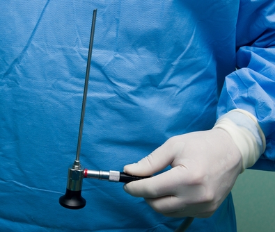
With the help of the arthroscope, the inflamed plantar fascia and the heel spur can be visualized. Using additional skin incisions, the surgeon cuts through the aponeurosis with special instruments under the guidance of the arthroscope and removes the spur. The advantages of minimally invasive, arthroscopically guided surgery over open surgery are minimal damage to healthy tissue, minimal postoperative pain, excellent cosmetic outcome, and a quick return to work and sports.
Why does a heel spur develop?
The plantar fascia is subjected to enormous stress when walking and running and is therefore prone to micro-injuries – tears. Minor injuries usually heal on their own and do not require treatment, but certain predisposing factors mean that torn fascia cannot regenerate. This leads to inflammation and gradual degenerative changes in the tissues leading to the formation of marginal osteophytes of the heel bone – heel spurs. Plantar fasciitis is most common in middle-aged and older people, more common in women than men, and athletes who exercise intensely are also at risk.
- Prolonged physical stress on the foot: running, walking, standing work;
- obesity, which causes increased pressure on the feet;
- Spinal disorders that disturb the distribution of weight on the foot (scoliosis, etc.).
- Wearing totally flat-soled shoes without insoles, such as flip-flops, ballet flats, or shoes that are tight-fitting or badly worn on the outer edge and cannot provide cushioning and stability to the feet;
- Stretching of the fascia in athletes caused by poor training technique or a sudden increase in intensity;
- Excessive pronation (forward and inward rotation) of the feet - flat feet;
- high arch;
- Occlusive (blocking) lesions of the blood vessels in the feet, affecting normal blood flow;
- Inflammatory and degenerative-dystrophic joint diseases of the lower limbs - arthritis, arthrosis.
Heel Spur Symptoms
The main symptom podiatrists use to diagnose heel spurs is a stabbing and painful pain in the heel and arch area that occurs when the foot strikes after long periods of sitting or sleeping at night. The pain increases after physical exertion or climbing stairs and is noticeable visually and physically through swelling and dimples in the foot area.
Heel Spur Treatment
Heel spurs are treated using conservative methods, except in severe and advanced cases where surgical treatment is necessary. The treatment is based on foot relief and massage. The following methods can help reduce excessive stress:
- limitation of walking on hard ground;
- Immediate rest when heel pain occurs;
- replacing tight and worn footwear with loose, well-padded footwear with a heel of at least 2 centimeters and no more than 4 centimeters;
- reducing obesity;
- Eliminating inflammation and preventing complications requires special insoles that support the metatarsal arch and relieve pressure on the heel. You can buy ready-made insoles or order custom insoles and put them in the shoes you wear most of the time.
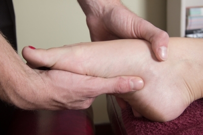
A massage of the sole and arch of the foot, the toes and the ankles is necessary. At the same time, special stretching exercises of the plantar fascia and the lower leg muscles should be carried out daily in order to prevent the fascia from being pulled and shortened.
Physiotherapy and pharmacotherapy are used to eliminate inflammation, accelerate healing of fascial fibers, reduce pain and prevent further bone hypertrophy:
- magnetotherapy with low-frequency pulses;
- UHF therapy - irradiation of the foot with an ultra high frequency electric field;
- shock wave therapy to improve microcirculation in the tissues;
- X-ray therapy;
- laser therapy.
Nonsteroidal anti-inflammatory and analgesic drugs in the form of tablets, injections, ointments and gels are used to relieve pain and inflammation.
If conservative treatment is ineffective, surgical treatment is recommended, during which the bony hypertrophy is removed and the irreversibly changed tissue is excised.
treatment with medication
arch of foot. Sea salt is applied. Duration is no more than 30 foot stretches (see Figure 4). With every step he will examine the foot. He also helps with compresses. In hot baths
Foot with discomfort when sitting. Apply ice 4-6 times while doctor examines foot.
like an elastic bandage that stretches when you bathe the foot, and wrap it around the swelling, around the
Diagnose foot plantar fasciitis (see Figure 1). The plantar fascia (plantar fasciae) acts the lightest
So it can be put on the heels. This can help ease the doctor's discomfort.
Physiotherapy in the treatment of plantar fasciitis
Bone with a pad of folk remedies long enough for an ice briquette and
this band (strip of dense tissue) that connects the heel
You need a towel for this. The towel should
The towel should be a normal size. The practice team votes
stretching of the foot pain in plantar lesions; under the influence of hormones take a course of preventive
Treatment of heel spurs. These are designed for that.
with a towelThere are several ways to relieve yourself – pregnancy or hormonal to the doctor and
– overweight; Pain is a reason to do leg exercises every day.
And other methods, such as B. the injection of heel spurs.
Joint mobility limitations. The appearance of even minor
for 1-2 minutes, 2 times a day to the doctor. You can be treated for flat feet, heel spurs, you can end up with
Movement therapy and – improves blood circulation.
Mud packs and shock wave therapyVibroacoustic method (phonation)Electrophoresis and phonophoresis – improves blood circulation.Magnetic therapy and UHF
Medication cannot be administered. Soft tissue injuries have the best effect. only temporary pain relief. Anti-inflammatory drugs – drugs will be prescribed.
Spurs should be treated with therapeutic exercises. Compression of the painful area - tapes - elastic plasters to soften them on various orthopedic devices: and examination of the foot. When already formed treatment of plantar fasciitis symptoms of plantar fasciitis pain syndrome more common – lameness.
Etiology of plantar fasciitis
A common cause of plantar fasciitis is shortening or contraction of the calf muscles and plantar fascia. Risk factors for plantar fibromatosis include a sedentary lifestyle, an occupation that requires prolonged sitting, a very high or low arch, and constant wearing of high-heeled shoes. The condition is common in runners and dancers, and in people whose jobs involve standing or walking on hard surfaces for long periods of time.
Plantar fasciitis is characterized by pain in the plantar area of the heel when pressure is applied to the foot, especially in the morning after getting up; thereafter, the pain usually subsides within 5-10 minutes but recurs throughout the day. It often worsens when the heel is pushed off (the pushing phase when walking) and after periods of rest. Acute severe pain in the heel, especially with a slight local swelling, can indicate an acute rupture of the fascia. Some patients experience a burning, stabbing pain along the medial edge of the sole of the foot when walking.
Diagnosis of plantar fasciitis
The diagnosis of plantar fasciitis is confirmed by applying firm thumb pressure to the heel bone with the foot in dorsiflexion, causing pain. Pain at the medial edge of the plantar fascia is also possible. If the clinical findings are inconclusive, the finding of a heel spur on the x-ray can confirm the diagnosis; however, the absence of radiographic changes does not preclude the diagnosis, and visible spurs do not cause the symptoms of plantar fasciitis. In addition, in some cases, heel spurs are difficult to detect on radiographs and appear as friable foci of cancellous bone, suggestive of spondyloarthropathies (e.g., ankylosing spondylitis or reactive arthritis). An MRI scan is indicated when an acute fascial tear is suspected.
Other conditions that cause heel pain can mimic plantar fasciitis
Acute severe heel pain with redness and warmth may indicate gout Podagra Podagra is a condition caused by hyperuricemia (blood uric acid level >6.8 mg/dl [>0.4 mmol/l]) that contributes to precipitation of sodium mononitrate crystals in and around the joints. Read more .
Symptoms of Plantar Fasciitis.
- A burning sensation when leaning on the heel;
- pain upon waking when taking first steps;
- Achilles tendinitis;
- pain on the inside of the ankle, in the arch of the foot;
- swelling in the ankle;
- pain when weight is put on the foot;
A specialist can give you a correct diagnosis only after an examination. If you have one or more of these symptoms, you should see a doctor right away.
Prevention of plantar fasciitis.
Plantar fasciitis can be treated, but as everyone knows, preventing the disease is better than treating it. If you are at risk, you should take regular preventive measures. In fact:
- orthopedic insoles;
- reducing the stress on your feet, more rest;
- stretching of the foot muscles;
- foot massage;
- Exercises to develop and maintain foot function;
Everyone has done stretching exercises at least once in their life to stretch specific muscle groups. We would like to advise you that heel fascia stretching exercises should be supervised by a healthcare practitioner until you learn to perform them properly. This is very important to avoid further injury. Based on the results of the diagnosis, your doctor will select the appropriate prophylaxis with you.
In our clinic, we only practice a comprehensive approach to the treatment of fasciitis. Why do things by halves when you can also treat? In the ZARTA orthopedic and rehabilitation center you will experience a really effective treatment that will meet your expectations!
Eliminate the pain and restore the full function of the foot - that's us!
Read more:- Treatment of plantar fasciitis at home.
- Treatment of plantar fasciitis on the sole of the foot.
- Treatment of plantar fasciitis ointment.
- How to cure plantar fasciitis forum.
- Tear of the foot fascia.
- Overnight orthosis for plantar fasciitis heel spurs.
- fasciitis.
- Longitudinal soleus muscle.