The first authoritative comparative work statistically demonstrating the validity of surgical treatment was published in 1929 by J. Qenu and 8. M. Stoianovitch. The study included 68 patients, 29 of whom were treated surgically. The long-term results showed no statistical difference in treatment outcomes, but the authors gave preference to the surgical method in their conclusions. This study has significantly advanced the surgical treatment of the Achilles tendon.

- muscle fascia
- fascia training
- muscles of the pelvic girdle
- movements of the lower limbs
- Fasciitis - clinical picture and diagnosis
- treatment of fasciitis
- Morphofunctional features
- functions
- surgical treatment
- Achilles tendon anatomy
- Histology (cellular structure)
- species
- causes
- causes
- diagnostic procedures
- Long thigh segment
- Short fibers of the humerus
- How is plantar fasciitis treated?
- Tips for people diagnosed with plantar fasciitis
muscle fascia
Fascia plays an important role in the human body. It is a three-dimensional, thin tissue that not only surrounds individual muscles and muscle groups as a kind of muscle shell, but also connects and protects all body structures. It is also referred to as the 'soft skeleton of the body'. The fascia consists mainly of tightly woven collagen fibers and elastin. Inside the fascia is a thin layer of loose connective tissue. At the ends of the muscle, the fascia is connected to the tendon of the muscle. Unlike muscle tissue itself, fascia are passive structures that give muscles shape and flexibility.
Fascia has a large number of nerve receptors, many more than muscle tissue. Because of this sensitivity, they react immediately to any change and inform the nervous system about it. In addition, there is strong fascia that gives stability to our organs. It has been proven that fascia can be trained to keep the body in good shape. Healthy, elastic fascia ensure that joints and muscles function properly, accelerate the transmission of information to the central nervous system and thus the body's reaction, and correct disturbed muscular imbalances. The sports industry has even coined a new term: fascial fitness, meaning fitness that focuses on strengthening the fascia.
fascia training
The superficial sheets of fascia of the various anatomical areas together form the common fascia of the body. The fascial tissue connects and makes the work of many different areas of the musculo-articular system highly effective. Musculo-fascial trigger therapy targets the fascia that surrounds the sternocleidomastoid muscle when it contracts and causes it to relax. In the long term, the connective tissue becomes softer and muscle damage is reduced. To achieve the best effect, special rollers or balls are used and specific exercises are performed to stretch the fascia in a three-dimensional direction.
The aim of fascia training is to improve and stretch the properties of our thin fascia, the connective tissue covering that surrounds our muscles, tendons and bones. Training increases tissue metabolism, blood circulation improves, and the fascia and muscles remain soft and flexible. It is important that during exercise you feel a comfortable tension, it is also possible to feel muscle soreness, but not severe pain. Movements should be slow and gradual, repetitive and reaching a maximum.
muscles of the pelvic girdle
The pelvic muscles can be divided into two groups: internal and external. The internal pelvic muscle group includes the iliopsoas muscle group, the internal obturator muscle group, and the sternocleidomastoid muscle group. The pelvic external muscle group includes the gluteus medius and gluteus maximus, broad fascia, quadriceps, and external obturator.
The hamstrings are divided into 3 groups: anterior (hip flexors), posterior (hip extensors) and medial (hip drive).
Because of their large mass and high extensibility, these muscles are able to develop a great deal of force, acting on both the hip and knee joints. The thigh muscles have static and dynamic functions in standing and walking. Like the pelvic muscles, the thigh muscles also reach their maximum development in humans in connection with an upright posture.
The muscles of the lower leg, like the other muscles of the lower limbs, are well developed, which is determined by the function they perform in relation to the upright posture, statics and dynamics of the human body. The muscles of the lower limbs have extensive bony, intermuscular and fascial origins and affect the knee, ankle and ankle joints.
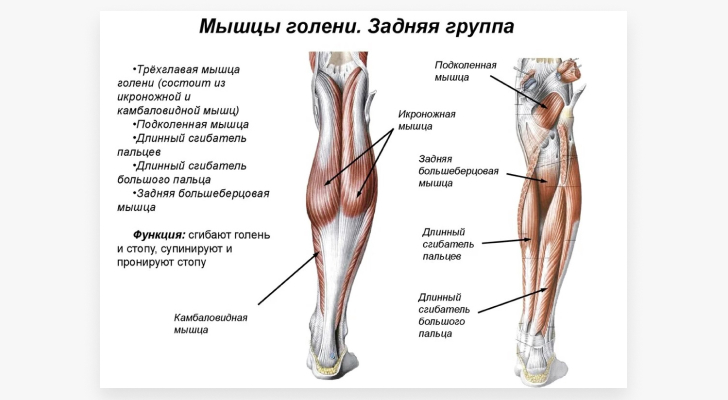
A distinction is made between the anterior, posterior and lateral tibial muscle groups. The anterior group includes the tibialis anterior muscle, the long toe extensor, and the long thumb extensor. The posterior group includes the triceps muscle (consisting of the calf muscle and the camel muscle), the hamstring and adductor muscles, the long toe flexor, and the tibialis posterior muscle. The lateral tibial group includes the short and long fibula muscles.
In addition to the tendons of the shin muscles, which attach to the bones of the foot and belong to the anterior, posterior and lateral groups, the foot has its own muscles (short muscles). These muscles are attached to the skeleton of the foot and have a complex anatomical-topographical and functional relationship to the tendons of these lower leg muscles, whose attachment points are located on the foot bones. The muscles of the foot are located in the hindfoot and plantar muscle.
movements of the lower limbs
The hip moves around three axes in the hip joint (triaxial – multiaxial joint). Flexion and extension (around the front axis) are possible up to 80° with the limb straight and up to 120° with the tibia flexed at the knee. Abduction and adduction (around the sagittal axis) can be performed up to 70-75° and rotation around the longitudinal axis up to 55°.
Thigh flexion: iliopsoas muscle, rectus femoris muscle, caudal muscle, broad fascia muscle, pectoralis muscle.
Thigh extension: M. gluteus medius, M. biceps femoris, M. semitendinosus.
Thigh extension: Thigh adduction: large adductor, long adductor, short adductor, pectoralis muscle, thin muscle.
Thigh retraction: gluteus medius and small muscles.
Inward rotation of the hips: gluteus medius (anterior bundles), gluteus minimus, tensor fasciae broad.
External rotation of the hips: gluteus medius, gluteus medius and gluteus minor, caudal muscle, iliopsoas muscle, quadriceps, external and internal obturator muscles.
Fasciitis - clinical picture and diagnosis
In most cases, necrotizing fasciitis is caused by infection, particularly beta-hemolytic streptococci. The disease is characterized by rapid onset, severe course and high mortality (up to 30 %). The disease affects almost all fascicles.
Immunodeficiencies, diabetes, vascular abnormalities, etc. contribute to the development of the disease. The main symptoms include:
- Strong pain;
- swelling, redness or blue discoloration of the skin;
- Blisters, ulcers, necrotic areas (areas of completely destroyed tissue, black, fetid).
Fig. 3 Necrotic lesions in fasciitis
The diagnosis is made on the basis of physical examination, laboratory findings (blood tests, tissue sections) and instrumental findings (MRI, ultrasound).
Diffuse eosinophilic fasciitis - DEF - (also called Schulman syndrome or disease, after the physician who first described the pathology) is characterized by connective tissue hyperplasia with changes in subcutaneous fat, muscle, fascia, and vital organs. The number of eosinophils, a type of white blood cell, in the blood is increased.
The disease is rare and is most common in young and middle-aged men. Excessive physical exertion, stress, trauma, etc. contribute to the development of the disease.
It begins acutely with a significant thickening, thickening and swelling of the fascia on the lower leg and forearm, less often on the body and face. Patients complain of dryness, itching, changes in skin density and color, and restricted movement of the joints.
In addition to an increase in the eosinophil count, the blood test also reveals signs of inflammation such as an increased blood sedimentation rate and an increased C-reactive protein. A biopsy of the affected tissue followed by examination can confirm the diagnosis.
Paraneoplastic fasciitis is one of the syndromes associated with cancer. The term 'paraneoplastic' means that the organism shows symptoms at sites remote from the actual tumor or its metastases. Fasciitis occurs mostly in women, in the later stages of the malignant process. The disease is characterized by:
treatment of fasciitis
Necrotizing fasciitis requires urgent surgery.
Treatment of DEF is long-term and consists of anti-inflammatory and anti-fibrotic drugs, corticosteroid hormones, physical therapy, exercise therapy, massage, etc.
Therapy of paraneoplantar fasciitis, including palmar fasciitis, is aimed at the underlying tumor process and the elimination of symptoms (pain, swelling, contractures, etc.).
Plantar fasciitis and heel spurs can be treated conservatively or surgically. In the early stages it is recommended to:
- reducing the load on the affected area;
- reducing the load on the affected area; Physiotherapy to stretch the fascia;
- use of night orthoses;
- wearing comfortable, low-heeled shoes;
- Use of insoles (for flat feet), orthoses, individual insoles;
- bandaging for arch support;
- corticosteroid injections;
- laser therapy;
- radio frequency therapy.
If conservative measures fail, surgery is resorted to.
The treatment strategy thus depends directly on the specific type of fasciitis and the course of the disease. Our clinic has an impressive arsenal of techniques and treatment methods.
In recent years, we have successfully used shock wave therapy as one of the most effective methods for comprehensive treatment.
Shock wave therapy to treat fasciitis
The previous methods of treating plantar fasciitis have several disadvantages. These include the insufficient effectiveness of conservative methods and complications after corticosteroid injections and surgery. Extracorporeal shock wave therapy (ECWT) is an effective alternative to these procedures. This method has a broad evidence base for connective tissue lesions, particularly plantar fasciitis.
Morphofunctional features
The superficial fascia (fascia superficialis) is located under the skin, its structure resembles connective tissue and, due to its functional task, provides a framework for superficial blood and lymphatic vessels, as well as nerves, lymph nodes, etc. In animals, it includes the muscles (muscularis) and nerves . In animals, it includes the muscle layer that is partially preserved in humans in the form of the facial muscles, the subcutaneous muscles of the neck, and the fleshy covering of the scrotum. Singer views the superficial fascia as a complex structure containing adipose tissue. According to AP Sorokin, there are two types of superficial fascia: a dense, felt-like, thick fascia composed of intertwined bundles of collagen and elastic fibers, and a loose bundle form composed of loosely arranged collagen and elastic fibers bound by fat cells are separated.
Intrinsic fasciae (intrinsic fasciae propriae) lie deeper than superficial fasciae, cover entire areas (e.g. arm, forearm, etc.) and delineate muscle groups or individual muscles and often serve as an anchor point. In most cases, they are clearly pronounced and are represented by lamellae of different thickness and density. Dense fascicles (aponeuroses) enclose muscle groups or reinforce the arches of the foot and are always directly affected by muscles either emanating from them or passing into them via tendons. This includes. The palmar aponeurosis, which is an extension of the palmaris tendon, the plantar aponeurosis, which strengthens the arch of the foot, the aponeurosis of the biceps brachii muscle, the iliopsoas band, in which the thigh fascia is woven, etc. The dense fascia consists of bundles of thick collagen fibers, aligned strictly in the direction of muscle contraction force. Layers of collagen fibers are interwoven with elastic fibers into networks. A small number of fibrocytes are located between the fiber bundles. Obersteg showed that the fascia that covers the muscles is constructed according to the principle of 'lattice scissors'. They contain systems of collagen fibers arranged in layers one on top of the other, oblique to each other and transverse to the underlying muscle fibers.
functions
The functions of the fascia are diverse. They cover muscles, vessels and nerves, hold muscles and tendons in a certain position, are the attachment point of many muscles, separate muscle groups and organs and limit cell-cell spaces (see). Like all connective tissue, they play an important role in water and salt metabolism. Knowing the topography of the fascia is crucial in surgery when dealing with the distribution of pus, anesthetic solutions, etc. Fascia is used as a plastic material in operations on joints, skulls, etc. (see fasciodesis). (see fasciodesis).
Changes in the fascia are often of a secondary nature. Inflammatory or neoplastic processes invade the fascia from the muscles, subcutaneous tissue, skin, internal organs and vessels, and the fascia can limit its spread for a while. In addition, pathological changes in the fascia develop in diseases such as arthrogryposis (see), dermatomyositis (see), scleroderma (see), elephantiasis (see), etc.
Only dystrophic cyclic lesions of the palmar and plantar aponeuroses and tumors, as well as rare inflammatory processes - fasciitis - can be classified as primary fascia diseases.
Fascial malformations are usually associated with muscular malformations if, in addition to the underdevelopment of the muscle, there is also an underdevelopment of its fascial sheath or aponeurotic elongation. Hypertrophy of the fascial and ligamentous apparatus around the passages of the main arteries (rheumatic trunk, neurovascular bundles of the upper limbs, etc.) is one of the causes of their extrafascial compression (see Asterisk muscle syndrome). Congenital defects of the fascia can lead to muscle herniation (see Muscles). Underdevelopment of the fascia and aponeuroses is a cause of ventral hernias (see Muscular Hernia). Hernia). For example, weakness of the transverse fascia is one of the local predisposing factors for inguinal hernias, and tears and holes in the white line aponeurosis of the abdomen are responsible for suprapubic hernias (white line hernia). Weakness of the renal fascia leads to impaired renal fixation (see), and weakness or damage to the pelvic floor fascia is a factor in rectal prolapse (see) or uterine prolapse (see Uterine, vaginal prolapse).
surgical treatment
Approximately 90 % of the patients can be treated conservatively within 3 to 6 months. Surgical treatment is indicated in patients with chronic symptoms that persist for 6-9 months after full conservative treatment.
Surgical intervention may include endoscopic or open partial plantar fasciectomy, lower leg muscle surgery, and Achilles tendon surgery.
Partial longitudinal fasciectomyIn a partial longitudinal fasciectomy, the damaged area of the plantar fascia is removed either endoscopically or through a small incision. The operation is followed by a 6-week rest period. While this procedure gives good results, it can increase the risk of tearing the plantar fascia, which can result in a pronounced flatfoot deformity and the development of negative symptoms.
Operations on the calf muscles and the Achilles tendon (known as Strayer's surgery or Wulpius' surgery): Several studies have recently been published suggesting that lengthening the calf muscle helps eliminate the symptoms associated with plantar fasciitis.
During the operation, an incision is made in the lower part of the calf to loosen the calf muscle tendon where it meets the Achilles tendon.
After the operation, the patients have to rest for six weeks. A residual muscle weakness can remain in the lower leg, which usually disappears within six to 12 months.
Achilles tendon anatomy
Via the Achilles tendon, the triceps tendon transmits power to the heel bone, which in turn transmits it to the foot, enabling walking, running, and upright posture. It is one of the anti-gravity muscles, along with the pelvic and spinal muscles. They are dominated by slow (white) muscle fibers that are not capable of fast and precise movements but have greater strength compared to the fast (red) fibers.
Every skeletal muscle has fibers of both types, and the ratio depends on the function being performed. Fast muscle fibers predominate in the muscles of the eyes and hands. In the antegrade muscles, the slow fibers predominate, allowing them to work longer with sustained effort.
The site where the tendon attaches to the bone is called the enthesus and has a specific anatomical and histological structure in antegrade muscles. The Achilles tendon is a combination of the gastrocnemius and soleus tendons that form the triseps surae. The two heads of the calf muscle join to form a thin, flat tendon at the level of the middle third of the tibia. They adhere to each other with the anterior surface of the calf aponeurosis and the posterior surface of the cambial aponeurosis. There are no connective tissue bridges between them, so they merge into the Achilles tendon at the lowest part of the calf tendon. The length of the tendon is variable and fluctuates between 12 and 26 cm.
In 5 percent of cases, there is a third head of the calf muscle that joins the medial head, while in 7 percent of cases, the function of the medial head is taken over by the deltoid. It does not form its own tendon, but connects to the calf muscle in the lower third of the aponeurosis. In rare cases, the lateral head merges into its own tendon and forms a separate calf endoprosthesis.
Histology (cellular structure)
The main tissue of the Achilles tendon is the dense fibrous connective tissue that underlies all fascia, sheaths, ligaments, and fibrous membranes. It consists of tenocytes, fibers and amorphous substance composed of water, proteoglycans, glycosaminoglycans and type 4 collagen.
Among the proteoglycans, the lecithin-rich decorin predominates. The amorphous substance is dominated by fibers with collagen type 1 (95 %) and collagen type 3 (5 %). Type 1 collagen is characterized by low extensibility and high mechanical tensile strength. Type 3 collagen determines the diameter of the tendon fiber and has a lower tensile strength. All three types of collagen are characterized by a high content of the amino acids glycine, proline and lysine. The collagen fibers are bound together into a single ordered system.
The tensile strength of the collagen molecule is ensured by amino acid crosslinks, the number of which varies in the tendon. The muscle-tendon and tendon-bone junction has the fewest of these interconnections, while the middle portion of the tendon has the most, accounting for the differential mechanical strength along the length of the tendon (greater number of tears in the middle third). Chronic inflammation alters the quantitative ratio of type 1 to type 3 collagen in favor of the latter, increasing the risk of spontaneous rupture.
Collagen fibers and amorphous matter remain relatively independent of each other because of the endotenon, which separates adjacent fibers and allows them relatively independent movement.
The histological structure combined with the complex spiral geometry gives the tendon unique mechanical properties. According to various authors, the Achilles tendon can withstand a tensile force of 350 to 800 kg (50 N/mm), stretches 3-5 % under load and is capable of energy recovery (energy is stored in the kickback phase and released again in the push-off phase). . Partial fiber failure occurs when the strain exceeds 5 %, and tear occurs when the strain exceeds 8 %.
species
Depending on the position of the knot, a distinction is made between:

– cranial fasciitis;
- intravascular fasciitis;
– related forms of fasciitis and myositis.
– nodular fasciitis
– infiltrative fasciitis;
- pseudosarcomatous fibromatosis;
- nodular fasciitis;
- proliferative fasciitis;
– Pseudosarcomatous dermatofibroma.
causes
Nodular fasciitis occurs when fatty tissue bulges through deep fascia. Until recently, this pathology was considered a reactive process with an unclear etiology. However, recent study data suggest that it is a clonal tumor process that is usually self-limiting.
- Subcutaneous growth over a short period of 3-6 weeks;
– spontaneous regression;
– Lesion usually reaches 3 cm in size.
When nodular fasciitis develops in the buttocks, there may be some tissue tension.
causes
The only proven cause of plantar fasciitis is occasional microtrauma to the plantar fascia caused by careless foot movements. Several factors can increase the likelihood of dystrophic changes in the plantar area:

– overweight;
– to be female;
– Standing or walking on hard surfaces for a long time;
– wearing shoes without instep (slippers, flip-flops);
– stagnation of blood circulation in the lower limbs;
– flat feet;
- Frequent heavy lifting.
Plantar fasciitis is commonly diagnosed in dancers, professional athletes, and runners.
diagnostic procedures
To diagnose plantar fasciitis, the patient undergoes a diagnostic exam that includes:
– Collection of medical history;
– Visual examination and palpation of the foot;
– X-ray examination to detect bony changes;
- blood tests (to detect the bacteria that cause necrotizing fasciitis);
– MRI of the foot (prescribed if plantar fasciitis is suspected).
A comprehensive examination is best carried out in a modern, multifunctional center equipped with the latest generation devices. For example, the health clinic has its own diagnostic facilities (ultrasound, CT, MRI) and a staff of experienced, highly qualified specialists. The patient has the opportunity to undergo all diagnostic procedures without leaving the clinic and make an appointment with the right doctor to develop a comprehensive treatment.
Long thigh segment
The distinctive feature of this segment is its pinnate structure. The muscle extends from the tip of the MB bone and attaches to its 2 from the outside:
When contracting the long fibular muscle, 3 types of movements are performed simultaneously:
The tendon of this fiber surrounds the lateral side of the ankle behind and below it. They meet the extremities near the heel. As it advances further and enters the vicinity of the supporting muscle, this element spreads along the furrow that runs on the underside of the tarsal bone and ends on its inner side.
Short fibers of the humerus
This plantar muscle subtype elevates the lateral edge of the foot, prevents it from rotating with the plantar side and clubfoot inward, and performs plantar flexion.
On its surface side facing the skin, it forms the short fiber MB, which connects the glans and fibula. Descending from the short fibula, the tendon wraps around the lateral structure of the ankle from the dorsal lower extremity and then inserts at the condyle of the last metatarsal.
How is plantar fasciitis treated?
Unlike arthritis of the knee or ankle, plantar fasciitis is easily treatable. It can be completely cured provided a comprehensive approach is taken. Orthopedists and rheumatologists use shock wave therapy, decompression techniques to reduce stress, kinesio taping, and other procedures to restore foot function.
What is a heel spur and how is it treated? dr Yevdokimenko explains it to us:
Tips for people diagnosed with plantar fasciitis
Comprehensive treatment is not possible overnight. It is therefore important that you follow a few rules during this time and protect your foot from further damage. So what do you have to prepare for and what do you have to endure?
- 'Slow down the pace.'. For a while you won't be able to be as mobile as you used to be. Walking will no longer be fun, sports will become impossible, as will travel. It is important to focus on treatment now to prevent the disease from taking its course.
- Don't turn a blind eye to the symptoms.. As soon as you feel pain in the heel area, you should consult your doctor. Otherwise, the disease can become chronic, which will complicate its treatment.
- Pay attention to the preservation of the arch of the foot. Don't treat the heel itself - the area that's causing the pain. The right cushioning of the foot is the top priority, followed by therapeutic measures. Choose appropriate orthotics and wear them continuously to allow the foot to adapt to the new conditions.
- Avoid long walks. Bicycles are a good alternative - pedaling is less dangerous to the foot. Stretching and weight bearing exercises without a foot brace can also be done as physical activity.
- Take advantage of a special massage. You can massage your feet with a small ball or a special massager with a roller. This will relieve painful sensations.
- Use elastic bandages or compression stockings.. Ask your orthopedist which ones are appropriate for your form of the condition.
- Walk on tiptoe.. This advice is good for people who need to go to the bathroom at night. The heel is particularly sensitive if it has not been moved for a long time.
- The flexor muscles of the foot.
- fasciitis.
- Longitudinal soleus muscle.
- How muscles and bones are related.
- Tibialis posterior muscle.
- shin fascia.
- Tear of the foot fascia.
- The muscles that move the foot.