shoulder and humerus.
- Physiology of the joints
- IMMOVABLE JOINTS
- Uniaxial joints
- Biaxial joints
- pets
- What does the respiratory rate depend on?
- The buttocks and the reins
- Which foods contain glucosamine?
- Useful foods with glucosamine
- What can I do if I don't have enough glucosamine in my diet?
- How does synovial fluid 'work'?
- How does a change in pH affect the synovial membranes?
- Why bone broth is recommended for joint problems
- What other benefits bone broth offers
- What are the functions of the joints?
- What are ribbons?
- What you can do now
- How do you treat hypermobility?
Physiology of the joints
The human body has over two hundred bony joints. In the adult body, many of these joints are only slightly or not at all mobile, so that we usually do not know them. This article describes the anatomy and function of the joints before discussing joint involvement in rheumatoid arthritis (RA).
Joints fall into three basic types, based on how much movement nature allows in each joint: immobile, slightly flexing, and freely flexing. Different joint types work in different ways to perform different functions.
IMMOVABLE JOINTS
The joints of the individual bones of the skull and pelvis are examples of rigid, immobile, or otherwise immobile joints. These joints are only mobile in early childhood, during growth or in special cases such as pregnancy. These joints are special because they are not affected by RA.
Some joints, such as those between the vertebrae in the spine, are limited in their ability to move normally. The bony structures of the vertebrae (spine and cervical spine) are separated by a cartilage cushion, the intervertebral disc. If these joints move more than is absolutely necessary, problems can arise. For example, there is the well-known herniated disc, which can occur when an easily movable disc moves further than it should. These joints are also unaffected by rheumatoid arthritis.
Uniaxial joints
1. Cylindrical joint, Trochoida articularis. A cylindrical articular surface, the axis of which is vertical, parallel to the longitudinal axis of articular bones or to the vertical axis of the body, allows movement around a single vertical axis – rotation, rotatio; such a joint is also referred to as a rotary joint.
2. Pulley joint, also called gum joint. block joint, ginglymus) (Example: interphalangeal joints of the fingers). Its articular surface is a transverse cylinder, the long axis of which is transverse, in the frontal plane, perpendicular to the long axis of the articular bones; therefore, the movements in the block joint are performed around this frontal axis (flexion and extension). The guide groove and scales on the joint surfaces prevent lateral slippage and facilitate movement around the same axis.
If the guide groove block not perpendicular to the axis of the joint, but at an angle to it, its extension results in a spiral line. This is called the spiral joint (e.g. the shoulder joint). The movement in a spiral joint is the same as in a pure block joint.
According to the scheme of the ligament apparatus ligamentsIn a cylindrical joint, the guide bands are perpendicular to the vertical axis of rotation, in a block joint perpendicular to the front axis and its sides. This positioning of the ligaments keeps the bones in place without impeding movement.
Biaxial joints
1. ellipsoidal joint, arulticatio ellipsoidea (Example: wrist). The articular surfaces have an elliptical shape - one is convex, oval with irregular curvature in both directions, the other is concave. They allow movement in 2 horizontal axes that are perpendicular to each other: flexion and extension around the frontal axis and abduction and reduction and adduction around the sagittal axis.
ribbons in ellipsoidal joints At their ends, the ligaments of the ellipsoid joints run perpendicular to the axis of rotation.
2. condylar joint, articulation condylaris (Example: knee joint).
condylar joint has a convex head of the joint in the form of a protruding elliptical, rounded process called the condyle, hence the name of the joint. The condyle corresponds to a depression on the articular surface of the other bone, although the difference in size between the two can be significant.
The condyle joint can be viewed as a variant of the ellipsoid joint, which represents a transition form from the block joint to the ellipsoid joint. The main axis of rotation is therefore directed forward.
From the block joint condyloid joint differs from the condylar joint in a greater difference in the size and shape of the articular surfaces. In contrast to the condyle joint, the condyle joint can therefore move about two axes.
From ellipsoidal joint It differs from the elliptical joint in the number of joint heads. In the condylar joint, there are always two more or less sagittal condyles that are located in the same capsule (e.g. (e.g. two femoral condyles in the knee joint) or are located in different joint capsules, such as in the atlanto-occipital joint .
Then In the condylar joint of the femoral heads Since the joint head is not elliptical in shape, the second axis does not have to run horizontally as in a typical elliptical joint, but can also run vertically (knee joint).
pets
NAVIGATION: Home Veterinary Medicine Textbook Basics of Animal Anatomy and Physiology JOINTS AND LIGAMENTS OF THE LIMBS
JOINTS AND LANDAGES OF THE LIMBS
Joints and ligaments of the forelimbs.
shoulder joint.It is formed by the shoulder blade and the humerus. It is a simple multiaxial joint and is held together by the joint capsule (Fig. 31).
The elbow joint. It is formed by the humerus and the bones of the forearm. It's a simple uniaxial joint. In addition to the joint capsule, it has external and internal collateral ligaments (Fig. 32).
carpal joint. It is formed by the radial, carpal, and metacarpal bones. It is a kind of complex uniaxial joint. In addition to the joint capsule and collateral ligaments, there are intercostal, interphalangeal and ligaments of the articular bone (Fig. 33).
talon joint. It is formed by the metacarpal bones, scaphoid bone and sesamoid bone. This joint is simply uniaxial. In addition to the joint capsule and collateral ligaments, the ligaments of the sesamoid bones are present here: the intercondylar ligament, the lateral ligaments of the sesamoid bone (external and internal), and the straight and oblique ligaments of the sesamoid bone (Fig. 34).
The coronoid joint. It is formed by the talus and coronoid bones. It's a simple uniaxial joint. In addition to the socket and collateral ligaments, it also has posterior ligaments.
coffin joint. It is formed by the cannon bone and the coffin bone. In addition to the joint capsule and the collateral ligaments, the uvoceps and pelviceps are also present (Fig. 34).
What does the respiratory rate depend on?
Another interesting point is that the horse's respiratory system and lungs are directly related to gait. If it's just one step, breathing is fairly calm and moderate. If the horse increases the workload and goes into the trot, breathing will change and intensify slightly during this trot. A little but not much.
In the galloping horse, on the other hand, the relationship between breathing and gait is much more pronounced. Normally, the breathing rate is in a one-to-one (1:1) ratio to the movement speed. That is, for a gallop there is a step and a breathing rhythm. As a result, breathing becomes stronger and deeper the faster the horse runs.
In general, a horse can breathe in and out about 15-20 liters of air during a fast run (a human can only breathe out 5 liters, so a multiple of that). This once again confirms how strong and resilient horses are. And how weak and helpless man is in comparison. However, man is able to control these beautiful animals, which gives him certain advantages.
The buttocks and the reins
The forelimbs are the area of the pastern bone on the horse's front leg. A horse's pastern joint should be dry and well developed. There is a lump of skin at its upper edge.
The hock and the head of the hock
The pronotum and labrum area should be dry with well defined tendons that are not thin. Thin femurs are an indicator of weak tendons, ligaments and weakness in the horse's legs.
Which foods contain glucosamine?
glucosamine in food is widespread, but before it enters our body, it is easily destroyed by various factors. This is because glucosamine and chondroitin are unstable. They are broken down when they are boiled, fried, or steamed. Glucosamine and chondroitin are found in foods mostly in the form of polymers – proteoglycans, collagen, proteins and other components.
Once you find out which foods contain glucosamine, do not rush into eating them, but first check if you are allergic to them. Better yet, talk to your doctor about it!
Useful foods with glucosamine
- poultry and beef. Since most of the glucosamine in food is broken down, it is recommended that you cook the broth over low heat and do not overcook.
- Pig ears, oxtails, chicken feet. These ingredients can be used to make sausages. However, such a dish has not only advantages, but also disadvantages: the excess cholesterol in it can be deposited on the walls of blood vessels.
- Cheese In its pure form, without heat treatment.
- sea and river fish. Especially salmon and salmon. Patients diagnosed with stage I arthritis are advised to take glucosamine with food – make fish jelly with gelatin at least once a week!
- seafood. mussels, crabs.
- Jam and 'marshmallows. Caution! People with diabetes should avoid it.
- eggs (hard boiled, not overcooked).
- Wheat germ, mushrooms, nuts, broccoli, seaweed.. These contain high concentrations of collagen-like compounds.
Danger!!! Even with regular consumption of products containing glucosamine, cartilage health is not guaranteed in the case of heavy joint stress at home and at work, metabolic disorders or age-related changes.
What can I do if I don't have enough glucosamine in my diet?
Although food contains glucosamine, many people cannot constantly eat jams or fatty broths for health reasons. In addition, pure glucosamine in food is scarce. If your joints are affected or you've noticed the first signs of deterioration - pain, crunching, limited mobility - a full regimen of chondroprotectors is required.
Artracam is a national drug from the group of chondroprotectors. You don't need to wonder which foods contain glucosamine, 1 sachet of Artrakam is its daily dose. Simply dissolve 1 sachet (bag of powder) in 200ml of water. The solution is odorless and colorless. 6 weeks cure. Noticeable improvement occurs within 3-4 weeks. It acts slowly, accumulates in the body, reduces the need for NSAIDs (nonsteroidal anti-inflammatory drugs) and painkillers. It can be taken prophylactically if you are at risk.
Benefits of Artracam Preferred to glucosamine in foods:
- 1 pack = daily dose.
- Effectively and in 1 step strengthens joints, relieves swelling and inflammation, eliminates pain. Especially in the early stages of the disease.
- Easily absorbable. The drug in liquid form is safer for the stomach.
- High bioavailability (a large amount of active ingredient reaches the 'site of action').
OfficiallyCurrently, most glucosamine is contained in pharmaceutical products - chondroprotectors.
Healthy joints for you and your family!
How does synovial fluid 'work'?
When a joint is under the lightest load, its cavity fills with lubricant from the deep layers of cartilage. If there is enough of it and it is of high quality, then the movement will be easy and will not cause discomfort. When movement stops (load is removed), the synovial fluid is reabsorbed into the cartilage tissue. This means that the joint is not constantly lubricated, but only during phases of activity.
The main functions of the synovial fluid are:
- avoidance of friction – the moving parts of the joint are not subject to injury;
- Ease of movement - each part moves as if by magic';
- Replaceable function. This includes saturating tissues with nutrients and getting rid of dead cells.
The efficiency of the individual functions decreases with age. Chronic diseases and infectious diseases play their part. Their causative agents and the drugs used to neutralize them significantly alter the composition of the body's fluid medium, including synovial fluid.
How does a change in pH affect the synovial membranes?
As acidity increases, joint lubrication deteriorates. 'Insufficiently lubricated' joints result in excessive friction, which can lead to injury and even deformation if the lubrication is persistently lacking or of poor quality. It is not so much the amount of lubrication that matters, but its quality. With the same amount, but increased acidity, the quality of the lubricant will decrease, as well as with insufficient production.
This reduction in quality is noticeable through the following symptoms:
- cracking when bending and stretching the joints;
- squeak when moving;
- painful sensations;
- tension in the joint area;
- Stiffness and limited mobility.
The joints are affected by both the amount and quality of lubrication. This becomes particularly clear in the function of the knee, elbow and ankle joints, which are subjected to the greatest stress.
Why bone broth is recommended for joint problems
With age, the amount of collagen in the body decreases and the joints lose their mobility. The cartilage tissue becomes thinner and breaks down. This is how osteoarthritis develops in the knees, shoulders and ankles. Gelatin, which can be obtained from bone broth, can help.
Gelatine is extracted from collagen-saturated joints in a long cooking process. It contains proline and glycine - substances that rebuild the connective tissue. If you consume bone broth regularly, the collagen layer in your joints will improve over time. This is especially true for people around 40 who are at risk of osteoarthritis, as well as for competitive athletes.
Bone broth can be prepared in a number of ways, such as with this recipe:
What other benefits bone broth offers
The glycine contained in this dish stimulates the synthesis of hydrochloric acid in the stomach, so the bone broth eliminates heartburn. But that's not all the advantages. Glycine is also a component of bile, which is essential for fat digestion in the small intestine. The broth also contains glutamine, which normalizes bowel activity. Their regular consumption reduces the severity of food allergies in many people.
The broth is also rich in collagen, which can restore intestinal permeability. It is therefore recommended for people with ulcerative colitis and Crohn's disease. It also improves the intestinal microflora and increases immunity, which means that the body can fight hip osteoarthritis or other chronic diseases more effectively.
What are the functions of the joints?
The human body has around 180 different joints that connect the bones of the skeleton. They are similar to joints, allowing bones to move smoothly and preventing friction and breakage. Thanks to the joints, the human body maintains a stable position in space and can move freely.
- The hip joint – is the strongest and most heavily loaded joint. Osteoarthritis of the hip and other hip abnormalities are at the forefront of joint diseases.
- The knee joint – The largest and structurally most complex joint. This is where the longest arm of the lower limbs, that is, the femur and the lower leg hand, which move with great effort when walking, meet.
- The shoulder joint – The shoulder joint is the most mobile joint. The joint capsule in it is very thin, which is why the shoulder joint is often injured.
The joints are covered with membranes that contain synovial fluid. If this is missing, the joints cannot fully fulfill their dampening function. The quality and quantity of this lubricant is affected by diet. If it is insufficient or too thin, the cartilage will wear down and become damaged. A synovial prosthesis can help, but before osteoarthritis develops, it is better to ensure proper nutrition in advance.
According to statistics, every second person between the ages of 40 and 70 suffers from joint pain.
What are ribbons?
Ligaments are the strengthening part of the joints. They are made of connective tissue and connect parts of the skeleton or internal organs with each other. Along with the joints, the ligaments form the skeleton of the musculoskeletal system and are where most injuries occur. Eating a healthy diet helps strengthen ligaments.
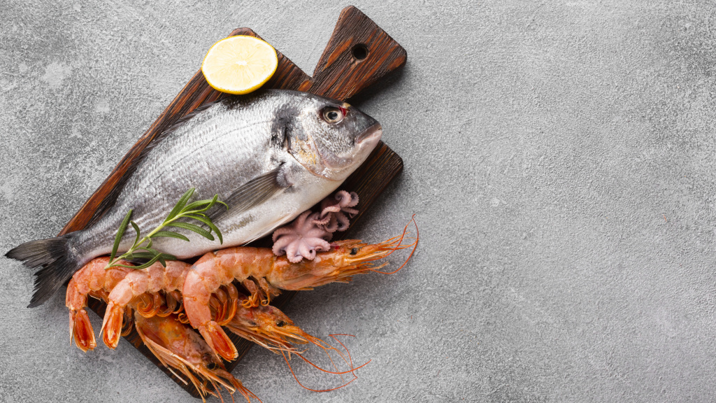
- Fish and seafood. They contain a lot of organic phosphorus, which is important for the strength of bones and joints.
- Lean red meat, by-products, tongue, and eggs are sources of iron. They help remove excess inorganic phosphorus from the body.
- Dates, plums, apricots.
- Oats and buckwheat groats.
- buckwheat honey.
- Nuts.
- Bitter chocolate and cocoa (contain magnesium).
- Green vegetables, figs, cherries, apricots.
- Dairy products, low-fat milk, hard cheese (source of organic calcium).
- Seaweed, mussels, crabs, bones and cartilage of fish, birds, casserole and jelly products (gelatine rebuilds damaged cartilage tissue in a similar way to chondroprotectors and promotes good synovial fluid consistency).
Cream and milk ice cream are surprisingly good for the joints
What you can do now
You can already check your joints for hypermobility yourself without having to see a podiatrist.
- Try stretching your pinky back without your other hand. If you are hypermobile you can do this more than 90°.
- Rotate your hand toward the inside of your forearm. Are you able to passively push your thumb back into your hand without exertion? With 'loose joints' this is not a problem.
- Extend the elbow joint and examine the arm line. Is it straight or is the elbow extended more than 10°? This is another worrying symptom.
- Repeat the same exercise with your knee. In hypermobility, it also stretches more than it should.
- Stand up straight, keep your legs straight. Bend towards the floor and touch your palms to a horizontal surface. Did you manage to keep your legs straight and not bend your knees?
Hypermobility is common in gymnasts and circus performers.
How do you treat hypermobility?
Joints that are too flexible do not always need to be treated. If they do not cause discomfort to the person, then it is enough to protect him from injury. However, when pain occurs, a comprehensive approach is required. An orthopedist may recommend the following:
- taking painkillers for a short period of time;
- physiotherapeutic measures to strengthen the ligaments (treatments are selected depending on the location of the joint, its mobility, the age and performance of the patient);
- a change of job if the job or hobby poses a risk to the musculoskeletal system and contributes to injury.
Hypermobility is not a serious condition. Although it does not lead to disabilities such. B. Osteoarthritis of the hip, but is dangerous under certain conditions in the long term. For this reason, it is often advisable to seek the help of a psychologist.
Hypermobile joints are more prone to osteoarthritis than normal joints.
Joint hypermobility is not a very common phenomenon. In the case of the upper limbs, the diagnosis is easy to make: perform simple movements and evaluate the results. Do you also experience these symptoms? Pay special attention to your joints and consult your orthopedist at the first sign of problems - pain, tingling, restricted movement. The most important thing: avoid injuries!
Read more:- tearing of the joint capsule.
- Pronation of the shoulders - what is it?.
- Those are the pluses.
- Orthopedic support for the foot.
- Pronation and supination of the shoulder.
- Joint doctor - what's in a name?.
- shoulder supinators.
- How much does shoulder dislocation surgery cost?.