Structure of the leg below the knee:

- limits
- The Bones of the Femur
- Fascia, ligaments, joints
- Cartilage, joint capsule, synovial fluid….
- What can hurt in a joint?
- In the event of a mechanical injury, the following happens:
- Treatment of leg pain (ankles, feet, toes)
- Bemer therapy
- Diseases
- ailments and symptoms
- Diagnosis
- our prices
- disease patterns
- leg muscles
- Front thigh group
- Posterior group of the thigh
- glutes
- Function and care of the lower limbs
- FOOT, ANKLE AND ANKLE.
- Anatomy. feet
- Movement
- shin
- Leg fracture - the concept
- causes of injuries
- Symptoms of a broken bone
- Classification of NSAIDs.
- Evaluation of non-steroidal anti-inflammatory drugs
- No. 1 – Ibuprofen (Borisov Plant, Republic of Belarus)
limits
Anatomically, the thigh sits below the oblique crease, beginning at the hip joint and ending at a line drawn 5 cm above the knee joint. This area is bounded at the top by the inguinal ligament and at the back by the gluteal ligament.
The special structure of the hip allows people to move. Due to its structure, this part of the leg is involved in:
- the flexion of the limb;
- rotation around its own axis by 180 degrees
- Raise and straighten the leg in a horizontal direction
- lowering the pelvis and squatting.
This is where the most important blood vessels and large nerves are located. The main components of blood - red blood cells, white blood cells, platelets - are made in the femur.
The Bones of the Femur
The large femur is located in this area. It is cylindrical in shape with a head at the top, a greater trochanter and a lesser trochanter on the outside where muscle fibers attach. There is an intercostal arch on the back.
The origin of the bone is connected to the hips. The lower (distal) end is elongated, forming a pair of outgrowths—the lateral and medial condyles, which attach to muscles and ligaments.
The bone's structure and mass is due to the fact that it carries most of the load that holds the torso together.
Fascia, ligaments, joints
The hip is covered by the broad fascia, which is divided into Scarpa's triangle:
The first has a loose structure, runs between the muscle fibers and carries lymphatic and blood vessels and nerves. The second is dense and tight, surrounding the hips from the outside.
The hip joint is supported by ligaments:
These elements ensure the stability of the joint and prevent buckling and injuries during movement.
Cartilage, joint capsule, synovial fluid….
Where the bones meet is their heads covered by cartilage.This protects them from friction and bumps. Inside the joint are. There are two meniscishaped like crescents. These are necessary to protect the cartilage and bones from mechanical stress.
The cartilage and the meniscus are located in the so-called 'joint capsule'. joint capsuleThe synovial fluid is inside the joint capsule. synovial fluid. Like oil, it lubricates all joint surfaces and prevents friction. This is the case when the amount of synovial fluid is sufficient and its viscosity is correct.
On the front of the joint are the kneecapor kneecap. It is held together by ligaments and tendons and is covered with cartilage on the inside. The kneecap is a kind of protective shield that protects the internal components of the knee joint from damage.
How is the knee joint constructed? A clear demonstration with detailed explanations:
What can hurt in a joint?
Cartilage and meniscus have no nerve endings and therefore cannot hurt. In fact, there is nothing in the joint that could hurt in the usual sense. The nerves do not reach the bone, but the periosteum - the thin membrane that covers the bone from the outside. This is why humans feel pain when a bone is damaged. What happens if there is no fracture?
If there is no fracture, the source of the pain may be the ligaments that surround the knee joint and hold the meniscus inside. In addition, nerve endings reach the joint capsule, which can also cause pain.
Cartilage doesn't hurt: it doesn't have nerve endings
In the event of a mechanical injury, the following happens:
- The joint is injured and the ligaments swell;
- The body begins the healing process - it pumps blood to the injured area;
- The skin around the joint turns red and the joint swells;
- The joint fluid can no longer circulate freely and presses on the nerves in the joint capsule, which leads to increased pain.
After a while, the small ligaments heal, the swelling goes down, the fluid starts to circulate again, and the pain goes away. This happens when only the small ligaments are damaged and the large ligaments and meniscus are intact. If the meniscus is damaged, the situation is different, since the affected person does not feel pain, but the structure continues to deteriorate.
Over time, the meniscus continues to be damaged and the surrounding cartilage also deforms from the adverse conditions it is exposed to. Their surface gradually wears away, causing bone friction, irritation of the nerve endings in the periosteum, and pain. This is how osteoarthritis of the knee develops, which is rarely diagnosed at an early stage.
By the time a person is prescribed treatment for osteoarthritis or osteoarthritis, the condition of their joints already leaves much to be desired. There is almost certainly not enough synovial fluid in the joint capsule, and when lubrication is lacking, the cartilage quickly dries out, cracks, and wears out. Intra-articular injections of Noltrex, a synovial fluid substitute, can help here.
Treatment of leg pain (ankles, feet, toes)
Treating leg pain at home often helps relieve not only the pain but other symptoms such as swelling, cramps, and discomfort. Treatment usually begins with combating the factors that cause leg pain and caused other ailments. For example, you should stop exercising, at least temporarily, if you experience pain in your legs (feet, ankles, or toes) while exercising. It is contraindicated to train 'through the pain'. It is important to wear quality and comfortable footwear. Supinators and other orthopedic aids help make walking more comfortable.
Cold application, rest, foot massage, gentle and light exercises (e.g. tendon stretching) can help with leg pain, leg swelling or cramps. Over-the-counter pain relievers can be taken to relieve leg pain.
For swollen feet, legs and ankles you can raise your swollen feet slightly above heart level and sit like that for a while. If you do a sedentary job, get up every hour and walk around for a few minutes. Limit your salt intake.
If home remedies for foot pain (feet, ankles, toes), swollen feet, or other problems don't work, see your doctor. Specialist advice is necessary also when pain and swelling increase, symptoms of infection appear, the skin becomes pale, tingling and numbness appear.
Bemer therapy
In our doctor's office Bemer therapy has proven to be the most effective way to treat all types of pain. The BEMER therapy – is an electromagnetic physiotherapy device from Switzerland, the main purpose of which is to improve blood circulation.
The BEMER device consists of three components: an induction mattress aimed at general regeneration, a reinforced applicator that makes it possible to act on a specific area, and a laser magnet that has the strongest effect on the painful area.
Read more about Bemer therapy see link, if you want advice or to make an appointment, you can do this via the feedback form or by phone: +7-495-212-08-85
Diseases 
- food poisoning
- abdominal pain
- nausea and vomiting
- Pain in the legs
- colds
- meningitis
- anemia
- weakness and fatigue
- insomnia
- flu
- muscle cramps
- catarrh
- Chronic Fatigue Syndrome
- Dry mouth
- motion sickness or seasickness
- Strong weight loss
ailments and symptoms 
- High body temperature
- Decreased immunity
- pain in different places
- Rapid weight loss or gain
- abnormalities of the stomach
- Frequent colds, acute respiratory infections
- Weakness, dizziness, malaise
If you have these symptoms, it could be a sign of illness, which is why we recommend seeing a specialist.
Diagnosis 
our prices
- Consultation with a GP – from R1,500.
- Biochemical blood test (standard, 10 tests) – 2470 rub.
- Biochemical blood analysis (extended, 14 indicators) – 3565 p.
- General blood analysis – 675 p.
- ECG (Electrocardiography) – 1500 p.
- Urine test – 320 p.
- Pulse oximetry - 500 p.
disease patterns
Unfortunately, the ankle can be injured or affected by disease.
- arthrosisOsteoarthritis is caused by calcium deficiency, trauma, cartilage and bone stress. Over time, this causes growths on the bones, so-called osteophytes, which limit mobility. The pain and stiffness are usually gone, but the ankle gradually begins to lose mobility. Treatment consists of medications combined with physical therapy and exercise. However, if the deformity is very severe, surgery is required.
- arthritisThe cause is an inflammatory process. It can be caused by rheumatoid arthritis, gout, or infections in the mouth. It is characterized by pain from morning to night. When you move, the pain is less noticeable. Diclofenac, Naise, Ibuprofen, ointments and gels can help with symptoms. Treatment should be carried out by a rheumatologist who is able to diagnose infectious arthritis, which can be dangerous if there is pus in the joint. When this diagnosis is made, the patient must be admitted to a hospital.
- injuries. Tendons, bones and ligaments can be injured. Symptoms are the same: swelling, pain, immobility, and an inability to put weight on the foot. First aid is to ice the affected area, rest, and see a doctor.
- tendon rupture (tendon). Occurs after a fall or sports overload. The foot can no longer be straightened and you can no longer walk on your toes. Swelling due to pooling of blood and pain with every movement. In this case, surgical intervention is required.
It is important to understand that the nervous system has a direct impact on muscle control. And when they are at rest, they begin to atrophy over time.
leg muscles
The anatomy of the leg muscles divides all muscle structures of the lower limbs into:
Overhanging the muscles of the upper limbs, these formations are considered the largest in the human body. That's because this area takes the brunt of the movement.
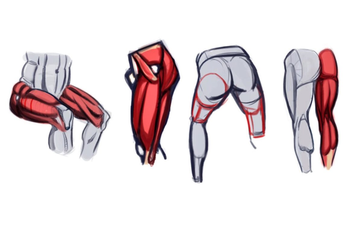
Front thigh group
It is formed by the quadriceps (the largest muscle in this section), which is responsible for straightening the limbs in the knee joint. It extends over the entire front surface of the thigh and is crossed by the oblique cutter.
- The rectus head (two-part, surpasses all others in length, extends to mid-thigh, then tapers to a tendon and attaches to the kneecap, the patella ;)
- inner (predominant in width, covers the rectus muscle on the front side and is covered by the sartorius muscle, runs obliquely to the thigh, where it forms a tendon) and the wide medial (flat and thin, lies on the front side, its upper part becomes covered by the rectus muscle);
- External extensor (flat on the anterior external surface; it is covered by the broad fascia muscle, which runs straight anteriorly; the muscles run obliquely downwards and cover the femur anteriorly, forming the rectus muscle tendon below).
Posterior group of the thigh
This part includes the biceps muscle (located on either side of the thigh) which folds together:

Its functional role is to cause flexion of the lower limbs at the knee joint and extension of the thigh.
glutes
Consists of the gluteus maximus, gluteus medius and gluteus minor muscles. The former extends over the entire buttocks region and determines its shape; it attaches to the iliac crest, the dorsal surface of the sacrum and the coccyx; it is responsible for the mobility of the hip joint, the straightening of the torso and the pull of the legs backwards.
Function and care of the lower limbs

The function of the lower limbs is to support the body and move it in space. The ability to move enables humans to lead full lives and actively participate in sports, running and dancing.
To properly care for feet and improve their health, one should:
- Avoid overloading the joints and distribute the load evenly;
- avoid injuries;
- always keep the limbs clean;
- Avoid hypothermia (many diseases are caused by cold and wet feet) and overheating in the sun;
- To eliminate skin diseases and maintain the health of the epithelium, use nourishing creams and masks (anti-inflammatory agents, anti-itch mixtures and anti-fungal agents; well-known names such as Vorozhea, DeoControl, etc.)
- massages (such muscular work prevents fluid stagnation in tissues, stimulates blood circulation, improves cell nutrition, acts on biologically active points and improves the health of the body as a whole);
- a healthy diet (sufficient intake of vitamins and trace elements from vegetables, fruits, fresh and natural products) and a healthy lifestyle (abandonment of bad habits, regular walks in the fresh air, hardening of the body);
- An important part of skin care in women is depilation (removal of unwanted hair with ointments and cosmetic creams or by mechanical removal);
- maintaining physical activity (regular gymnastics, strengthening the musculoskeletal system, swimming) is important.
FOOT, ANKLE AND ANKLE.
Let's talk a little bit about the human body…. What is considered in the human body Foot? Nowadays we refer to the foot from the beginning of the thigh over the sole to the tips of the toenails. But Originally the foot is just called the top of the foot.. Only the surface of the foot from the tips of the toenails to... is so called. just above the ankle and….

... And that's where everything needs a special explanation. People have now forgotten what the body is called in Russian and why it is called that. The phrase is well known, but its exact meaning is largely forgotten. When it comes to the leg, there are many words for it in Russian. In the science of anatomy, today it is common to refer to both the thigh and the ankle by the same name - ankle. The calcaneus and ankle are the protrusions of the lower ends of the tibia above the foot. These protrusions are called lateral ankle and medial ankle in anatomy, which means something like outer ankle and inner malleolus. The Latin word 'lateral' means 'side' and the word 'medial' means 'medial'. However, the meaning of the Russian words 'ankle' and 'ankle' is different. Otherwise there would be no different Russian words! In Russian there are no so-called synonyms. Original Russian called ankle only outside protrusion The bone above the foot. Only on the outside! The malleolar bone is the protuberance at the end of the outer malleolar bone. A The ankle is called only is the inner protuberance of the bone above the foot.. Inside only! The ankle is a protuberance on the large bone of the foot.

The outside of both feet is the calcaneus and the inside is the hock. The outside of the foot is called cheek of the foot. They are also called them cheek accented with an 'A' but now pronounced as 'Shweda'. The inside of the foot is called cheek of the foot.. Also known as the kida or Keda with the stress on the 'A'. …ankle and shoulder are almost on the same level, one directly opposite the other. The point where the extension of the tibia to the ankle and heel bone begins is one of the body lines. At the back of this line are the lower ends of the calcaneus muscles and roughly where the Achilles tendon begins. This body line has several names: candala or in other words. Kaidana, Knifebreaker, Kova, Poota or Uza… … But in Russian there are no synonyms. If there are different words, they have different meanings. This is how the Russian language works. The shin line, which is called knocker, puta or uza depending on the case, is covered by the elastic band of the sock in shoes. …Hence the upper part of the foot, from the tip of the nail to the line of Puta or Uza, is called in Russian as aleHa. And all other parts or surfaces of the leg were not called a leg. They all have their own names - thigh, knee, shin, and so on. Nowadays we call the leg the drop foot. But The leg is not only that the lifting of the foot, rather but also the distance from that elevation to the pooty or tie.. This is above shin .. That's another way of putting it The foot consists of the arch and the ankle.. The arch of the foot is the elevation of the foot that sometimes prevents us from putting something on our foot, and the ankle joint is where the arch of the foot and the shin meet. …By the way. The leg length of an adult male with a foot size of about forty-two to forty-three centimeters is given as twelve inches. In reality, it's about twelve inches. Twelve inches is one foot.. And the foot is not called a foot in Russian. Hence the name of the game football. Soccer is mainly played with the foot, which is called foot in Russian.
Anatomy. feet
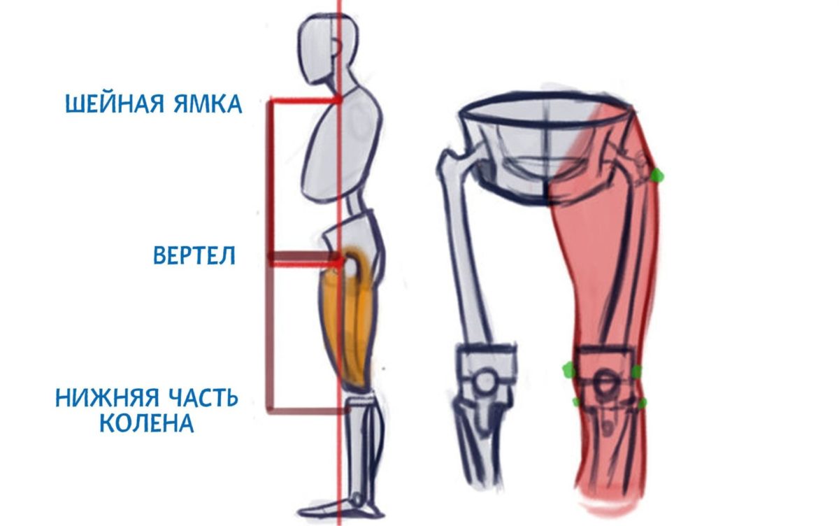
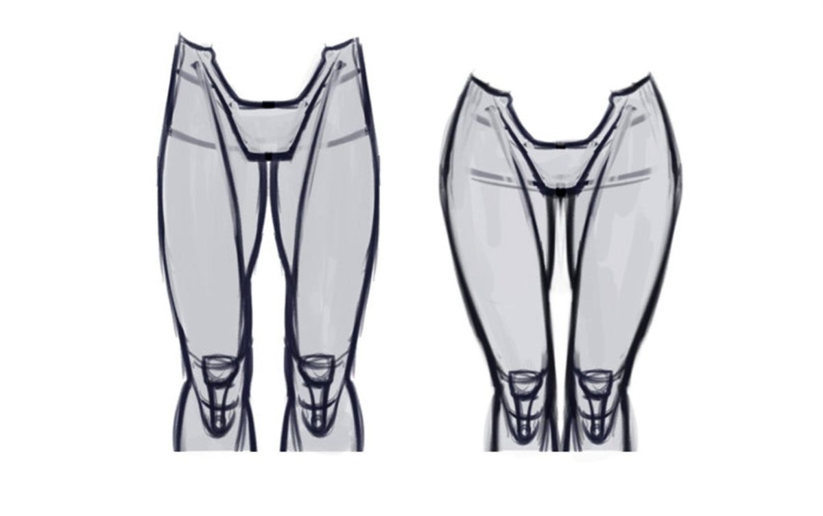
Contrary to what I encourage artists to do, which is to draw more detail, I find that they over-complicate the hips. Remember that the hip contains more fat than the lower leg, so complex anatomical details are less noticeable. Attempting to depict a heavily muscled thigh is doomed to fail unless the artist fully understands its structure. So I'll show the basic shapes first, then move on to the more complex shapes:
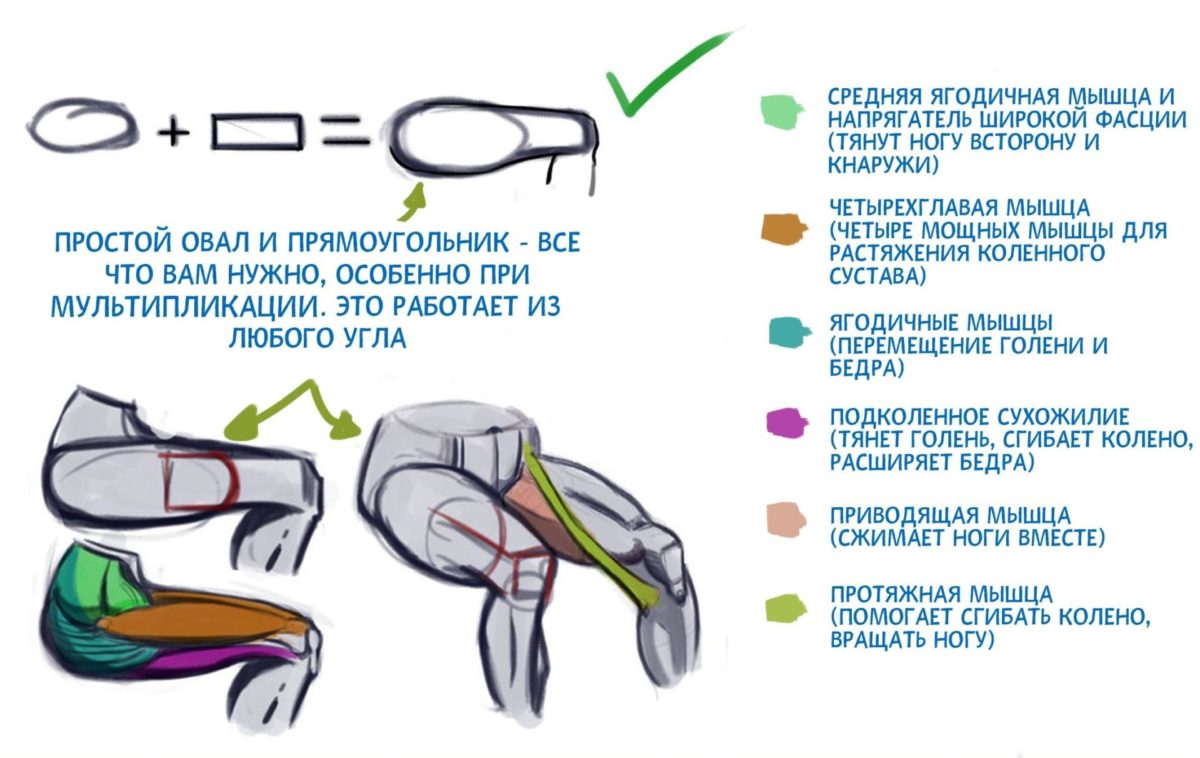
Formally, the femur begins with a bulb at the upper end of the thigh (trochanter). It is the superior external condyle of the thigh equidistant from the jugular fossa and popliteal fossa.
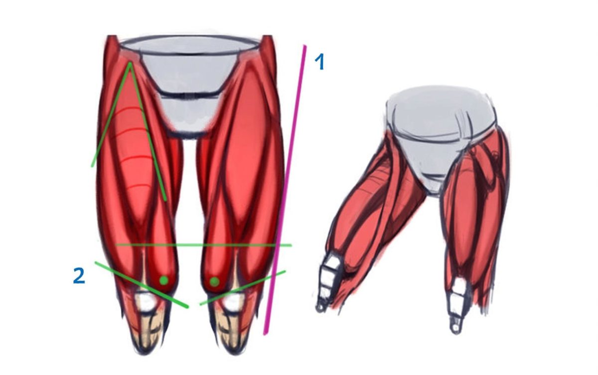
2. Quadriceps muscle of thigh. It runs at an angle. The vastus femoris medialis is located beneath all of these muscles.
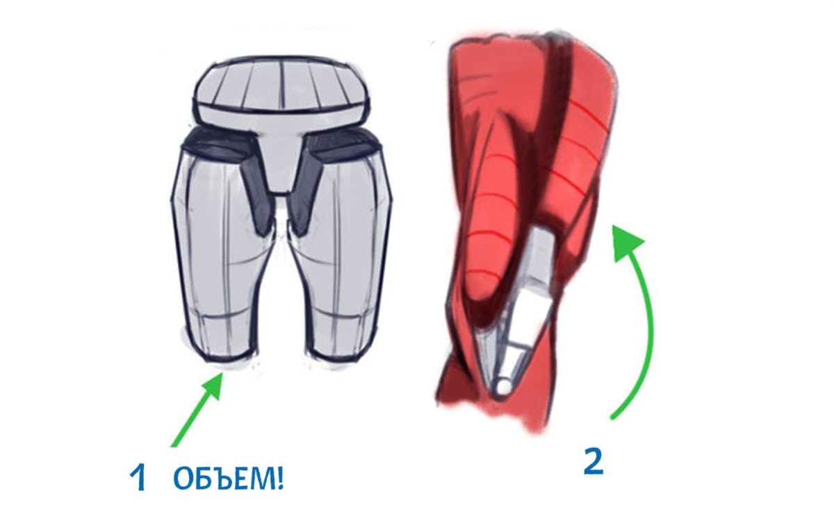
If you want to detail the thighs (remember the character is wearing pants), think about large and small volumes and how to include them.
Each bone and muscle occupies a specific place, is connected to each other and casts a shadow.
They are the same from all angles: start with the basic figures and then move on to the details. Try to hold back, after all, the skin will cover the muscle. After all, we can't see every muscle fiber.
An example: The structural pattern can be seen on the left. On the right the general pattern of the muscle (as it looks together with the skin).
Male thighs are straight. In contrast, female thighs are more angulated, which is beneficial for reproduction.
Movement
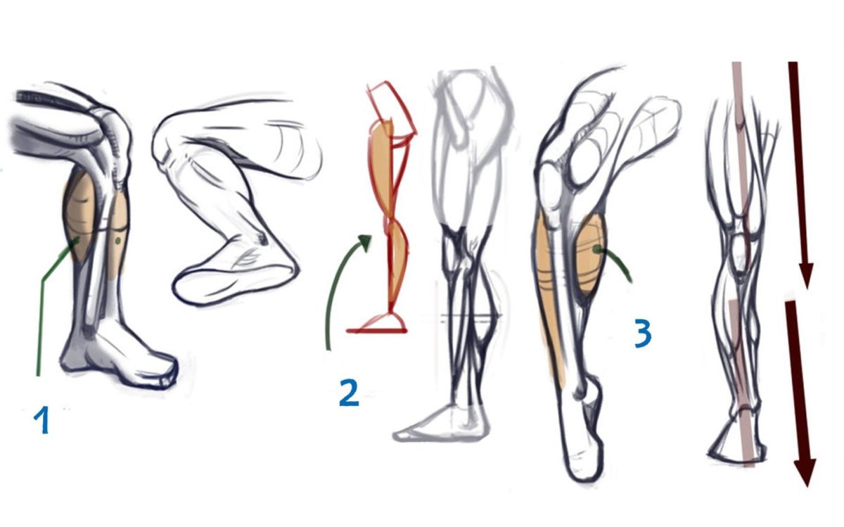
shin
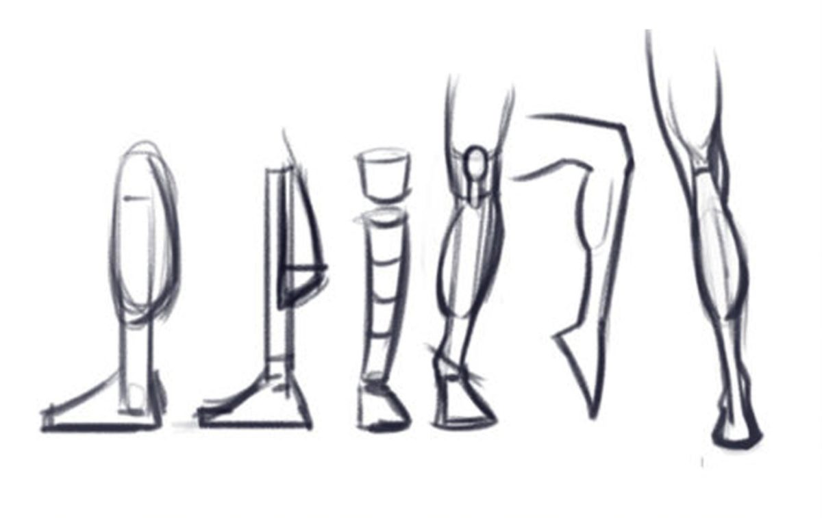
The shin appears to be a fairly simple body part, but it's even more complicated when you look at the details. For example, the bulk of the tibia sits just below the femur but is offset outward. This is a common mistake. Other important anatomical details are listed below. It takes a long time to memorize them all, but it's worth it.
Please refrain from trending and draw the lower leg as a simple inverted cone:
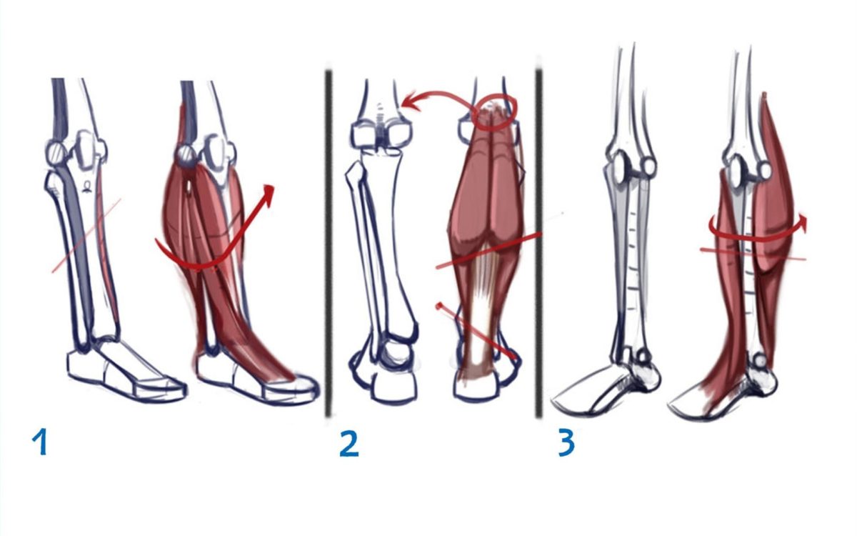
1. The tibia is mainly cylindrical, but notice how it is aligned on the front. This is due to the protruding flat, hard edge on the shin. You can feel them right under your skin! There is no muscle there.
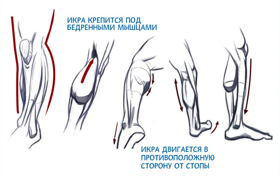
The 2nd calf muscle consists of two nodes that come out of the femur. On the other side is the ankle.
3. The tibia is visible from the front. Exposed bones cause a lot of pain when I get hit in the leg?.
1. The calf muscle is located posterior, on the opposite side (anterior) to the tibialis muscle and toe extensor.
2. Profile: Note the flatter front of the shin and the curved back. It is opposite the femur, creating balance.
3. The calf is stronger than the shin because it is used more; you pull the leg down more than when you lift it up.
Note that the shin is not in line with the thigh. It is slightly tilted to one side. As the lower leg broadens, balance improves.
Leg fracture - the concept
The main symptom of a fracture is damage to the bone, leading to a breakdown in its integrity. Such an injury requires repositioning and a cast. Depending on the classification, fractures can be transverse, oblique, longitudinal, or spiral. There are also shrapnel injuries where splinters form. The latter require a high level of medical skill to restore limb integrity.
causes of injuries
Not only onlookers and stuntmen can severely injure limbs during their work. Babies suffer leg injuries (birth injuries) before they are born. People can suffer bone damage from falls from a height, from exercise, and from accidents.
Altitude alone is enough to cause this. People often break bones when they fall backwards (slipping on ice in winter). Any part from the waist to the foot can be damaged.

Symptoms of a broken bone
Pain is the most obvious symptom of a broken bone. In some cases it is strong and stinging. A brief loss of consciousness may occur. Pain shock is not uncommon. The main causes of bone injuries are.

– failure to step on the foot,
– The formation of an extensive hematoma, a temporary swelling,
– a crunching sensation under the skin (in the case of a dislocation) and movement of the fragment (in the case of a displaced fracture),
– severe dizziness (sometimes)
– the feeling that the muscles in the limbs have shortened,
– increase in body temperature to 38-39 degrees,
– nausea,
– Disorder of tissue integrity (the most pronounced is an open fracture, in which the broken bones protrude from the skin).
In such cases, a doctor is essential. They can perform surgery to reposition the bone, immobilize it with a cast, and prescribe treatment.
It is important to understand that the response to fractures is individual. It depends on many additional characteristics. If you can walk on your leg without a cast, it doesn't mean there isn't a fracture.
In the first hours, the clinical picture is different:
Classification of NSAIDs.
Nonsteroidal anti-inflammatory drugs are classified according to whether they are COX-2 selective or not. On the one hand there are non-selective NSAIDs and on the other hand there are COX-2-selective NSAIDs.
Table - conditional division into 4 groups depending on the mechanism of action
Predominantly COX-2 selective inhibitors
Highly selective (specific) COX-2 inhibitors
Alternatively: chondroprotectors, which are prescribed in the event of severe contraindications. These are also effective against joint pathology and osteochondrosis without affecting the internal organs and systems. Chondroprotectors reduce the need for paracetamol and NSAIDs.
Evaluation of non-steroidal anti-inflammatory drugs
New generation NSAIDs are used as therapeutic agents in various areas of medicine. They are prescribed for the treatment of pathological processes in the joints, since the active substance and auxiliary substances do not destroy cartilage structures.
Here is a list of new generation nonsteroidal anti-inflammatory drugs (NSAIDs) that have a selective effect. They are effective but can cause side effects. Therefore, prior consultation with a doctor is recommended. Do not take NSAIDs for more than 3 consecutive days without medical supervision.
No. 1 – Ibuprofen (Borisov Plant, Republic of Belarus)
Opened the list of nonsteroidal anti-inflammatory drugs for joints. The phenylpropionic acid derivative ranks first among the NSAIDs. The tablets are used to treat all types of pain, including those associated with arthritis or osteoarthritis. According to the World Health Organization, the drug is available worldwide and is considered one of the safest among aspirin, paracetamol and others.
The active substance blocks an enzyme called cyclooxygenase, which converts it into prostaglandins and thromboxanes (substances that cause inflammation in muscle tissue). It reduces the release of these substances and thus effectively relieves the discomfort caused by the inflammation.
'Ibuprofen relieves the symptoms of fever and many types of pain: headache, neck, muscle, tooth and arthritis pain. It relieves cramps in the abdomen that occur with menstruation.
Read more:- What parts of the leg are called.
- parts of the human leg.
- Ligaments of the human leg.
- Ankle injury, ICD code 10.
- Photograph of a person's leg with a description.
- Can orthopedic shoes be returned?.
- outward rotation of the leg.
- The flexor muscles of the foot.