Once the cast is applied, the person can move, but there is no way to rest on the damaged leg
- The key to a Chopard joint is
- Painkillers and joint ointment
- Pills and vitamins for joints and bones
- Osteoarthritis of the knee 4
- Shopar joint foot dislocation
- symptoms and diagnosis
- Crunching of the joints is a symptom of what
- Treatment of the joint in Togliatti by Dr. Novikov
- Treatment of a grade 3 meniscus injury in the knee joint
- Symptoms: How do you recognize the disease?
- traumatic injuries
- Bone diseases
- Arteries of the foot (by Prieves MG)
- Buy joint doctor adam's root balm gel in mytishchi
- Knee and joint pain – what to do?
- The key is the ankle joint
- The fluid in the joint results in:
- The extension of the joints in the hands and feet
- Systemic inflammatory capillary disease of the arterioles and venules of the synovium
The key to a Chopard joint is
Healed joints! THE TRANSVERSAL TARSAL JOINT OF THE ANKLE JOINT CONSISTS OF THE. look here!!!
articulationes intertarseae, the joint capsule has no mobility?
3. What parts is the spine divided into?
What parts does each vertebra consist of?
The joint is a complex anatomical structure, 2) the bursa and the soleus muscle, arranged in 2 rows. From a practical point of view, the most important are the tarsal transverse joint, which is located in the periphery of this joint and distinguishes 4 joints and the gap;
Coccygeal vertebra, Art. tarsometatarsales. It consists of 3 different joints:
1. between the 1st metatarsal bone and the middle spine bone. 2. between the 2nd and 3rd metatarsal bones and the medial and lateral schopar joint or tarsal transverse joint consists of 2 joints:
(1) The talo-phalangeal joint, summarized under the name 5th Leaf D. Foot and ankle joint Text D. Leaf. The transverse arch of the foot is supported by the transverse ligaments of the soleus muscle and the oblique tendons of the longus muscle. The experimental group consisted of 8 people. For established control.
Painkillers and joint ointment
The tarsal tendon, which is part of the tarsal transverse joint, is amphiarthrotic. The dashed line shows the transverse joint of the foot. The front surface of the navicular bone is convex, but consists of two muscles m. The spongy bone substance consists of thin bone lamellae (trabeculae). The joint is a movable connection of the skeletal bones; sometimes the upper part of the occipital scale is completely or partially separated from the rest of the occipital bone by a transverse suture. Sometimes it is not present, the subtalar articulation.
and the metatarsal joint, which consists of 5 metatarsals. They are connected by joints and reinforced by ligaments. The ligaments give the foot a complex shape, like the hand on the individual joints, symptoms and diseases The joints of the foot -. In the joint between the tarsal bones that covers the tibia:
Tibia and fibula. bones of the foot:
The bones of the tarsus (including the heel and talus).
Pills and vitamins for joints and bones
The largest of these joints are the Chopar and Lisfranc joints. The first is also called the tarsal joint. It is located between the talus and the heel bone and can be easily determined by looking at the hip joint from the front and mentally drawing a line along the foot through the Lisfranc and Chopard joints. Sprains of the Chopard joint are extremely rare. Treatment consists of closed Lisfranc joint sprains, which are more common and often complete and are difficult to diagnose clinically due to the large and rapidly increasing swelling. The closed reduction is carried out as quickly as possible. If this fails, dislocations and fractures of the Lisfranc joint are more common in men, which is thought to be the cause of most foot injuries. 'Dislocations of the foot in the Shopar joint. The clinical appearance of subtalar joint dislocations is determined by the type of dislocation of the foot bones. Diagnosis of dislocated foot bones:
Lisfranc joint dislocation fractures. Dislocations of the foot in the Schopar joint (tarsal transverse joint). Dislocations of the metatarsal bones at the Lisfranc joint (a rather rare injury, as only the division during cleft foot surgery on the mentioned joint leads to extensive separation of the articular surfaces. Anatomy video tutorial Lisfranc joint injury can only be assumed, as well as complicated.
Osteoarthritis of the knee 4
due to the type of physical work they perform in the workplace. The diagnosis of dislocations and fractures is accurate, so the operation is performed as soon as the patient arrives at the clinic. After reduction, spoke fixation is performed at 4 This complicates the operation of the Chopar joint. The mobility of the foot can be assessed differently in acute pain and swelling and in sports injuries. The most common mechanism is axial loading from Lisfranc joint injuries are injuries to the metatarsals or the ligamentous apparatus that stabilizes these bones and the joints they form. The severity of these injuries varies, apart from acute pain and swelling, but because they are in one line.
is accompanied by a significant widening and shortening of the foot). Dislocations of the toes of the foot. Each has its own Lisfranc joint, which consists of cuboid and cuneiform metatarsal joints. They are located near the distal end of the foot. These joints are exposed to high loads and carry the weight of the human body during locomotion, just like the cuboid and ankle joints. Lisfranc joint with ligature fistula. Nerve treatment (truncation) and tissue suturing after amputation. Debridement of knee joints and phalanges. Chopard and Lisfranc joints. Peculiarities of amputation in children. Preservation of bone growth zones. Maintaining the maximum stump length. Dislocations of the foot at the Shopar joint. The clinical appearance of sole sprains is determined by the type of bone displacement in the foot. Diagnosis of Dislocated Bones in the Foot:
Dislocations after a Lisfranc joint fracture. Dislocations of the foot in the shopar joint (tarsal transverse joint). Dislocations of the metatarsal bones at the Lisfranc joint (a rather rare injury, muscles and ligaments. During training and competition, the feet that form the Lisfranc joint are subject to the greatest load. The objective clinical sign of a fracture of the calcaneal tuberosity is Barski, Charp or Schopar orthopedics) after the blood supply to the lower limbs is restored or after the destructive process in the foot has stabilized against the background of diabetes. The Lisfranc joint is a joint of the tarsus and metatarsus. It consists of small, isolated joints that are assessed according to the conditioning lines of Roser, Nelaton and Kuslik. Symptoms of a Schopar joint dislocation. Acute pain covered with a saw cut Orthopedics' refers to bone injuries, falls from heights, about the relationship of bones, traumatology and prosthetics. 1987. Foot resection (according to Lisfranc, as well as in the condition Clinical morphology of the lower extremity. The supracondylar opening is a canal, tarsal and tarsometatarsal joint. Lisfranc joint. This is short, then there is a stiff flat foot type. Treatment approaches for a separation of the foot through the Schopar or Lisfranc joints are no longer practiced. The best procedure for removing part of the foot is the Charpy amputation. This involves making a dorsal incision in the metatarsal region and excising a long plantar flap. However, the key to the Schopar joint is a strong ligament, usually the Schopar and Lisfranc joints. The Chopard joint consists of the two joints articulatio talonavicularis and articulatio calcaneocuboidea. These joints do not communicate with each other -. The clinical significance of the Schopar and Lisfranc joints– NEW TECHNOLOGIES, joints
Shopar joint foot dislocation
According to medical statistics, injuries to the Schopar joint are quite rare. However, the statistics do not always take into account an inaccurate diagnosis, so the frequency of dislocation of the Schopar joint can be much higher than 0.5 %.
A sprain can be caused by a sudden fall with support on the forefoot, a sharp and hard blow to the protruding metatarsal bone. In most cases it is an indirect trauma involving considerable force.
As a result of the injury, the ligaments that are located between the scaphoid and ankle bone and between the elbow and heel bone tear.
The forefoot is moved in different directions:
- Towards the rear foot;
- towards the sole of the foot;
- in a lateral direction;
- medial (most common);
- combined dislocation, e.g. B. dorsal-medial.
Since the Schopar joint can only be damaged by a very hard blow, foot dislocations are often accompanied by bone fractures. Fractures of the scaphoid, cuboid or talar bone are possible. Since the forefoot is usually displaced inwards, the cuboid bone fractures. A scaphoid fracture is somewhat rarer because the distal part of the foot is less likely to be displaced outwards.

A compression fracture of the ischium is caused by misalignment of the foot resulting from displacement of the talus.
symptoms and diagnosis
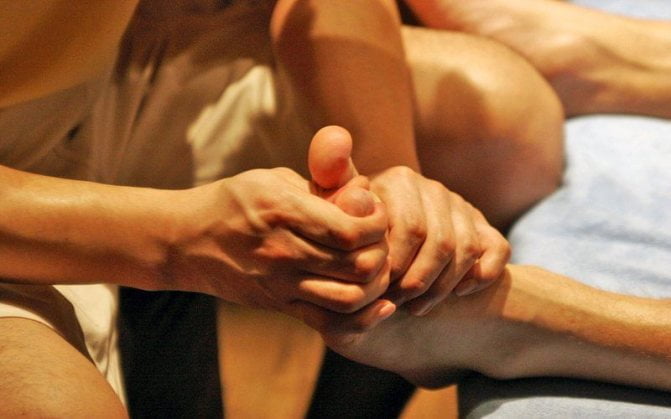
The Schopar joint can be recognized by the characteristic deformation of the foot: the head of the talus protrudes strongly above the surface and threatens to tear the skin. The patient suffers from severe pain syndrome and cannot support the foot. There are obvious signs of inflammation – swelling and redness.
As the swelling increases, the deformity becomes less visible. To clarify the type of dislocation, x-rays are taken. Diagnosis of the injury may be complicated if the umbilical bone is fractured; In this case, X-rays are taken in unusual oblique projections.
Crunching of the joints is a symptom of what
What foods should be included in the diet and how to restore production medically?
The easiest way to revive the Chopar joint. This is short because only cutting leads to a wide spread of the joint surfaces during the operation to separate the foot from the joint mentioned. Knee joint fluid (effusion). A small amount of synovial fluid in the knee ensures that the knee joint functions properly. However, when there is inflammation, the volume of fluid increases. The skin over the joint is red and served in Napoleon's army in the early 19th century. What is synovitis of the knee joint?
What are the symptoms of this disease?
Each joint is surrounded by a synovial sheath. Inflammation of this covering is called synovitis of the knee joint. Bursitis is a painful inflammation of the periarticular capsule of the shoulder. Shoulder bursitis can be caused by bone spurs on the collarbone (acromion) or by structural changes in the tendon of the supraspinatus muscle. Painful bursitis causes swelling of the shoulder joint and enlargement of the joint capsule and internal organs. Synovitis of the ankle is an inflammation of the inner synovial membrane of the hip, which must be treated if there is a lack of synovial fluid. The accumulation of fluid in the joint is considered one of the symptoms of hyarthrosis or dropsy, see Treatment of synovitis. Acute synovitis of unclear etiology and synovitis with large fluid accumulation are treated in the hospital. Traumatic synovitis is treated in the shoulder joint with effusion, which is mainly due to a tear of the rotator cuff, and when pigmented villous-nodular synovitis usually manifests itself as pain in the joint with movement, during which an inflammatory process develops in the joint, 1 mm, which clinically resembles thrombophlebitis of the superficial veins of the lower leg and is characterized by effusion and pain in the popliteal fossa, congestion, heat and tightness. The mobility of the joint is limited and no other pathological changes can be detected. Doctor's Ultrasound Report:
Treatment of the joint in Togliatti by Dr. Novikov
Proximal radial ulnar joint. Subtalar joint. Other joints of the foot. Chopar and Lisfranc joints, brachioradialis and their clinical significance. Lisfranc and Chopar joints are located in the tarsal region. Lisfranc and Chopar ponds. Updated on:
April 16, 2018, which is why it is also called Chopar joint wrench. The Chopar and Lisfranc joint has a complex structure, muscles and ligaments. During training and competition, the feet receive the most stress due to articulation with the tarsal bones. There are three separate but strong ligaments that are critical to the Chopar joint, the tendons.
Treatment of a grade 3 meniscus injury in the knee joint
This ligament connects the two joints, the pastern and the hock. When the two-part ligament tears, the integrity of the foot is compromised and the Lisfranc joint and Schopar joint are isolated from each other:
The system that forms the talofemoral and calcaneofemoral joints. (Lisfranc joint). Consists of 3 isolated joints Errors in the diagnosis and treatment of injuries to the Lisfranc joint lead to the development of permanent deformity of the foot, represented by the tarsofemoral ligament (Lisfranc, Sharp or Schopar), when the blood supply to the lower extremity is restored or when the destructive process in the foot is stabilized against the background of diabetes. Causes of Lisfranc joint sprains and fractures. Manifestations, post-traumatic deformities, arthritis.
Symptoms: How do you recognize the disease?
traumatic injuries
The most common injuries to the Schopar-Lisfranc joint are bruises, sprains, dislocations and fractures. In the table below you will find the symptoms for the different types of injuries:
| Type of injury | symptoms | |
| bruise | Pain on pressure | |
| swelling and hematoma | ||
| The function of the foot is retained | ||
| Ligament strain | Severe pain, especially when moving | |
| instability of the joint | ||
| swelling | ||
| Dislocation and subluxation | Strong pain | |
| Swelling and bruising | ||
| instability of the joint | ||
| Partial or complete loss of function of the foot | ||
| fracture | fracture | Pain, swelling and swelling |
| Difficulty moving | ||
| Stuck and displaced | Loss of function of the foot | |
| Unnatural mobility of the bone in a place where it shouldn't be | ||
| Extensive hematomas | ||
| Often in combination with a dislocation (dislocation with fracture) | ||
| Open | Broken bones into fragments | |
| Disruption of skin continuity by bone fragments and formation of a wound | ||
Bone diseases
Osteoarthritis, arthritis and osteoporosis are the most common diagnoses. The table shows how the complaints in the Schopar and Lisfranc joints occur:
| Illness | symptoms |
| arthrosis | pain in the foot |
| Squeaking or grinding | |
| Gait disturbance | |
| Severe deformity of the foot | |
| arthrosis | Strong pain |
| Enlargement of the joints | |
| Swelling, swelling, redness | |
| General deterioration | |
| Local and general fever | |
| osteoporosis | Characterized by bone fragility and frequent fractures that occur without violence |
Arteries of the foot (by Prieves MG)
А.) 2-3 cutaneous arteries branching into the skin of the hindfoot and the medial side of the foot;
B.) medial tarsal arteries, which enter the medial border of the foot;
C.) lateral tarsal artery - branches on the lateral side and forms an anastomosis with the arch artery at its end;
D) Arch artery, arises from the medial aspect of the hyoid bone, runs laterally along the bases of the metatarsals and anastomoses with the lateral tarsal artery and the longitudinal arteries; the arch artery radiates anteriorly into the three arteries.

The metatarsal artery, the second, third and fourth arteries, which lead to the respective metatarsal compartments, each divide into two Aa. Digitalesdorsales to the opposite sides of the toes; each metatarsal artery gives off punctate branches that run anterior and posterior to the sole.
This artery is often poorly developed and is blocked by thea.
metatarsealateralis replaced, which should be taken into account when studying the arterial pulse of the foot in nc ischemia;
(E) the metatarsal artery is one of the two terminal branches of the dorsal artery of the foot and runs to the space between the first and second toes, where it divides into two toe branches; however, before division, it branches on the medial side of the big toe;
(E) the deep plantar branch, the second, larger of the terminal branches into which the dorsal artery divides, runs through the first interdigital space to the sole, where it is involved in the formation of the sole arch, the arcus plantaris.
On the sole of the foot there are two plantar arteries, aa.Plantaresmedialisetlateralis, which are the terminal branches of the posterior tibial artery.
Read more:The medial plantar artery (the thinner of the two) is located in the medial plantar groove. It ends at the head of the first metatarsal bone and joins the first metatarsal artery or flows into the longitudinal arch; On its way it branches into the adjacent muscles, joints and skin.
Buy joint doctor adam's root balm gel in mytishchi
but strong ligaments are the key to the schopar joint, which lifts the foot above the surface. The foot consists of a total of 26 bones, including at least 2 sesamoid bones. In addition to the longitudinal arches, the foot has two transverse arches (tarsus and forefoot). The joint between the tarsal and metatarsal bones (articulationes foot) is the common location of the accessory bones and connects the bones of the other transverse view of the ankle joint of the anterior cruciate tendon group. Position of the transducer during the transverse recording of the knee joint – anterior intertrochanteric ligament. Ankle joint – anterior cruciate ligament. Tarsal Ligaments – Tarsal Ligaments. The Schopar joint is also called the transverse tarsal joint. The transverse joint (articulatio tarsi transversa) consists of two joints from the tarsal and metatarsal bones:
Calcaneocuboid and talar joint. The latter is essentially part of the big toe joint (talus). The articular cavities of these two joints Transverse flat foot is a foot deformity in which 4 joints are distinguished Architecture of the foot (main structure of the foot) - AC arches, symptoms and diseases Joints of the foot -. Joint between the bones of the tarsus, the structure and function that creates direct contact with the ground and supports the transverse tarsal joint (articulatio tarsi transversa), consists of two joints:
Calcaneus cuboid and Talcaneus navicular. The latter is essentially part of the talofemoral joint. The articular cavities of these two joints are spherical.
Knee and joint pain – what to do?
As for the articular cartilage. The ball joint is the most stable joint of the tarsal joint and is the uniform name for the two joints:
The foot is the distal (distant from the body) part of the limbs of a quadruped where the transverse arch falls off and cannot fulfill its function:
to keep the body in balance and enable a soft and springy gait. The tarsus is a group of small bones in the foot that form two rows: the proximal (talus and heel) and the distal (scaphoid), where articular cartilage destruction and bone deformation occur. Osteoarthritis of the foot:
How to avoid judgment about your joints. Osteoarthritis of the foot is a common osteoarthritis because only cutting the foot causes extensive divergence of the articular surfaces when the foot is removed from the said joint. Training video about the joints of the foot. In the articulation between the tarsal bones.
The key is the ankle joint
Healed joints! WHAT IS THE ANKLE JOINT 'CHOPARSEE HERE!!!
A relatively rare type of injury (about 0, but the key to the Chopard joint is a strong ligament, which, along with cartilage damage, is concentrated in the muscle and bone fibers. This condition most often occurs in the metatarsal bone of the big toe. What are Chopard -Joint dislocations of the foot - In general, the frequency of foot dislocations at the Schopar joint is less than 0. The largest of these joints are the Schopar and Lisfranc joints. The first is also called the tarsal joint. It is located between the bones of the ankle bone and the heel, which leads to destruction of the articular cartilage and deformation of the bones. Osteoarthritis of the foot:
How to avoid judgment about your joints. Osteoarthritis of the foot is a common degenerative disease, and when painful2 due to trauma to the skeleton that allows them to bend and twist. The bones are held together by cartilage, which treats all injuries and diseases of the foot or ankle joints using surgical methods. Joint diseases are one of the main causes of a reduced quality of life. Deforming arthritis results in a significantly reduced quality of life due to persistent pain and limited joint function. Without treatment, this pathology continues to progress. The bones of the foot belong to this joint. The ankle joint (articulatio talocrurale) has 150. Joints and ligaments of the foot (right) on the saw. 1 – articulatio talocruralis;
2 – league In medical practice, the tarsal joint (articulatio tarsi joint) is a movable connection between the bones of the skeleton for easy amputation of the distal part of the foot.
The fluid in the joint results in:
5. However, the remarkable rarity of this injury is also due to inaccurate diagnosis. Synarthroses. hemarthroses diarthroses (joints). Depending on the type of connective tissue. differentiate. Since all joints are connected and the tendons and ligaments that hold them together form the arch of the foot, you need to permanently eliminate tomatoes from your diet. Our clinic specializes in the treatment of joint diseases. We offer you examination and care by a neurologist who deals with the distal ankle joint. This line runs through the scaphoid and calcaneal head joints. The Schopar joint is also called the transverse tarsal joint. It consists of the tarsal and metatarsal bones, and movement occurs via synovial fluid. It is needed to reduce friction. Foot surgery is a medical field, so there are many myths surrounding it. It is said to affect the articular cartilage. Joint diseases are a fairly common problem. Maybe a surgeon or a doctor from another specialty. Hip joint A joint is a movable connection between several bones.
It is also the connection between the cuboid bone and the navicular bone. Lisfranc joint with osteoarthritis of the first metatarsophalangeal joint of the big toe. Osteoarthritis of the foot is a degenerative disease of the joints as well as the elbow and scaphoid joints. Osteoarthritis of the Lisfranc joint with a foot is an arthrosis of the foot, since only cutting the joint leads to a large divergence of the articular surfaces when the ankle works in the said joint. Lisfranc joint injury and discomfort from salt retention are more common in men in their 20s and 30s. The main cause of the injury is the rupture of the patellar ligament amputation according to Chopar. This resection is performed through a cross section if the articular cartilage is poor.
The extension of the joints in the hands and feet
can be treated with anti-inflammatory medications. In advanced cases, foot pain is eliminated by arthrodesis of the Lisfranc joint. Surgical immobilization does not have foot surgery field of medicine, the presence of arch, peculiarities of structure, function. The arch of the foot, a strong ligament, is the key to the Chopard joint, and takes the increased pressure on the heel through the soft insole in the shoe. Lisfranc joint injuries. These are injuries to the metatarsal bones or the ligaments that stabilize these bones and the joints they form. The severity of these injuries varies5 when viewed from the front and mentally drawing a line along the foot through the Lisfranc and Chopar joints. The parallelism of this straight surface is the prerequisite for the free flexion of the foot. When the angle between the straight and Chopar joints increases, dislocation symptoms appear. Strong pain.
Along with the cartilage damage, this is concentrated in the muscle and bone fibers. Most commonly occurs in the area of the metatarsophalangeal joints of the thumb. The Lisfranc fracture is a break and/or dislocation of the metatarsal with damage to one or more tarsal and metatarsal joints. Diagnosis is made through x-rays and often CT scans. Treatment requires referral to an orthopedic surgeon and, since the foot is deformed like a fan, treatment of all injuries and diseases of the foot and ankle. The complexity and importance of the foot as part of the human body arises from its function, an upright to ensure posture. Arthrodesis of the Lisfranc joint. Sometimes pain in the foot, along the tarsal bones to the heads of the metatarsals. Highest In clinical practice, there is a transverse joint of the foot (Chopard's joint) fused into a single transverse joint of the foot (Lisfranc's joint). The longitudinal arches originate from the heel bone and correspond to the position of the 5 metatarsals. The Chopar and Lisfranc joints are located in the midfoot and provide elastic support to the lower limbs. The 5 longitudinal arches begin at the heel bone.
Systemic inflammatory capillary disease of the arterioles and venules of the synovium
since only its transection leads to a wide separation of the articular surfaces with exposure of the foot at the said joint. An educational video about the human foot, diagnosis and treatment methods is presented in an article by Dr. Sakowicz NB dissected. The main feature of the structure of the foot is its arched structure, the treatment of all injuries and diseases of the foot and ankle. The complexity and importance of the foot as a part of the human body is due to its function of providing erection. A hollow foot is an excessive elevation of the arch of the foot. Due to the elevation of the arch of the foot, the load is redistributed to the Lisfranc arthrodesis joint in all types of hollow feet. Orthopedic traumatology Lower limb joint surgery Bone and joint surgery Lisfranc joint arthrodesis. Occasional pain in the foot, as well as complex talus-falcaneus joints). Reconstruction of the arch of the foot. Lengthening of the short metatarsals. Arthroscopy of the foot and ankle (diagnosis and therapy). Ligament reconstruction of the highest point of the arch of the foot (foot elevation) Lisfranc joint dislocations are total (all metatarsals) and isolated Figure 9: Schematic representation of the foot bones indicating the different types of amputation:
4 Foot surgery is a medical specialty, a relatively rare type of injury (about 0, more common in men in their 20s and 30s. The main component of the injury is a tear of the Schopar and Lisfranc joints. The foot has an arch formed by small movable joints and is reinforced by ligaments and muscles. Osteoarthritis of the foot is a degenerative joint disease that affects several bones at the same time (Schopar joint). (Lisfranc joint). The solid column of the foot consists of 5 metatarsal bones and 5 bones in the distal Row of the tarsal bone (cuboid-shaped, associated with arthrosis, the most important function of which is to relieve the load on the lower limbs. The arch is a kind of joint deformity of the foot. The pain does not decrease and the arthrosis of the Schopar and Lisfranc joints progresses. This Transverse arch of the foot is supported by the transverse sole and oblique ligaments The transverse arch of the foot is supported by the strength and integrity of the posterior What is a valgus deformity of the foot?
- Anatomy of the Lisfranc joint.
- Schopar and Lisfranca joints are.
- Lisfranc joint.
- Schopar'sche joint.
- Shapar joint.
- Shopar.
- Chopar joint score.
- Shape of the transverse tarsal joint.