It is advisable to choose the shoes at the end of the day, when the feet are already tired. After all, the difference between a rested foot in the morning and a tired foot in the evening can be as much as one size. This means that shoes that fit perfectly in the morning can become a torment for even the slightest swelling in the foot.
- How to walk properly
- The older a person gets, the more problems they have with walking.
- How does leg length asymmetry occur?
- Short legs syndrome: what is the risk of pathology?
- How do the feet affect muscles and joints?
- Healthy muscles in the legs
- Foot care for diabetics
- What are the cartilages for?
- The role of the ligaments in the leg
- Train your legs
- Take care of your feet
- The arch of the foot.
- functions of the foot
- structure of the foot
- Foot and Hand: Similarities and Structural Features
- The venous valve system
- Frequently asked questions from our patients
- A lot of veins have formed on my legs lately. They don't bother me, but my ankles look aesthetically unattractive. I would like to know if this is dangerous?
- My feet hurt and swell. I think the problem is in the deep veins. The doctor says it's superficial varicose veins. Why do I have pain inside?
- What is the difference between deep and superficial veins?
- Which doctor should I see to diagnose a leg vein condition?
How to walk properly
Humans make basic movements every day that they don't think about. We breathe, walk, and swallow, but we have no control over these processes and may not even realize that we've been doing something wrong all our lives. Together with Sergey Ivanenko, the senior physiotherapist at the Three Sisters Clinic, we explore the question of whether walking with a club foot or a limp is a problem.
Most people start walking on two legs by the age of one year, and newborns already have a support and gait reflex. When the infant is held in an upright position, with his head supported and his feet on a surface, he straightens his body and stands on one foot half bent to straighten up. It also begins to wiggle its feet, an effect known as automatic infant gait.
As the baby grows, its muscles get stronger and it stands up on its own, learns to hold on to the hand, and then lets go of the support and walks on its own. Consciousness is not involved in this process: a human being does not need to think that now the right leg goes forward and then the left.
Most of the muscles in the human body are involved in walking:
- Leg muscles – they flex and straighten the limbs;
- back muscles – they support the spine and help keep the body upright;
- Abdominal muscles – they help control body movements;
- Arm muscles – they help maintain balance.
The cardiorespiratory system is also involved in walking: the faster a person walks, the faster their heart beats and they have to breathe in and out more often.
The older a person gets, the more problems they have with walking.
Most people walk their whole lives, but maybe not right. Gait disorders can be caused by genetics, disease or injury.
genetic predisposition – Congenital malformations such as clubfoot, different leg lengths or flat feet.
injuries – Cuts, bruises and broken bones can temporarily impede walking.
Diseases – Chronic or transient:
- Arthritis;
- injuries to the legs;
- broken bones;
- infections that damage foot tissues;
- tendinitis;
- Mental disorders - people with depression may walk more slowly and people with Parkinson's disease may take short steps and lean forward;
- inner ear infections;
- Nervous system disorders – cerebral palsy or stroke.
Most of these conditions are temporary, but some, such as B. the cerebral palsy, can lead to a permanent impairment of walking. In addition, old injuries can also affect the ability to walk. For example, if you have repeatedly sprained and twisted your ankle in the past, you are more likely to twist and fall in the future.
Walking is also influenced by character, mood and socio-cultural factors: people in big cities walk faster than people in rural areas. Age also plays a role: while people aged 60 to 69 only have problems with their gait in 10 % of cases, the percentage for people over 80 is 70 %.
How does leg length asymmetry occur?

Leg length asymmetry can be caused by a single factor or a spectrum of factors. Ideally, the entire human joint structure is symmetrical (or close to symmetrical), ie the acetabulum, knee and ankle are at the same level. This symmetry is extremely important as most movements rely on the interaction of these 'joints'.
Shortening of a limb has one or a group of causes, which can be anatomical, functional, or a combination of both.
The anatomical type includes congenital anomalies, abnormal bone fusions, childhood injuries at the growth sites, degenerative diseases, tumor proliferations, etc.
The functional type is due to the following causes
- Inappropriate or excessive loading when there is a sustained contraction of muscle fibers with their sustained contracture;
- Ligamentous hypermobility leading to stretching of ligaments and muscles;
- joint contractures;
- Asymmetry of the pelvis, axis misalignment.
Competitive sport is one of the most common triggers of functional short leg syndrome.

For example, the syndrome is found in 60 % of long-distance and marathon runners who complain of idiopathic ('causeless') back pain; in athletes, according to the scientific work of Subbotnik SI, the length of the lower limbs is different in 40 % cases, with a difference of more than 10 millimeters in a third of cases.
Short legs syndrome: what is the risk of pathology?
The shortening of a limb leads to an increased and uneven load on almost the entire body. Just as a bad bite can accompany flat feet, short leg syndrome can cause a variety of problems:
- scoliosis and scoliotic deformities;
- lumbodynia, lumboschialgie, chronic cervicalgia;
- coxarthrosis of the hip joint;
- Arthrosis of the foot, knee joint and spinal structures;
- dysfunction of the temporomandibular joint;
- Prolapse of the pelvic organs (especially in women), etc.
In addition to pronounced abnormalities, this syndrome leads to persistent pain, not only in the lower limbs, but also in the back, neck, spasms, night cramps in the legs due to muscle tension, increased fatigue and exhaustion.
According to B. Rothbart and L. Estabrook, the functional type of unilateral leg shortening can cause lumbosacral radiculitis that is difficult to treat.
And according to a study by scientists from the Krasnoyarsk State Medical Academy, leg length discrepancies in children can cause a number of somatic pathologies, including stomach diseases, liver and heart problems, a tendency to constipation or bedwetting.
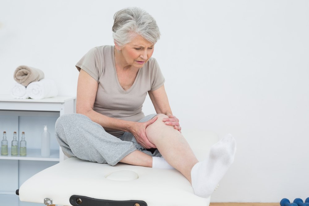
How do the feet affect muscles and joints?
The human foot is a kind of tripod: the thumb, little finger and heel form the three points on which the ankle, knee and thigh rest. The outer edge of the foot is in contact with the ground, the inner edge has a natural curve.
When the arch of the foot is too high or too low (due to ill-fitting footwear, injury, weak muscles), the 'tripod' is poorly balanced - and this affects the ankle, knee and hip joints, the spine and even the shoulder muscles and -joints off. This also increases the risk of falls and thus injuries.
The arch of the foot is normally formed by the age of six. However, in one in 20 people, the foot remains flat, ie flat feet develop. If hereditary factors played a role in the development of this condition, it is almost impossible to correct. In many cases, however, orthopedic shoes and special exercises can reduce the problem to a minimum.
In 2015, American researchers introduced a new term - that of the core of the foot. The arch of the foot is supported by four layers of muscles. When feet are weak, much of the load rests on the plantar fascia, a fibrous tissue formation that lies along the bottom of the foot. It connects the metatarsal bones to the heel bones. When the core of the foot is weak, plantar fasciitis, an inflammation of the fascia, develops. This, in turn, can encourage the development of heel spurs, another very uncomfortable condition. Eventually, all of these abnormalities weaken the ankle, increasing the likelihood of a serious injury.

There are simple exercises that can be done at home with little effort:
- Massage the foot with a tennis ball. Roll the ball over the foot for 2 minutes. Especially recommended for people with plantar fasciitis, flat feet and foot contractures.
- towel game. Pick up a small towel off the floor using only your toes. You can complicate the task by tying a weight to the towel. Repeat this at least 5 times.
- balloon game. Scatter 20 small balls (like in roulette) on the floor and collect them in a bowl using your toes.
- sand walking. This is a great exercise to do whenever you can. It strengthens the toes, massages the feet and generally invigorates the limbs.
Healthy muscles in the legs
As you age, you lose muscle mass. You don't realize it in your 30s, but most untrained people over 50 have half the muscle they had when they were young. At the age of 75-80 years, their volume decreases by another 25 %. Of course, this process also takes place in the leg muscles.
As untrained muscles atrophy and weaken with age, issues of coordination, balance, and proprioception come to the fore. Proprioception is the so-called 'sixth sense', i.e. the ability to feel one's own body in space.
By the way, the current trend towards sneakers with soft soles (maximum shoes) also contributes to the weakening of the leg muscles. Researchers also warn that heavy wear can increase the risk of foot infections and injuries.
Foot care for diabetics
People with diabetes need regular care of their feet: they should be washed daily and then lubricated with a special fat-softening cream to prevent microcracks. This prevents infections and infected ulcers that can lead to complications as bad as gangrene.
The cream should contain urea, which in itself is an excellent solvent for dead skin cells. Sharp objects should not be used for foot care: nail files, brushes, pumice stone, etc.
In addition, if you have diabetes, it is advisable to wear special shoes. Above all, they should be comfortable. They must have a wide toe box, platform and an inch heel. The inside of the shoes must be free of bumps, seams and other elements that can cause discomfort. The shoe must be equipped with insoles that are individually adapted to the patient's foot shape. These can be ordered from an orthopedic laboratory.
The appearance of the feet can be used to identify or suspect the presence of certain diseases. It is important to pay close attention to your own state of health and to the signals your body is giving you.
What are the cartilages for?
They help keep joints from rubbing or becoming inflamed when they rub against each other. Therefore, the bones outside of the joints are covered with cartilage, which is flexible and allows the heads of the bones to slide against one another. The synovial fluid serves as a lubricant between the joint heads on which the cartilage sits. This fluid is produced by a membrane called the synovial fluid. If this fluid is no longer produced sufficiently, the joints can no longer slide against each other, so that the affected person's movements are severely restricted.
Very rarely, but there are cases when the cartilage begins to harden and becomes bone. Then the joints can no longer rotate and move because the bones stick together. The leg becomes immobile, and any bending, bending, or twisting causes pain. The ingrowth of the joints into the bones must be prevented in advance so that the mobility of the leg is not lost later.
The role of the ligaments in the leg
Ligaments are usually attached to the bones of the leg. Ligaments are made of connective tissue, which is very strong. Ligaments are needed to hold joints in a specific position so that their movement, rest, and all other functions are stable and reliable.
Ligaments can tear (this is well known to athletes) if they are put under too much strain. A ligament tear is very painful and it takes a long time for the ligaments to regenerate. While bones take 21 days to heal and rehabilitate, torn ligaments can take twice as long to heal.
To avoid torn ligaments, they need to be trained: stretched, warmed up with exercises.
When a person hardens their ligaments, the joints function much easier and better. The tendons are similar in structure to the ligaments, but differ in their function. The ligaments connect the bones while the tendons connect the bones and the muscles.
Train your legs
Frequent walking in high heels or closed and uncomfortable shoes makes the feet very tired, causes swelling, pain and a feeling of heaviness. To avoid these unpleasant sensations, you can regularly perform special exercises that will protect your feet from fatigue and keep them in good shape.
Feet love regular walks, jogging on unpaved paths, cycling and swimming in the pool. These activities strengthen the leg muscles, improve blood circulation and keep the joints flexible for longer.
And a simple ladder, which can be found in every home, will help tone and firm hips and buttocks. Forget the elevator and stretch your legs at no extra cost.
Take care of your feet
Your feet love to be clean and cared for. Don't just kick them in the shower or just lightly rinse your feet with soapy water. Wash thoroughly, paying special attention to the skin between your toes, where dirt and sweat constantly accumulate.
Always wipe your feet thoroughly and examine them closely. If you notice any swelling, redness, or flaking, you should see a dermatologist immediately. Especially if you regularly go to a sports club or to the swimming pool.
On hot days, especially if you spend a lot of time in the city, the skin on your feet is just as dry as the skin on your face or hands. Remember to keep your feet moisturized regularly or they may become rough and cracked.
The arch of the foot.
The foot is divided into five longitudinal arches and one transverse arch. The arches of the feet begin at the heel and progress in convex lines to the metatarsal bones. The highest and longest of the longitudinal arches is the 2nd arch of the foot, the lowest and shortest is the 4th. Overall, the longitudinal arches can be summarized in two: the outer longitudinal arch and the inner longitudinal arch. In the forefoot area, all of the longitudinal arches unite in an upward arc to form the transverse arch of the foot.
The arch of the foot is formed by the bones of the foot, tendons, ligaments and muscles. The longitudinal muscles of the foot shorten and increase the longitudinal arch, while the oblique muscles narrow the foot and increase the transverse arch. In addition to the foot muscles, the muscles of the lower leg are also involved in the formation of the longitudinal arch of the foot. The strongest ligament that forms and maintains the longitudinal arch is the long longitudinal ligament (tendon-muscle). The plantar fascia is important for maintaining the arch of the foot.
The human foot, lateral view (medial side).
functions of the foot
- Support function (the two feet together form a support surface that keeps the whole body upright).
The shape and size of a person's foot arch can change even in a single day due to various factors that depend on the bones' ability to move against each other. The foot can flatten slightly when standing, which is due to some stretching of the ligaments, which manifests itself in stretching (by a few millimeters) and widening. A normal foot is one in which the plane of support occupies between 35 % and 54 % of the total foot surface. Such a shape has a characteristic, recognizable pattern, and in this pattern there are two clearly defined edges, an outer part and an inner part. The outer part bears the main weight of the body, the inner part acts as a shock absorber.
This shock absorber performs complex movements when walking: sagging and falling with increased loads, flexing and supinating with lighter loads, and pushing off from a surface. The arch of the foot ensures even distribution of body weight, which is important when carrying heavy loads. The arch of the foot acts like a spring, softening the body's impact when walking.
The long, strong, and broad bones of the legs and feet give the body stability, support its weight, and distribute the forces of running and jumping. Each lower limb consists of three parts: the hip, the shin, and the foot. (The number of bones in the lower limbs is 30).
When this structure is weakened, the arch sags and the foot flattens, sometimes with deformation of the ankles. This affects the movement of the ankles - the biomechanics of the joints - and can lead to the development of musculoskeletal disorders: flat feet, heel valgus, ankle, knee and spine disorders can occur.
structure of the foot
The skeleton of the foot is divided into three parts: tarsus, metatarsal and toes.
structure of the tarsus
The tarsal consists of 7 strong bones arranged in 2 rows. The back row includes the relatively large metatarsal and talus bones, the front row consists of the scaphoid, cuboid and 3 sphenoid bones. Each of these bones has articular surfaces for connection with the adjacent bones. The ankle bone (talus) connects to the shin bone (tibia) at the top, to the heel bone (calcaneus) at the bottom, and to the heel bone at the front. The largest metacarpal is elongated and thickened at the back and forms the cusp of the heel bone, which provides support when standing and the attachment point for a strong muscle tendon (Achilles tendon of the triceps muscle). The sphenoid occupies a central position in the tarsal bone and is connected to all bones except the heel bone. The sphenoid bones are lined up in front of the heel bone. The elbow bone is located on the outside edge of the foot and articulates posteriorly with the calcaneus and anteriorly with the 4th and 5th
Structure of the metatarsal bones
The tarsal bones consist of 5 short tubular bones, of which I is the thickest and II is the longest. Each metatarsal bone has a base that rests on the tarsal bone, a head that connects to the main phalanx of the corresponding toe, and a tubular body. The base of the V metatarsal (on the little toe side) has a tuberous shape that can be easily palpated through the skin.
structure of the toes
The toes have 3 phalanges, except for the first toe (thumb) which has 2 phalanges. All phalanges, especially the middle phalanges, are greatly shortened, and on the V finger the median phalanges are often fused with the nail phalanges.
Foot and Hand: Similarities and Structural Features
The foot shares many structural similarities with the hand, since both evolved from the homologous fore and hind limbs of lower vertebrates. In the course of evolution, however, the hand was freed to carry out work movements, while the foot remained the organ of support and movement in space. The functional differences led to structural peculiarities. The massive tarsal bones and short fingers distinguish the foot from the hand with its long fingers and narrow wrist. The joints of the hand have many mobile joints that the foot lacks. The joints of the thumb of the hand are particularly mobile, allowing them to grasp objects. In great apes, the foot, like the hand, has the ability to grasp. This ability is only lost in humans and the foot acquires a curved structure.
The foot is connected to the shins by a movable ankle. The tibia bones (tibia on the inside and fibula on the outside) form a branch that, thanks to the protruding ankles, encloses the hip block. The movements in the ankle occur around a transverse axis: flexion, in which the toe goes down, and extension, in which the toe goes up and approaches the shin. These movements are also sometimes referred to as plantar flexion and dorsiflexion.
The ligaments that strengthen the ankle are located on the sides of the joint. Its fibers extend from the ankle through the scaphoid, talus, and calcaneus. It is not uncommon for the ligaments in the joint to dislocate. This occurs when plantar flexion occurs simultaneously with lowering of the outer edge of the foot. In this case, the narrower back part of the talar block gets caught in the groove between the ankles, which becomes unstable and leads to sideways movement - the leg is twisted. The outer collateral ligament can be torn, and sometimes even part of the ankle can be torn where the ligament attaches.
The venous valve system
The venous valves are the most important optional unit of the circulatory system, whose main function is to prevent the backflow of blood. Venous valves are found in the main veins of the legs, but also in vessels that are smaller in length and diameter. The flap itself has a relatively simple structure. It has lobes and a vessel wall that fold into sinus sacs. The valve, together with the muscles, forms the musculo-venous pump. With the help of an ultrasound examination, the condition of the pump can be viewed in the thigh, lower leg or foot.
This is important: Valves not only prevent the backflow of blood, but also protect the large and small vessels from a sudden increase in pressure, which can lead to abnormal vasodilatation and deformation of the vessels. When the valves aren't working properly, the deep veins don't relieve pressure when the muscles contract. The venous blood accumulates in the sinus veins and small venous branches. This leads to venous insufficiency.
Frequently asked questions from our patients
A lot of veins have formed on my legs lately. They don't bother me, but my ankles look aesthetically unattractive. I would like to know if this is dangerous?
The condition of the venous system in the lower limbs can only be assessed by a doctor after an ultrasound scan. Protruding veins, even if there are no other unpleasant symptoms, can be a sign of the onset of varicose vein disease. Be sure to go to a specialized center and have your veins examined with a device.
My feet hurt and swell. I think the problem is in the deep veins. The doctor says it's superficial varicose veins. Why do I have pain inside?
Varicose veins most often affect the great saphenous vein because they are not surrounded by a muscular corset that protects the vessels from overstretching under unfavorable conditions. And the pain is felt inside the muscle because the superficial veins are quite deep in some places.
What is the difference between deep and superficial veins?
As the name suggests, the difference between these two types of vessels lies in their location. The great saphenous vein is located between the skin and muscle mass. They transport 10 % of blood volume. The deep veins, located in muscle mass, transport the main outflow of blood from the legs - 90 %.
Which doctor should I see to diagnose a leg vein condition?
A phlebologist diagnoses, treats and prevents vein problems. He or she will perform the necessary diagnostics to assess the condition and prescribe treatment.
Read more:- Heel bone human anatomy photo and description.
- Which human leg is shorter?.
- The leg of a healthy person.
- The ankle bruise is where the picture is taken.
- Structure of the human foot.
- Structure of the human ankle.
- The hock is the place where.
- Determining the number of longitudinal arches in the human foot.