We found that the K factor is the same in boys and girls during the first period of childhood. In the second period of childhood, in both asthenics and hypersthenics, the K factor decreases to the same extent in girls and boys (Table 1). It decreases more significantly in normosthenics. The differences between these adjacent age groups are statistically significant.
- Examination of the longitudinal arch of the foot in children using computer tomographic plantography
- Purpose
- The high arch of the foot and everything related to it
- What is the cause of this?
- What are the risks?
- What can I do if my feet have high arches?
- Why shouldn't you forget to make an appointment with your orthopedist for a check-up?
- Prevention of flat feet in children
- Treatment
- prevention
- What arises from the problems with
- Possible complications
- Main symptoms of longitudinal flatfoot in adults
- Symptoms of flat feet depending on the change in the arch angle of the foot
- CONTACT
- Classification of flat feet according to the cause
- Factors contributing to static flat feet:
- CONTACT
- Poor posture and flat feet
- Overview of the lesson 'Bad Posture and Flat Feet'.
- Specificity of foot function in children
Examination of the longitudinal arch of the foot in children using computer tomographic plantography
The goal of the Our study aimed to determine the dynamics of the anatomical parameters of the foot during the development of the longitudinal arch in children. methods. A total of 1561 children of both sexes with asthenic, hyperstenic and normosthenic physique were examined; Of these, 199 were first-generation children and 394 were second-generation children, 252 were teenagers and 716 were adolescents. The longitudinal arch of the foot was examined using computed tomographic plantography based on the K factor. Results. In the first phase of childhood, the K factor has the same value for boys and girls. In the second period of childhood, the K factor decreases by the same value in both asthenic and hypersthenic girls and boys. It decreases more in normosthenics. During puberty, the K factor increases in boys of all somatotypes compared to the previous period and becomes slightly higher than in girls. Among girls, the coefficient increases in normosthenics (p0.05), while it decreases in asthenics (p>0.05). In adolescence, the K factor continues to increase compared to the previous period in both sexes for all body types: in girls by 4.54 (p<0.05), 15 (p0.05) and in boys by 2.3, 12.8 and 16.3 %, respectively (p<0.05), remaining highest in boys.
AI Perepelkin SBEE HPO Volgograd State Medical University, Ministry of Health of the Russian Federation, Department of Human Anatomy, Prof. Dr.-I. Krayushkin; AI Krayushkin – Volgograd State Medical University, Ministry of Health Protection of the Russian Federation, Department of Human Anatomy, Head of the Department, Professor, Doctor of Medical Sciences ES Atroshchenko – Volgograd State Medical University of the Ministry of Health Protection of the Russian Federation, 6th year medical student.
Purpose
Determination of the dynamics of the anatomical parameters of the foot in the formation of its longitudinal arch in children.
We led the development of a research method that uses a hardware-software system to assess foot condition. The work was carried out in three phases: Phase 1 involved the development of a hardware-software system for analyzing the anatomical and functional condition of children's feet. In phase 2, children of different age groups and university students were examined. In Phase 3, a treatment program was developed and implemented based on 18 different foot conditions. For each child and adult, a consent protocol for the study was completed, which was approved by the local independent bioethics committee (Protocol No. 87 dated November 26, 2008).
Many of the existing foot examination methods are costly and quite time-consuming. Therefore, there is a need for a qualitative change in the most 'popular' and technically simplest methods, taking into account the specifics of doctors, teachers and educators, which will facilitate the solution of the tasks set. We have proposed a computer-aided plantographic complex and methods for determining the anatomical and functional state of the foot (Patents of the Russian Federation for inventions No. 2253363, 2309663, 2331360, 2358650). The computerized plantographic complex (OOO 'Ortoped', Volgograd) has a registration certificate from the Federal Service for Supervision in Health Care and Social Development № FSR 2011/10105 dated February 11, 2011, which approves its production, sale and use in the Russian Federation approved. The method aims to increase the resolution of flatbed scanning, the depth and quality of diagnosis of foot pathologies and the integral assessment of the final results through the creation of special diagnostic algorithms.
The Plantograph is a specially reinforced flatbed scanner that can support the weight of a human body. The image of the foot was displayed on a computer monitor and processed using a graphical computer method. Various forefoot, midfoot and hindfoot measurements were calculated, including 1st and 5th toe flexion angle, K-factor and heel angle, and the software generated an individual report for each child's foot.
The high arch of the foot and everything related to it
A person's foot can have a normal arch, a low arch, or a high arch. The first option is not associated with abnormalities, pain or gait disorders. In the second case, a low arch means that you have flat feet. It causes pain, swelling, and gait disturbances that can lead to valgus foot deformities and ingrown toenails.
Like flat feet, the hollow arch of the foot is a pathology. With a high arch, the ability to turn the foot is significantly limited. The foot sinks toward the outer edge and the load is distributed unevenly from the big toe to the little toe and ring toe.
What is the cause of this?
The high elevation of the athlete's foot type can be seen with the naked eye. When the foot is placed on a flat surface, you will notice that the middle part of the arch of the foot rises above the surface. This is often seen in ballerinas. It can be the result of muscle hypertonicity caused by certain neurological diseases. Spinal cord tumors, cerebral palsy, polyneuropathy and poliomyelitis can also be triggers for the development of this condition.
What are the risks?
The disadvantages of a buckled foot are quite numerous. It may be that the foot does not fit into the shoe but is pressed against the upper part of the shoe. Women may experience discomfort or even pain when wearing high-heeled shoes or sandals.
People with this peculiarity have a significantly restricted gait and increased pressure on the ankles. Calluses form on the soles of the feet and the feet shrink. The toes are particularly affected, as they become deformed and clawed over time. In severe cases, a person with high heels may suffer physical disabilities and partial inability to work.
What can I do if my feet have high arches?
In children, foot abnormalities are detected during preventive examinations. In adults, gait problems and pain when walking may indicate that the heel is too high.
Only a doctor can make an accurate diagnosis. If problems are identified, the affected person can be prescribed various physiological treatments and massages. Wearing special orthopedic shoes and insoles plays its own role in correcting arch deformities.
Vibration insoles have a positive effect on such pathologies. They are considered to be the salvation of people with high arches as they eliminate all the discomfort associated with it. They can be worn by anyone. They are used for both therapy and prevention.
If you think the information on this page will be useful to your friends, acquaintances or colleagues, please share the post on your social network. Simply click on the corresponding symbol:
Why shouldn't you forget to make an appointment with your orthopedist for a check-up?
Although the diagnosis is not made until the age of 5, the doctor can already determine whether the child has a predisposition to the development of any pathology. Based on regular observations, he can determine what needs to be done to prevent flat feet from developing. The doctor will tell the parents what needs to be done individually and to what extent so that the child's feet develop properly.
Three visits to the orthopedist in the first year of life and every six months thereafter, even if everything is normal.
Do not trust the advice of sellers in children's shoe stores. They are more interested in sales than in the health of their little customers. In some stores there are special devices that are supposedly designed to examine children's feet for flat feet. Do not rely on the readings of these simple devices. As a rule, they indicate a certain degree of flat feet in all children, since they are designed not for diagnosis, but to increase the heel of children's orthopedic shoes. A diagnosis can only be made by a doctor and only after a comprehensive examination. It is also best to seek advice from an orthopedist when choosing footwear.
Prevention of flat feet in children
Our ancestors hardly had flat feet. This is because they mostly walked a lot and ran barefoot. Flat feet are rarely observed in rural children and barefoot Africans. But in sedentary city dwellers this pathology is everything and more.
- Sedentary lifestyle, lack of physical activity;
- excess weight, which increases the load on the foot;
- Wearing the wrong shoes or another child's shoes;
- rickets in childhood;
- injuries;
- Trauma; Inherited predisposition.
To reduce the risk of developing this disease:
- Foot and toe massage. This should first be done by a professional, then the parents themselves will learn the proper techniques.
- Warm foot baths with sea salt.
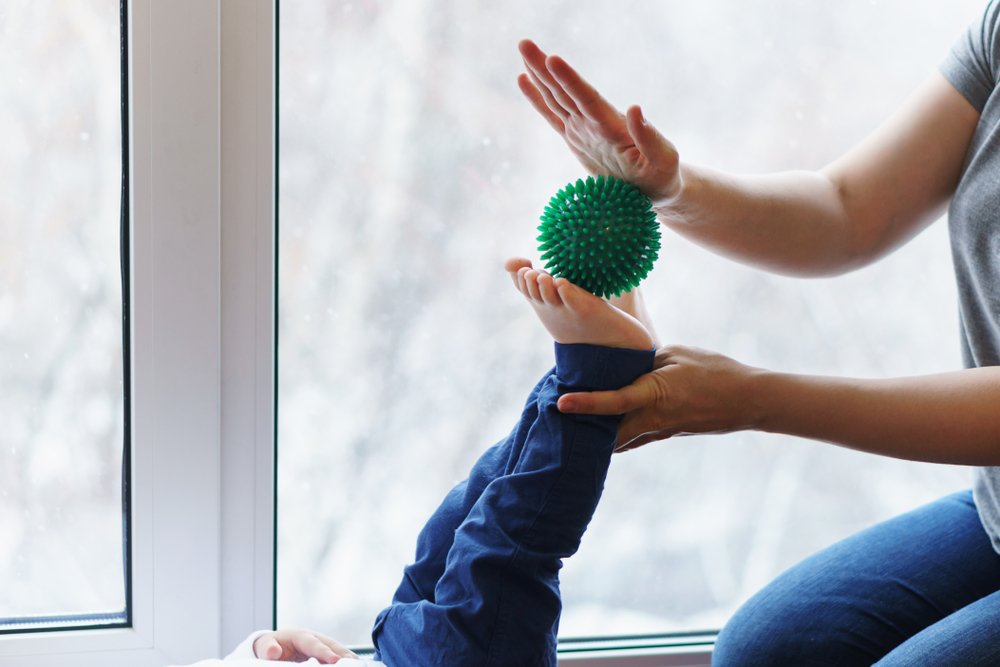
- Walking barefoot on grass, pebbles, seabeds or special massage mats. You can create a special path at home using stones, chestnuts, round sticks or logs.
- Special exercises that children like to do in the form of games. This includes rolling different sized balls or massage rollers, walking on your toes, on your heels, on different sides of your feet. The doctor selects the exercises, their sequence and the number of repetitions, and this should be done individually.
- Application of paraffin and ozokerite, electrophoresis.
- Avoid obesity, eat a healthy diet and get plenty of exercise. However, avoid putting too much strain on the foot and lifting heavy weights.
- Swimming, ice skating and inline skating.
- wearing suitable footwear and, if necessary, special orthopedic insoles.
We will address the last point separately.
Treatment
Treatment is mostly conservative, with physical therapy being the mainstay. The exercises strengthen the muscles that support the arch of the foot and contribute to the tension of the ligamentous apparatus, correct the misalignment of the feet, train the stereotype of the correct posture of the entire body and lower limbs when standing and walking, have a restorative effect on the body, improve the metabolism and activate the motor drive. Special exercises to correct foot deformities play an important role. At the beginning of the course, the exercises are performed while sitting or lying down to prevent the body weight from acting on the arch of the foot if the muscles are not yet strong enough (Fig. 3, 1-8). In the future, standing or walking exercises will be prescribed, which, in addition to muscle training, also enable the correction of the arch of the foot and the valgus position (Fig. 3, 9-18). It is advisable to incorporate all of these exercises into the children's daily routine in various combinations (morning exercises, physical education classes at school and at home several times a day). To strengthen the muscles involved in maintaining the correct height of the arches of the feet, in addition to physiotherapy, exercises can be performed in natural conditions, such as: E.g. walking barefoot on loose soil, sand, logs, climbing on a rope, pole, raking sand with your feet, swimming, etc. Massages and self-massage help to strengthen the muscular system of the lower limbs and foot, while the muscles of the forelegs and inner surface of the lower limbs and soles of the feet should be treated. The massage is particularly suitable for foot pain and fatigue at the end of the day, after standing or walking for a long time. Massages are carried out in courses of 1.5-2 months, 10-12 minutes per session.

Physiotherapeutic treatments are used to improve tissue trophics. In cases of pronation, insoles (supinators) that cover the transverse arch or both transverse and longitudinal arches are recommended. For transverse P. Use bandaged rubber cuffs with arch padding (Fig. 4). For pronating wounds, an orthopedic shoe is indicated.
prevention
Prevention consists in organizing appropriate physical education for children of all ages, in strengthening the musculoskeletal system of the lower limbs and feet, in equipping children with shoes with a rational back and elastic at the level of the sole of the metatarsophalangeal joints, with a small heel and lacing .
Attention should be paid to the child's correct gait: Children should not walk with their feet and toes spread wide apart to avoid overloading the inner foot and supporting ligaments. In daycare centers, kindergartens and schools, targeted exercises should be carried out to strengthen the muscles and ligaments of the lower limbs and feet. If you are predisposed to P., swimming is recommended, sports such as weightlifting, long-distance running, skating, which are associated with overloading the lower limbs, are excluded.
flat feet Rachitic flatfoot is a result of rickets, in which the bones become soft and flexible and easily deformed under stress. Treatment should consist of plastering, vitamin therapy and physiotherapy. For non-fixed, that is, manually corrected forms of P. Remedy, plaster casts, orthoses, orthopedic shoes are indicated, for fixed forms – orthopedic shoes.

Traumatic flat feet (traumatic) results from abnormally healed fractures of the ankle, tarsal, and metatarsal bones (Fig. 7) and damage to the soft tissues that strengthen the arch of the foot. Treatment includes physical therapy, orthotics and orthopedic shoes.
If there is a combined valgus deviation, sometimes after an ankle fracture, an ankle osteotomy with immobilization of the foot in a corrected position is indicated.

What arises from the problems with
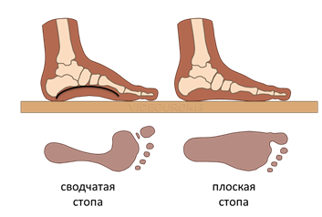
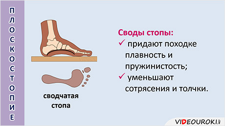
Podometry using the Friedland method calculates an index that is usually between 29 and 31. When developing hollow and arched feet, the index is over 31; if the number is below 29, one can speak of the development of a flattened area, a valgus curvature or a flat foot.
With the help of computer technology, the overload zone, the degree of curvature can be accurately determined to the millimeter and the location of the lesion can be clearly determined, which makes it easier to choose the correction scheme.
X-rays show the curvature or flattening of the arch, and if the image is unclear, CT or MRI scans are also used.
During the 'wet examination' you can visually see the structure of the foot - the longitudinal arch is usually not impressed. If the metatarsal, toes, and heel are not molded, high copular vault is diagnosed, and if all plantar structures are molded, flat foot is diagnosed.
If there is a flattening or curvature, the patient should have a correction made immediately. Otherwise there is a risk of damage to the ankle joints and many structures of the musculoskeletal system.
Possible complications
If the pathological curvature of the arch of the foot is not treated, complications can occur:
- Increased pressure on the metatarsophalangeal joint provokes the formation of a heel nodule in the first toe, which causes pain when walking;
- Increased pressure on the heel bone and prolonged trauma lead to bone spurs in the heel bone. Concomitant metabolic disorders;
- Muscle soreness, fatigue when walking weaken the musculoskeletal system and increase the risk of injury: sprains, bruises, formation of exudative laxity in the ankles, tightness in the feet, muscle weakness, motor imbalance;
- frequent blisters and corns cause great inconvenience to the patient;
- Joint stress causing inflammation and degeneration, exacerbation of chronic inflammation: bursitis, synovitis, toe arthrosis. In children, chronic processes lead to valgus deformity, gout, arthritis, impaired production of synovial fluid.
The importance of arch disease for humans is underestimated. The development of the curvature is moderate. It changes the anatomy of the feet and leads to a redistribution of the load on the musculoskeletal system. A low arch is less dangerous because of the risk of disability.
It is important to detect the pathology early and consult a doctor to avoid impairment of freedom of movement.
Main symptoms of longitudinal flatfoot in adults
A longitudinal flatfoot occurs when the curvature of the foot on the inside changes. It usually occurs in conjunction with other foot deformities (30 % cases) and is more common in young women under 25 years of age. Patients complain of rapid fatigue when walking or standing for long periods of time and sudden buckling of the foot. There are problems with choosing shoes - old and once comfortable shoes become tight and uncomfortable as the foot enlarges due to the stretching. The attentive patient will notice that the inner edge of the sole wears out more quickly. This is because clubfoot occurs when the patient unconsciously rests on the inside edge of the foot.
Transverse flatfoot is more common in older people, and some experts say everyone over 55 already has it. In transverse flatfoot, the arch of the foot that supports the toes gives way, causing them to become deformed and hammer-shaped. The patient feels pain in the forefoot area. Horned nails and ingrown toenails form. The bone enlarges, which is especially noticeable in the big toe, which begins to turn to the side and has a bump at the base of the toe. The first toe is often traumatized and the likelihood of an ingrown toenail is high. The other toes stick out and bend upwards, which is also painful. Depending on the change in the arch angle of the foot, there are 3 degrees of flat foot.
Symptoms of flat feet depending on the change in the arch angle of the foot
The shape of the arch of the foot is weak. The longitudinal vault has been preserved, its height is not less than 25 mm. The toes spread slightly and begin to rub against the shoes when walking. Horned nails appear. In the evening your feet are tired and swollen. The pace changes. However, a short rest quickly relieves the pain.
The arch of the foot decreases and is no more than 17 mm. The pain is quite persistent. It is not possible to walk long distances or stand for long periods of time. The big toe may become stiff and curved.
CONTACT
For comprehensive information on the treatment and prevention of orthopedic, rheumatological or neurological diseases, please contact us:
 Tel. +7(495)120-46-92
 Email: [email protected]
return form
 Send us a message on Telegram
 Contact us on WhatsApp

Our address is 11 Trifonovskaya St., Moscow, Russia.
Classification of flat feet according to the cause
Depending on the cause, acquired flatfoot is divided into the following types:
- Paralytic type. It usually occurs after osteoarthritis as a result of paralysis of the tibialis and/or foot muscles;
- Traumatic type. Occurs after trauma, e.g. B. after a fracture of the foot bone or a fracture of the ankle joint, if ligaments are torn or the muscles supporting the foot are strained;
- Rachitic type. Rickets leads to bone fragility and mineralization disorders; the bone becomes soft and deforms under the weight of the body.
- Flat feet as a result of an illness. Some diseases affect the bones and joints and cause flat feet..
- Static type. This type occurs when the muscles and ligaments cannot cope with the strain.
Static flatfoot is the most common (over 80 % cases). It is not caused by illness or injury.
Factors contributing to static flat feet:
- Standing for long periods of time (e.g. salespeople, hairdressers, surgeons);
- Unsuitable footwear – uncomfortable, tight, high heel, flat sole, too soft or too stiff;
- sedentary lifestyle;
- Excess weight - every additional kilogram puts additional strain on the foot, that is, the higher the weight, the more severe the flat feet;
- Age (over 55) – the ligaments become less flexible and at this age osteoporosis develops;
- congenital ligament weakness or muscle pathology;
- long periods of strict bed rest – muscle wasting minimizes arch muscle support;
- lifting heavy weights;
- participation in professional sports;
- Pregnancy - due to the surge of hormones during this time, the bones become softer, and the pounds that a woman quickly gains increase the load on the feet.
CONTACT
For comprehensive information on the treatment and prevention of orthopedic, rheumatological or neurological diseases, please contact us:
Tel. +7(495)120-46-92
Email [email protected]
feedback form
Send us a message on Telegram
Contact us on WhatsApp
Our address is 11 Trifonovskaya St., Moscow
Poor posture and flat feet

In this lesson, students will become familiar with the human musculoskeletal system, learn about poor posture and flat feet, look at the signs of good and bad posture, learn how to develop good posture, and how poor posture affects the human body. The lesson also contains information about flat feet: signs, causes and prevention.
Overview of the lesson 'Bad Posture and Flat Feet'.
Every person has a habitual posture when at rest and when moving, or to put it another way, a posture.
The posture – is the posture that people have become accustomed to when standing, sitting, walking and working. It depends on the development of the skeletal system and muscles. A person with good, correct posture has a straight back, straight shoulders, a head held high and a chest that protrudes slightly above the stomach. Such a person looks slim and good. With good posture, the curves of the spine are moderate and evenly undulating. The shoulder blades are symmetrical and the shoulders are straight. The muscles of a person with correct posture are lean and the movements are well defined.
Good functioning of all organs and high performance are only possible with correct posture.
A person with bad posture looks completely different. with bad posture. He / she has. the head stretched forward, the chest flattened, the shoulders pulled forward, the stomach bulging, and the chest is stretched forward.
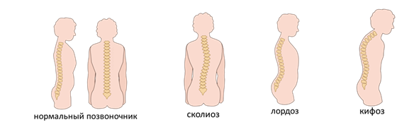
Various spinal curvatures occur due to poor posture curvature of the spine: lateral curvature (scoliosis), excessive lumbar lordosis or excessive thoracic kyphosis ('hunched back' or crooked leg). On scoliosis The shoulders, shoulder blades and pelvis are positioned asymmetrically.
On the side of Bad posture The vertebrae and intervertebral discs shift. This impairs the functioning of the heart, lungs and digestive system. The result is a reduced metabolism, headaches and increased fatigue.
Over time this can lead to the development of osteochondrosis (This is a disorder that affects the cartilage and intervertebral discs in the joints.). This makes it difficult to walk and bend over, and your back and limbs hurt at night, making it impossible to sleep.
Specificity of foot function in children

Clubfoot is a foot deformity that manifests itself as a lowering of the longitudinal and transverse arches. According to various authors, between 20 and 40 children suffer from flat feet. The foot performs a number of important functions:
- support
- suspension (shock absorption)
- locomotion
- Balance
The musculoskeletal system in the body takes on the spring function:
- physiological curves
- Band washers
- Longitudinal and transverse arch of the foot
The most severe form of flat foot is valgus flat foot, which is characterized by heel pronation and forefoot abduction. This type of foot deformity is a consequence of rickets in infancy (2 to 4 years), while in older preschool age (5 to 7 years), longitudinal foot without valgus component occurs more frequently and gradually decreases in severity. The type of foot, like the type of posture, is formed by school age (up to the age of seven), so that the preschool age is the most favorable for correcting existing musculoskeletal deformities. In recent years, a close connection has been established between flat foot deformity and neurological microsymptoms in children, as well as with symptoms of dysplastic skeletal development.
From a biomechanical point of view, the foot has a functionally adequate anatomical structure, so the suppleness and ease of gait depends on its condition. The base consists of 3 vaults. The outer arch of the foot runs from the outer surface of the calcaneal tuberosity to the head of the fifth metatarsal bone. The inner arch runs from the heel bone to the head of the first metatarsal bone; the apex of the inner longitudinal arch is the tubercle of the calcaneus. The transverse arch runs from the head of the first metatarsal to the head of the fifth metatarsal. The inner longitudinal arch and the transverse arch have a cushioning function for the foot, the outer longitudinal arch has a supporting function. When the internal longitudinal arch is lowered, a longitudinal flatfoot is created, and when the transverse arch is lowered, a transverse flatfoot is created.
Read more:- Shape of the transverse tarsal joint.
- At what age do girls' legs grow?.
- Longitudinal and transverse arches of the foot.
- Which ligaments strengthen the transverse arch of the foot?.
- flat feet (valgus foot).
- Which doctor treats flat feet?.
- The longitudinal arch of the foot is.
- How do you tell if it's an ankle fracture or a dislocation?.