Without my emphasis it looks like this:
- 'Foot fins': Detect and correct transverse flat feet
- Why does transverse flatfoot occur?
- The human foot
- risk factors
- Old
- Cosmetic foot surgery
- Gender
- Occupational Injuries
- pregnancy
- sports and dancing
- Causes of foot pain.
- Foot
- External morphology of the human foot
- literature
- Why does the foot go numb?
- neuropathies
- polyneuropathies
- traumatic injuries
- diagnosis
- plantar muscles
- Plantar fascia (Aponeurosis plantaris)
- What are the risks of trauma?
- Features of lower limb injuries
- Human Foot Artery Anatomy Information:
- Which doctors should you go to to have your foot arteries checked?
- Symptoms of pes cavus
- How to make a diagnosis
- orthopedics of the foot
- The most common operations to correct foot deformities
'Foot fins': Detect and correct transverse flat feet
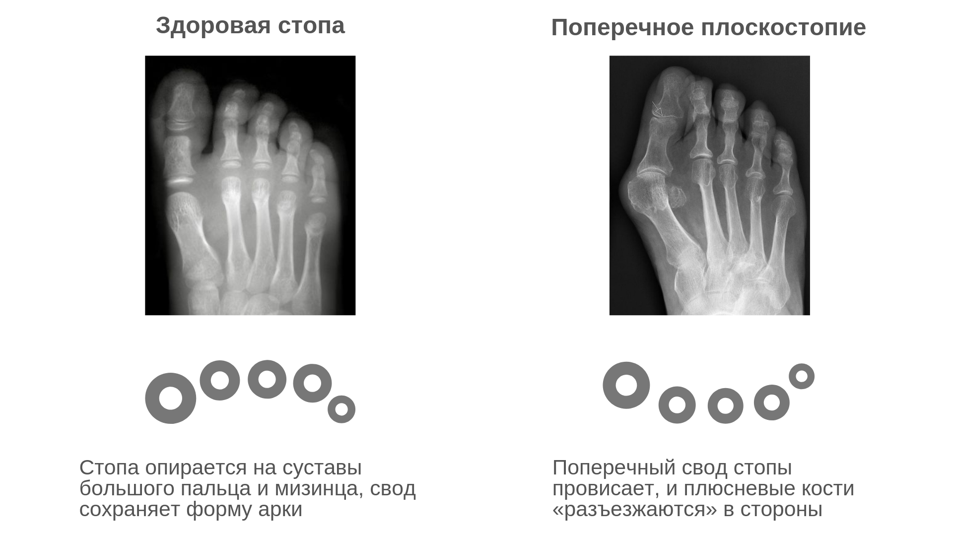
The longitudinal and transverse arches of the foot spring like springs and distribute the load that weighs on the feet when walking. In AS, the metatarsal bones loosen and the foot feels like goosebumps. This causes the transverse arch of the foot to 'bend'. We will try to find out what side effects this can have on your foot and how you can live with it. Try it!
In transverse flatfoot, the metatarsal bones pull apart and the forefoot widens. This causes the foot to lean on the second, third, and fourth metatarsal bones, rather than the first and fifth as it normally should. While the heads of the 2nd, 3rd and 4th metatarsals used to be relaxed, in flat feet they begin to bear the full load. As a result, the thumb moves sideways due to muscle failure and the joints grow and become a 'lump'. This leads to a valgus deformity of the big toe, or as we used to say, a 'knuckle' on the foot.
Why does transverse flatfoot occur?
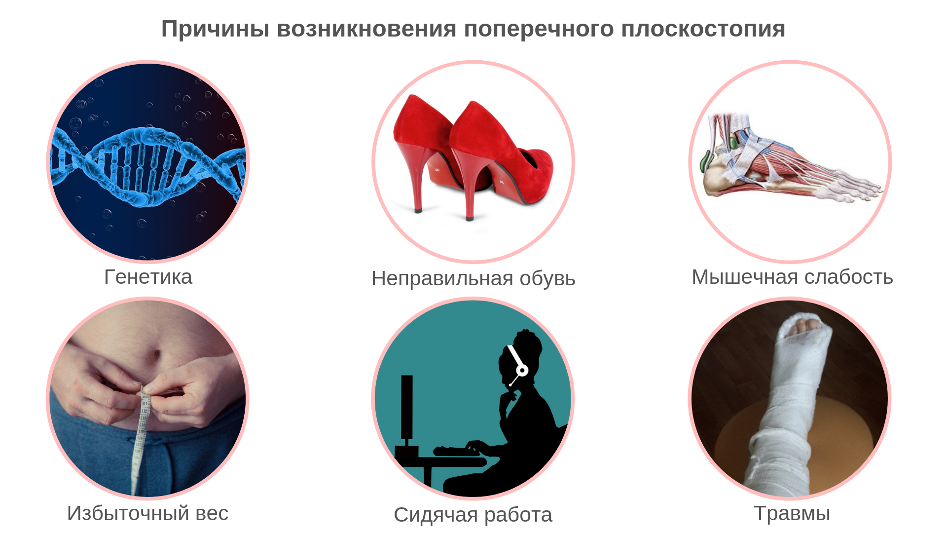
Transverse flatfoot can occur for a variety of reasons – inherited or acquired. The most important of these are described below.
- Genetically conditioned. A genetic weakness of the musculoskeletal system is most commonly inherited in women. In men, inherited flat feet are less common. So women are at risk!
– Unsuitable footwear. If you do not want to develop or worsen transverse flatfoot, avoid wearing high-heeled shoes, shoes with narrow toes, or shoes with stiff, inflexible soles. Instead, choose comfortable shoes with a low heel. Clean out your shoe closet too: replace uneven-soled shoes as they keep your feet in the 'wrong' position and slow down their recovery.
- muscle weakness. Weak metatarsal muscles – caused by a passive lifestyle and sedentary work. Keep moving!!! Then your leg muscles will become stronger and more resilient.
– Excessive weight. Excess weight puts too much strain on the legs, preventing the muscles from performing their main function.
- Stand. Constant standing leads to a static load on the legs. This leads to muscle fatigue and increases the risk of developing flat feet. Standing work also decreases blood flow to the legs, leading to pain and tension.
– injuries. If the arch of the foot is originally normal, flat feet can develop as a result of trauma…. This is usually due to a fall from a height, a car accident, or a sports injury.
The human foot
The human foot is the lowest part of the lower limbs. The part of the foot that is in direct contact with the ground is called the foot or sole. The foot has three bony supports, two in the forefoot and one in the hindfoot. The back part of the foot is called the heel; the front part of the foot, which includes the five toes, is called the toes. The toes enclose the phalanges of the foot skeleton. The bones of the foot extend from the tips of the toes to the heel and together form the body of the foot. The tarsal and phalanx bones are similar to the metatarsal and phalanx bones of the hand, but are less developed because they are less mobile. When walking, the heel touches the ground first, followed by the lateral edge of the foot, the ball of the sole and the big toe.
Depending on the length of the toes, three types of feet are distinguished:
- 'Greek guy'. The thumb and third toe are shorter than the second toe, followed in descending order by the fourth and little toe.
- 'Egyptian guy. The longest thumb, followed in descending order by the second, third, fourth, and pinky fingers.
- 'Rectangular guy. All fingers are about the same length. The thumb is the same length as the second, followed in descending order by the third, fourth, and little fingers.
It is common in most cultures (including European and Japanese) to cover one's feet with shoes when outdoors, mainly to protect them from injury. Shoes have a protective function: sandals, sneakers, loafers, boots and ankle boots. There are differences in preferences for wearing shoes indoors. In some European countries, Canada and New Zealand, it is customary to remove shoes when entering a home; there is no such rule in most US homes. In Japan, the tradition of removing your shoes when entering a room is so widespread that soft floors are intentionally designed so that shoes cannot be walked on.
In some cultures, showing bare feet is considered indecent or even offensive. For example, in the Arab world and Thailand, uncovered feet are a sign of poverty or unconventional religious beliefs.
risk factors
Risk factors that contribute to foot pain include.
Old
Older people are at a higher risk of developing foot pain. With age, the foot expands and becomes flatter. The skin on the feet becomes thin and dry. Foot pain in old age can be a sign of age-related degenerative diseases such as arthritis, diabetes and heart disease. In addition, problems in the feet themselves can affect the balance of the musculoskeletal system in old age.
Cosmetic foot surgery
In the name of fashion, some women have undergone surgery to improve the shape of the foot so they can wear heeled shoes. Surgical techniques include clipping the toes, shrinking the foot, or injecting silicone into the balls of the feet. Over time, these procedures lead to foot pain, and surgeons discourage foot surgery unless indicated.
Gender
Women are more prone to severe foot pain, which is likely due to wearing high-heeled shoes. In the older age group, severe foot pain is a common factor leading to disability.
Occupational Injuries
Approximately 120,000 work-related injuries occur in the United States each year. About a third of this is due to foot injuries. Most foot disorders such as arthritis, tunnel syndrome and plantar fasciitis are due to excessive stress on the foot (standing and walking for long periods of time).
pregnancy
Pregnant women are at increased risk of foot problems because they gain weight and release hormones that make the ligaments more flexible. These hormones aid in childbirth but weaken the foot.
sports and dancing
People who exercise regularly are at higher risk for conditions such as plantar fasciitis, heel spurs, osteoarthritis, Achilles tendonitis and bone fractures. Women are more prone to fractures.
Causes of foot pain.
Foot pain can affect different parts of the foot. Bones, ligaments, tendons, muscles, fascia, nerves, blood vessels and the skin can be the source of the pain. The most common causes of foot pain are the following conditions:
- plantar fasciitis.
- osteoarthritis
- Podagra
- athlete's foot
- Rheumatoid arthritis
- Condylar deformity of the foot (hallus valgus)
- Achilles tendon injury
- Diabetic foot
- calluses on the foot
- heel spur
- clubfoot
- metatarsalgia
- fractures
- Morton's neuroma
Foot
Foot (Latin. pes ) is the distal (front) limb of the quadruped and the arch that is in direct contact with the ground and provides support when standing and moving.

The human foot is the lowest part of the lower limbs. The part of the foot that is in direct contact with the ground is called the foot or sole[1], the top of the foot is called the hindfoot. The entire foot has an arched structure that is not immobile but has flexibility and elasticity due to the joints. The bony structure of the foot is divided into the tarsus, the metatarsal bones and the phalanges [2].
External morphology of the human foot
The external morphology of the foot reflects the bony structure and is divided into the forefoot, midfoot and hindfoot. In the forefoot, a distinction is made between, among other things. the toes and on the sole side. The forefoot is the sole of the foot and the midfoot is the sole of the foot.and the metatarsal is the arch of the foot. arch of footand the hindfoot on the sole side forms the heel.
The arch of the foot is the part of the foot that does not normally touch the ground on the sole side and forms the instep on the back side. instep of the foot.. The convex part of the arch of the foot consists of the five metatarsal bones in the body of the foot, the outer processes of which form the toes and are called phalanges [3]. The sole of the foot is at the lowest part of the arch of the foot in front of the toes and protects the joints from impact. The extreme toes of the human foot, by analogy with the fingers of the hand, are called big toe (hallux) i little toeand the other three toes, starting with the big toe, are called toes II, III, and IV. The general area of the arch and heel of the foot can be referred to as the forefoot, the toes as the bunions or toes.
The skin of the sole is thick, rough, hairless and rich in sweat glands. The skin of the posterior surface is elastic and easily displaced, so any inflammatory process leads to swelling in the posterior part of the foot. The sole surface only partially replicates and reflects the underlying bony structure. This is because there are numerous fat deposits on the surface of the foot and the surface of the foot is covered by thick skin. The oval pads represent the plantar ends of the toes. Their [Cheo?] Appearance is due to the presence of fat pads on the sole that [?who?] are in contact by the lateral edge of the foot (when the toes are not laterally extended). The balls of the big toe are flatter, wider and separated from the foot by a clear crease. The thumb is separated from the other toes by a deep seam, crowned by a strong nail, and the axis of the toe is slightly shifted to the side. The thumb is flat, the others are arched. The fingers are progressively shorter from the thumb to the little finger. Sometimes the second finger is the longest.
literature
- Biological Encyclopedic Dictionary / edited by MS Gilyarov; Editors: AA Bayev, GG Winberg, GA Zavarzin et al. – Moscow: Sov. Encyclopedia, 1986. – С. 611. – 100 000 copies.
- M. Ц. Rabinovich, Plastic anatomy of man, four-legged animals and birds.
- Giovanni Civardi, Plastic representations in the anatomical drawing
- ↑Ushakov dictionary
- ↑ Biological Encyclopedic Dictionary / edited by MS Gilyarov; Editors: AA Bayev, GG Winberg, GA Zavarzin, et al. – M.: Sov. Encyclopedia, 1986. – C. 611 – 100 000 copies.
- ↑Spleen (metatarsus)
- ↑Department of Internal Affairs of the North-Western District of Moscow.
- ↑Muscles of the Foot - Anatomy - Medical Encyclopedia
- ↑ Michel Strogoff, Dubai, 2007: 'Don't sit with your feet facing other people.'
Why does the foot go numb?
neuropathies
Numbness in the feet can occur with peripheral nerve neuropathies of the lower limbs. There is usually a localized sensory disturbance in a specific area of the foot. Timeliness depends on the damaged nerve:
- Neuropathy of the sciatic nerve. The numbness covers almost the entire foot or occurs in parts of the foot. In piriformis muscle syndrome, the toes are more likely to be affected.
- Neuropathy of the tibial nerve. Injury at the popliteal level is manifested by almost complete numbness of the foot, tarsal syndrome with hypoesthesia at the medial and lateral edges, median nerve involvement at the plantar with numbness at the medial edge, and calcaneodynia with hypoesthesia at the heel.
- Fibular nerve neuropathy. When the general trunk or deep branch is affected, numbness is noted on the dorsal surface of the foot. When the superficial branch is affected, only the medial part of the dorsum of the foot is affected.
- Neuropathy of the femoral nerve. Hypoesthesia occurs at the medial edge of the foot.
polyneuropathies
In contrast to mononeuropathies, in which only one extremity is affected, polyneuropathies are characterized by symmetrical involvement of the legs and arms with sock- and glove-like sensory disturbances. Multiple nerve damage can occur in the following diseases:
Neoplasms, severe liver or kidney diseases can form the background of nerve damage. Polyneuropathy of digestive, toxic-infectious origin can sometimes be found. The most common cause of toxic polyneuropathies is alcoholism.
traumatic injuries
Nausea in the feet occurs with spinal cord injuries and injuries to the peripheral nerves. In patients with spinal cord injury, the severity of the injury determines the severity of the symptom and the extent and type of neurological impairment. In victims with peripheral nerve damage, the area of sensory loss corresponds to the area of hypoesthesia in neuropathy of the affected nerve trunk.
diagnosis
The neurologist is responsible for determining the cause of the numbness in the foot. Patients with vascular disease are referred to a vascular surgeon. An endocrinologist examines patients with diabetes. The doctor determines when and under what circumstances the sensory disturbance occurred, how the symptom has changed over time and what side effects there are. The following procedures can help verify the diagnosis:
- Physical examination. The specialist assesses the appearance of the foot, the color and temperature of the skin, and the pulsation of the arteries. In the process, signs of swelling and inflammation, hyperkeratosis, cracks, abrasions, trophic ulcers are noted.
- The 'neurological' examination. The doctor examines the reflexes, determines the limits of abnormal sensitivity, the presence of muscle wasting, neurogenic contractures.
- Examination of the vessels .. The condition of the arteries is examined by ultrasound, duplex, reovasography, capillaroscopy, thermography and peripheral arteriography.
- Electrophysiological Methods. Electromyography and electronography are used to differentiate neuropathies and determine the extent and severity of lesions.
- visualization techniques. Soft-tissue ultrasound, spinal X-rays, CT or MRI of the brain, and other investigations may be recommended to clarify the origin of the pathology.
- laboratory tests. Laboratory tests include determining the level of sugar and cholesterol in the blood. Tests for specific markers are done to determine the nature of the underlying pathology in Raynaud's syndrome.

plantar muscles
The plantar muscles are further divided into three groups: the muscles of the thumb, the muscles of the little finger, and the medial muscles. This is a real treat for me - I love it when muscles are divided into different groups and layers, such as the neck muscles. I populate our spreadsheet with this information:
Before we move on to the soleus muscles, I want to say a few words about the plantar aponeurosis, literally. We'll cover them in more detail in the articles on topographical anatomy of the lower limbs (when I get to that point with the upper limbs), but you should know the basics as it's quite an important anatomical entity
Plantar fascia (Aponeurosis plantaris)
The plantar muscle is a strong, dense, thick band of connective tissue that inserts proximally at the tuber of the heel bone and distally at the toe phalanges. The plantar muscle originates from the superficial fascia of the sole of the foot.
The plantar fascia is direct below the . The plantar aponeurosis lies directly under the subcutaneous fatty tissue and is firmly connected to it by connective tissue bridges. The spaces between the ligaments are filled with fatty tissue.
In the following, the M. soleus is presented in various figures and tables. In order to see the soleus muscle, the skin, subcutaneous fat and aponeurosis of the soleus muscle must be removed, which is why the aponeurosis is not visible on the images of the muscle. Let's look at them now (without skin and subcutaneous fat):
And this is what the plantar aponeurosis looks like in my favorite atlas by YL Zolotko:
What are the risks of trauma?
There is no doubt that the ability to walk, run, stand safely and firmly on one's feet is very valuable and important for every human being. When a person loses the ability to move, they are unable to enjoy life to the fullest, perform routine household chores, and engage in jobs that require some level of physical activity.
Leg and pelvic injuries are one of the causes that can contribute to a person's disability and loss of the ability to walk, move, or run. Lower limb injuries are mechanical conditions in which the integrity of muscles, ligaments, joints, bones and skin is compromised and parts of the joint system are dislocated and unable to communicate properly with one another.
Lower limb injuries are generally divided into two groups - closed and open. The former are the most dangerous for a person because they are not visible and cannot be assessed visually - the injury is under the skin, which remains intact. It can involve tissue damage, fractures, dislocations, and, less commonly, muscle and ligament strains.
Open wounds and lacerations mean that the skin at the site of the injury is also torn, cut or bruised and internal injuries may be visible. This principle of separation is particularly important for those who need to provide first aid in the event of an accident, natural disaster or accident, because even the absence of obvious external signs of injury is no guarantee that the person is well.

What are lower limb injuries? They are all differentiated by the type of disorders they cause in the body.
Features of lower limb injuries
A contusion, accompanied by swelling, bruising, and pain, is considered the most minor of all lower-limb injuries. It can occur after a fall or a violent impact. The integrity of the blood vessels in the skin is compromised, resulting in a bruise that discolors over time. Soft tissue contusions can also be associated with more serious injuries such as fractures and lacerations.
A dislocated joint is a pathological condition in which parts of the joint, most commonly the condyle, move out of their usual 'place', e.g. B. the head protrudes beyond the hip socket.
These injuries can be caused by sudden and abnormal movements that are outside the joint's functional range of motion, or by impact and heavy weights.
A bone fracture is the occurrence of a crack or break in the bone, which can result in the spread of bone fragments in a wound. Fractures can be closed or open - in the first case, the skin remains intact, in the second case, the skin is crushed or cut by protruding bone splinters or by mechanical impact. The affected person feels severe pain, the fracture site is swollen and blood circulation is impaired. The pain makes it extremely difficult to move and step on the sore leg.
Tears and dislocations of tissues, ligaments, and muscles are caused by sudden, violent movements that exceed the joint's capacity. These injuries are common in athletes, but even ordinary people are not immune to painful and dangerous injuries. Overtired people who train in the gym without observing the training and safety rules are particularly vulnerable.
Tissue crushing occurs as a result of a violent impact, disrupting the normal structure and vitality of tissues, blood circulation and metabolic processes. Such wounds are dangerous, prone to infection and inflammation, and slow to heal.
Human Foot Artery Anatomy Information:
Run on the back of the foot. Dorsalis pedis, the dorsal artery, which is an extension of the anterior tibial artery, attaches to the bones in the ligaments, and carries the tendon of the long thumb extensor medial and the medial abdomen of the short finger extensor laterally. Here, at the A. dorsalis pedis, the pulse can be determined by pressing against the bones.

In addition to the 2-3 cutaneous branches that open into the skin of the dorsal and medial side of the foot, the dorsal pedis artery gives off the following branches:
- A. tarseae mediates, Arteria tarsalis medialis, to the medial border of the foot.
- A. tarsea lateralis, artery of the lateral tarsal; it arises laterally and joins at its end with the next branch of the foot artery, namely the foot arch artery.
- Arcuate artery, arising from the medial aspect of the spleen bone, runs laterally along the metatarsal bone and anastomoses with the lateral tarsal artery and the soleus artery; the arch artery runs anteriorly in three Aa. metatarseae dorsales, the second, third and fourth arteries leading to the respective metatarsal spaces and each dividing into two Aa. leading digital dorsales to opposite sides of toes; each metatarsal artery gives off hollow branches that reach the sole anteriorly and posteriorly. Often the arcuate artery is weak and is replaced by the lateral metatarsal artery, which should be considered when examining the arterial pulse on the arteries of the foot in endarteritis.
- A. The primal dorsal metatarsal, the first metatarsal artery, is one of the two terminal branches of the dorsal artery and runs to the space between the first and second toes, where it divides into two toe branches; even before the division, it branches to the medial side of the big toe.
- The ramus plantaris profundus, the deep plantar branch, the second, larger of the terminal branches into which the dorsal artery divides, runs through the first interdigital space to the sole of the foot, where it participates in the formation of the arch of the foot, the archus plantaris.
Which doctors should you go to to have your foot arteries checked?
Is there anything that worries you? Would you like to learn more about the arteries of the foot, or do you need an examination? You might want to make an appointment with dr. – Clinic Eurolaboratory is always there for you! The best doctors will examine you, advise you, provide the necessary care and diagnose the problem. You can also doctor at home. clinic Eurolaboratory is open for you 24 hours a day.
How to contact the clinic:
The phone number of our clinic in Kiev is: (+38 044) 206-20-00 (multichannel). The clinic secretariat will find a suitable day and time for you to visit the doctor. Click here for our coordinates and directions. For more information about all of the clinic's services, visit the clinic's website.
If you have been examined before Be sure to bring the results with you to your doctor's office. If you have not yet done any examinations, we will carry out the necessary work in our clinic or with our colleagues in other clinics.
It is important that you take a very close look at your general health. There are many diseases that do not initially make themselves felt in our body, but unfortunately are treated too late. To do this, it is simply necessary to be examined several times a year Get a medical check-up several times a yearYour doctor should not only know how to prevent a serious illness, but also how to keep your body and organism as a whole healthy.
If you have questions to ask your doctor, you can find the answers to your questions in our online consultation area. Self Care Tips. If you are interested in reviews of clinics and doctors, you can find information on the forum. You can also register on the physician portal Eurocoolto keep up to date with the latest pes cavus news and information, automatically sent to your mailbox.
Symptoms of pes cavus
Those affected complain about a changed gait and poor resilience, especially on long walks. There is an excruciating pain in the lower part of the foot. The complaints often extend to the ankle. The elevation of the foot complicates the choice of footwear and insoles. The normal position of the instep in the shoe does not provide comfort as it is too low and too narrow for patients. Anterior instability of the forefoot is another characteristic external sign.
Other clinical signs and symptoms:
- Limited mobility of the lower limbs
- Persistent swellings, calluses, corns
- Weakening of the leg muscles
- weakening of the tendon reflexes
Other symptoms depend on the cause of the deformity.
How to make a diagnosis
The doctor examines the lower limb, assesses the type of anomalies in its structure and determines the severity of movement disorders. A planogram is created. Other symptoms and risk factors are looked for to identify the underlying disease. Previous trauma, malignant tumors, congenital syndromes and other pathologies must be excluded. A diagnosis can only be made after more detailed investigations.
Additional diagnostic procedures:
X-rays. Photographs show the position of the bones, previous traumatic influences.
Computed tomography or magnetic resonance imaging. Scanning provides the doctor with high-resolution images that show different anatomical structures layer by layer.
MRI is a more precise and modern imaging procedure without the disadvantage of exposure to radiation. The detailed images thus obtained allow the doctor to find the cause of abnormalities in the skeletal system. Specialists in MRI clinics use this type of examination to diagnose pes cavus.
orthopedics of the foot
The term 'foot orthopedics' takes on a whole new meaning in modern medicine, which differs markedly from the dominant doctrine in Soviet medicine. For many decades, there was virtually no systematic approach to the comprehensive management of skeletal deformities of the foot. There was a small list of operations performed according to standard indications and without sufficiently thorough diagnostics. As a result, there were few positive results, and the medical community and patients commented negatively on the severity and pain of the postoperative period and the small number of successful procedures. All this was due in large part to the underdeveloped diagnostic capabilities, the lack of knowledge of new data on the pathogenesis of the process and outdated surgical techniques.
On the basis of multi-centre studies, the orthopedic treatment of foot deformities is now comprehensively treated:
In modern orthopedics, on the basis of a large amount of material, new principles of surgical treatment of the foot have emerged, which are presented below:
- Maximum correction of osteoarticular deformities
- Stable bone fixation through special techniques
- Complete correction of the entire arch of the foot.
The principle of maximum correction involves the simultaneous correction of all elements of the foot deformity with the restoration of normal foot arches and the ability to wear normal footwear in the post-operative period without the need to remove internal fixation structures.
The most common operations to correct foot deformities
The approach to surgical choice is always individual to each patient, but there are some universal approaches.
- Elimination of the 'thickening' itself
- Restoration of the anatomical structures at the base of the 1st toe (correction of the ligaments)
- Alignment of the axis of the 1st finger.
Important in the operation is the technique of performing the osteotomy (cutting the bone) and fixation (fixing the newly formed bone in the right direction with removal of the connective tissue that has grown due to the inflammation). During the osteotomy, the orthopaedist/traumatologist correctly aligns the axis of the I finger. The osteotomy procedure itself is the secret of a successful surgery.
In the past, a transverse osteotomy was performed, followed by precarious fixation with thin spokes or a massive metal structure. After such an intervention, the foot is relieved for 3 to 3.5 months. During this period, the ligaments and muscles of the foot atrophy, the foot becomes unsightly, rehabilitation is delayed, and the ability to wear normal footwear is severely reduced (the presence of a massive metal structure on the bone). The metalloplasty is then removed in a second operation. All this is extremely annoying, painful and uncomfortable, especially for the elderly. The effects are usually very unpredictable. Later, there are difficulties in fitting shoes and scarring on the foot. The entire recovery process takes 6-7 months.
The modern methods of longitudinal or transverse osteotomy, the so-called chevron or SCARF osteotomy, have a completely different effect. This type of surgery is performed at ANDROMEDA Clinic by leading orthopedic and trauma surgeons using a minimally invasive oscillating saw that is only used in our clinic. When special Baruca screws are used to fix the bone fragments, the result of the operation exceeds all expectations.
Read more:- The tarsal bone hurts from above - what to do?.
- The structure of the human foot and diseases.
- Metatarsal tarsal bones.
- The key to a chopper joint is.
- Structure of the human foot.
- structure of the toes.
- structure of the toes.
- bones of the foot.