The stretches should be held for about 30 seconds and repeated two to three times on each side. They can be done daily or three times a week to gradually improve mobility. If you work long hours at a desk, try to stretch in short breaks throughout the day.

- Hip flexors are weakened by sitting for too long
- What happens when the hip flexors become weak or stiff?
- Human Lower Limb Muscles Anatomy Information:
- Which doctors should you see for an examination of the muscles of the lower limbs?
- Anatomy of the flexor muscles of the human lower limbs - information:
- Which doctors should you see to examine the flexor muscle of the lower limbs?
- Anatomy of the Human Foot Muscles – Information:
- Which doctors should you see for a foot muscle examination?
- Soft tissue diseases of the foot
- muscles of the pelvic girdle
- Movement of the lower limbs
- muscular structures
- front group.
- KNEE MUSCLE
- Muscles of the anterior surface of the thigh
- Muscles of the lateral surface of the thigh
- FOOT MUSCLE
- Fitness exercises for slim legs
- Read more
- Immunodietology - not for everyone, but for you
- A child's first cold: what to prepare for?
- Teething: A Guide to Baby Teething at All Stages
- High-quality care: what modern diapers can do
- Which scalp peeling should you choose: advice from a dermatologist
- Agonists, synergists and antagonists
- Definitions
- Examples of antagonistic muscles
- Upper limbs.
- Lower limbs.
- My videos about synergists, antagonists and agonists
- Specificity of antagonist function.
Hip flexors are weakened by sitting for too long
Everyone knows that you should get up from your desk as often as possible or use a standing desk if possible. One of the main benefits of this is the activation and stretching of the hip flexors. But what are the hip flexor muscles and why are they so important – and what happens when we allow them to become weak and stiff (rigid)?
The hip flexors – are powerful muscles on the front of the thigh. They include:
The hip flexors are activated when you pull your knees toward your chest. They are important for walking and running. They are also important in sports because they flex the hip and work with the quadriceps to extend the knee when you need to run or kick. An athlete whose hip flexors are damaged will have great difficulty running or pedaling. The hip flexors also work with the glutes and other core muscles to stabilize the spine and are therefore important for posture.
What happens when the hip flexors become weak or stiff?
Weak hip flexors can make climbing stairs, running, or even walking on a level surface difficult or painful. This can also cause other hip muscles to have to work harder to compensate. This changes your gait pattern. The hip flexors weaken when we sit too much, but simple stretching and strengthening exercises can make the muscles less stiff.
A tight hip flexor can make walking and standing difficult because it pulls down the spine. This causes you to lean forward, which strains your lower back muscles (which work in the opposite direction to keep you upright). An imbalance between the hip flexors and the opposing muscles that pull the trunk in the opposite direction can lead to lower back pain.
Stiffness in the hip flexors can reduce the range of motion in the knee joint. This can lead to a stiff gait where the knee doesn't bend as much as it should. Over time, this can lead to pain in the knee joint. In general, weak or tight hip flexors can cause joint or muscle dysfunction, which can lead to injury.
Human Lower Limb Muscles Anatomy Information:
Legs or lower limbs The legs or lower limbs bear the brunt of the burden as they must perform support and movement functions. They are the strongest of all the muscles in the human body. The lower limbs are divided into the pelvic muscles and the free muscles of the lower limbs. The latter group is divided into the muscles of the thigh, shinbone and foot. The pelvic muscles arise from the pelvic bone, sacrum and lumbar spine and are attached to the femur. Their job is to keep the body upright, flex and extend the hip joint, and move the hips.

The hip muscles are divided into three groups: the anterior, posterior and medial (adductor) muscles. These muscles are also involved in maintaining the body, but their primary role is movement. The lower leg muscles are also divided into three groups: anterior, posterior and lateral (lateral) muscles. They are responsible for the dorsiflexion and plantar flexion of the foot, the support function of the foot and its alignment.

The muscles of the foot are similar to the muscles of the hand and are divided into 3 groups. Due to the upright posture of humans, they have a special task. The main function of the foot muscles is to provide stability in the various foot positions, flexion, extension and adduction of the toes. The muscles of the lower limbs are divided into the muscles of the edge of the lower leg, the thigh muscles, the lower leg muscles and the foot muscles. The muscles of the lower limbs are innervated by the lumbosacral plexus, lumbar plexus et sacralis.
Which doctors should you see for an examination of the muscles of the lower limbs?
Are you worried about something? Would you like to learn more about the muscles of the lower limbs or do you need an examination? You can make an appointment with your doctor – Clinic Eurolaboratory is always there for you! The best doctors will examine you, advise you, provide the necessary care and diagnose the problem. You can also doctor at home. clinic Eurolaboratory is open for you 24 hours a day.
How to contact the clinic:
The telephone number of our clinic in Kiev is (+38 044) 206-20-00 (multichannel). The clinic's reception staff will arrange a suitable day and time for you to see a doctor. You can find our coordinates and directions here. For more information about all of the clinic's services, please visit the clinic's website.
If you have been examined before Be sure to take the results with you to your doctor's office. If you have not yet done any examinations, we will carry out the necessary work in our clinic or with our colleagues in other clinics.
It is important that you take a very close look at your general health. There are many diseases that do not initially make themselves felt in our body, but are unfortunately treated too late. This simply means that you have yourself examined several times a year to be examined by a doctor several times a yearIt's important to get checked a few times a year, not only to prevent a serious illness, but also to keep your body and mind healthy.
If you would like to ask your doctor a question, find answers to your questions and read in our online advice section Self Care Tips. If you are interested in reviews of clinics and doctors, you can find the information you need on the forum. Also register on the medical portal Eurocoolto stay up to date with the latest news and information about lower limb muscles, which will then be automatically sent to your mailbox.
Anatomy of the flexor muscles of the human lower limbs - information:
The anterior group (flexors) of the muscles of the lower limbs. Has an insertion on the lesser trochanter; includes M. iliopsoas (M. psoas major and M. iliacus) and M. psoas minor.
M. iliopsoas – iliacus muscle (m. iliopsoas-lumbar)consists of two heads.
One is the psoas major muscle, the large lumbar muscle, which originates from the lateral surface of the vertebral bodies and intervertebral discs of the 12th thoracic vertebra and the four upper lumbar vertebrae as well as from the transverse processes of the lumbar vertebrae. Running downwards and slightly laterally, it approaches the iliacus muscle.

The second, the iliacus muscle, is the iliac muscle that arises from the iliac fossa of the hip bone and the anterior superior and inferior iliac spines. On the medial side it is somewhat covered by the psoas muscle; between its edge and it a deep groove forms in which the femoral nerve lies. The fibers of the iliopsoas muscle descend to the psoas major muscle and unite to form the iliopsoas muscle; This lies on the anterior surface of the hip joint, emerges under the inguinal ligament through the lacuna musculorum and attaches to the lesser trochanter. Features. Causes flexion and supination of the hip at the hip joint. Due to the strengthened lower extremity, he can flex the lumbar spine. (Inn. L2-L4. Lumbar plexus.)
M. psoas minor, the small muscle of the lumbar spine (psoas minor)The small muscle of the lumbar spine (psoas minor), which is adjacent to the psoas major, is not always present. It passes into the iliac fascia and ends in the iliopubic eminence. It stretches the iliac fascia and can flex the lumbar spine. (Inn. Z1-Z2. Lumbar plexus.)
Which doctors should you see to examine the flexor muscle of the lower limbs?
Is there anything that worries you? Would you like to receive more information about the flexor muscles of the lower limbs or would you like to be examined? You can book an appointment with dr. – Clinic Eurolaboratory is always there for you! The best doctors will examine you, advise you, provide the necessary care and diagnose the problem. You can also doctor at home. clinic Eurolaboratory is open for you 24 hours a day.
How to contact the clinic:
The telephone number of our clinic in Kiev is (+38 044) 206-20-00 (multichannel). The clinic's secretariat will find a suitable day and time for you to see a doctor. You can find our coordinates and directions here. Further information about all of the clinic's services can be found on the clinic's homepage.
If you have already been examined previously, Make sure that you take the results of these tests with you to the doctor's office. If you have not yet done any examinations, we will carry out the necessary work in our clinic or with our colleagues in other clinics.
It is important that you take a very close look at your general health. There are many diseases that do not initially make themselves felt in our body, but are unfortunately treated too late. This simply means that you have yourself examined several times a year to be examined by a doctor several times a yearIt's important to get checked a few times a year, not only to prevent a serious illness, but also to keep your body and mind healthy.
If you would like to ask your doctor a question, find answers to your questions and read in our online advice section Self Care Tips. If you are interested in reviews of clinics and doctors, you can find the information you need on the forum. Also register on the medical portal Eurocoolto stay up to date with the latest news and information about lower limb corset muscles on the website, which will then be automatically sent to your mailbox.
Anatomy of the Human Foot Muscles – Information:
FootAs with the hand, in addition to the tendons of the long muscles descending from the lower leg, there are also short muscles on the foot, which are divided into dorsal and plantar muscles.
M. Extensor digitorum brevis – short extensor of the toes, is located on the back of the foot below the tendons of the extensor longus muscle and originates on the heel bone before entering the tarsal sinus. It splits anteriorly into four thin tendons to the toes I-IV, which attach to the lateral edge of the tendons of the extensor digitorum longus and the extensor hallucis longus and together form the dorsal extension of the toe tendons. The medial calcaneus, which runs obliquely with the tendon to the thumb, is also referred to separately as the extensor hallucis brevis muscle. Function. Performs extension of fingers I-IV along with a small lateral extension of fingers. (Inn. L4-S1. Deep peroneal nerve).


Plantar muscles (Plantar muscles). Forms three groups: medial (muscles of the thumb), lateral (muscles of the little finger) and medial, lying in the middle of the soleus muscle.
The muscles of the medial group are the three:
- M. Abductor hallucis muscle, muscle adductor hallucis of the big toe, lies on the uppermost medial edge of the soleus muscle; arises from the medial process of the calcaneal tubercle, retinaculum mm. flexorum and tiberositas ossis navicularis; attaches to the medial sesamoid bone. (Inn. L5-S1 N. plantaris med.).
- M. flexor hallucis brevis, the short flexor muscle of the big toe, borders the lateral edge of the preceding muscle and attaches to the medial sesamoid bone and the plantar calcaneocuboid ligament. Immediately anteriorly, the muscle divides into two heads, between which the tendon of the flexor hallucis longus runs. The two heads attach to the sesamoid bones at the first metatarsophalangeal joint of the big toe and to the base of the basal phalanx of the thumb. (Inn. S1-S2. Nn. plantares medialis et lateralis).
- M. adductor hallucis, the muscle that drives the big toe, is deep and consists of two heads. One (oblique head, caput obliquum) has its origin at the base of the proximal ankle joint and the ligament. The other head (caput transversum) arises transversely from the joint capsules of the metatarsophalangeal joints II-V and the ligaments of the soleus (plantaris). The second head (transversum, caput transversum) arises transversely from the joint capsules of the metatarsophalangeal joints II-V of the big toes and the plantar ligaments; it runs transversely to the length of the foot and, together with the oblique head, attaches to the lateral sesamoid bone of the thumb. (Inn. S1-S2. Lateral plantar nerve.) Function. The muscles of the medial soleus group, in addition to the activities indicated by the names, are involved in strengthening the arch of the foot on the medial side.
Which doctors should you see for a foot muscle examination?
Are you worried about something? Would you like to learn more about the foot muscles or do you need an examination? You can Book an appointment with dr. – Clinic Eurolaboratory is always there for you! The best doctors will examine you, advise you, provide the necessary care and diagnose the problem. You can also call the doctor at home. clinic Eurolaboratory is open for you around the clock.
How to contact the clinic:
The phone number of our clinic in Kiev is: (+38 044) 206-20-00 (multichannel). The clinic secretariat will find a suitable day and time for you to visit the doctor. Click here for our coordinates and directions. Further details on all of the clinic's services can be found on the clinic's homepage.
If you have been examined before Be sure to bring the results with you to your doctor's office. If you have not yet done any examinations, we will carry out the necessary work in our clinic or with our colleagues in other clinics.
It is important that you take a very close look at your general health. There are many diseases that do not initially manifest themselves in our body, but unfortunately are treated too late. It is simply necessary to undergo a medical examination several times a year Go for checkups several times a yearThe doctor should know not only how to prevent a serious illness, but also how to keep the body and organism healthy as a whole.
If you have questions to ask your doctor, you can find the answers to your questions in our online consultation area. Self Care Tips. If you are interested in reviews of clinics and doctors, you can find information on the forum. You can also register on the physician portal Eurocoolto stay up to date with the latest foot muscle news and information on the site, automatically delivered to your inbox.
Soft tissue diseases of the foot

MMuscles, tendons, tendon sheaths, ligaments, fascia, aponeuroses and the joint capsule play an important role in joint stability. Periarticular soft tissue diseases can be considered as diseases and disorders associated with joint inflammation. When describing soft tissue pathology, the following terms are commonly used:
- Tendinitis – inflammation of the tendon tissue;
- Tenosynovitis/Tendovaginitis – inflammation of the tendon tissue and tendon sheath;
- Enthesitis/Enthesopathy – Inflammation of the tendon tissue where the tendon attaches to the bone;
- Bursitis – Inflammation of the synovial capsules, thin-walled cavities lined with synovial membrane that facilitate the movement of tendons and muscles over bony prominences.
pThe pathology of the ankle and foot, as well as the periarticular soft tissues of this region, is the most common reason for a visit to the doctor and, according to Russian and foreign literature, accounts for from 6 to 21 % of all pathologies of the musculoskeletal system.
pSoft tissue abnormalities of the ankle and foot can be caused by both external and internal factors. External factors include overload (change in movement pattern), trauma (single or repeated microtrauma), local injection of corticosteroids (ICS) into the tendon, which can cause degeneration of tendon tissue, intrinsic – congenital anomalies of the joint structures, leading to a disrupted Biomechanics lead, imbalance of the muscles surrounding the joint, hypodynamia (immobilization), disturbed blood supply to certain tendon zones, age-related regression of the musculoskeletal system. There is often a combination of several factors.
muscles of the pelvic girdle
The pelvic muscles are divided into two groups – the internal and the external. The internal pelvic muscle group includes the iliopsoas muscle group, the internal obturator muscle group and the sternocleidomastoid muscle group. The external pelvic muscle group includes the gluteus medius, medius and minimus: the latissimus tenosus, the quadriceps and the external obturator muscle.
The hip muscles are divided into 3 groups: anterior (hip flexors), posterior (hip extensors) and medial (hip drive).
Because of their large mass and high extensibility, these muscles are able to develop a great deal of force, acting on both the hip and knee joints. The hip muscles have static and dynamic functions in standing and walking. Like the pelvic muscles, the thigh muscles in humans have reached their maximum development due to upright posture.
The muscles of the lower leg, as well as the other muscles of the lower limbs, are well developed, which is determined by the function they perform in connection with the upright posture and the static and dynamic functions of the human body. The lower leg muscles, which originate from the bones, the intermuscular septa and the fascia, affect the knee, ankle and ankle joints.
A distinction is made between the anterior, posterior and lateral shin muscle groups. The anterior group includes the tibialis anterior muscle, the long extensor muscle of the fingers and the long extensor muscle of the thumb. The posterior group includes the triceps (consisting of the calf muscle and the multifidus muscle), the longitudinal and biceps muscles of the thigh, the long toe flexor muscle and the posterior tibialis muscle. The tibialis lateralis muscle group includes the short and long fibula muscles.
In addition to the tendons of the lower leg muscles, which attach to the bones of the foot and belong to the front, back and lateral groups, the foot has its own muscles (short muscles). These muscles attach to the foot skeleton and have a complex anatomical, topographical and functional relationship to the tendons of these lower leg muscles, whose attachment points are on the foot bones. The muscles of the foot are located on the hindfoot and the plantar muscle.
Movement of the lower limbs
The movements of the hip are carried out in the hip joint and take place around three axes (triaxial - multiaxial joint). Flexion and extension (around the front axis) is possible within a range of 80° when the limb is extended and up to 120° when the lower limb is flexed at the knee. Adduction and adduction (around the sagittal axis) can be performed in a range of 70-75°, and rotation around the longitudinal axis up to 55°.
Thigh flexion: iliopsoas muscle, rectus thigh muscle, tailor muscle, broad fascia muscle, pectoralis muscle.
Thigh stretch: gluteus medius muscle, biceps femoris muscle, semitendinosus muscle.
Thigh Extension: Adduction of the thigh: large adductor, long adductor, short adductor, pectoralis muscle, small muscle.
Retraction of the thigh: gluteus medius and small muscles.
Inward rotation of the hips: gluteus medius (front balls), gluteus minimus, tensor fasciae broad.
The following muscles externally rotate the hip: gluteus major, gluteus medius and gluteus minor, gluteus caudalis, gluteus iliopsoas, quadriceps, externus and internus obturator muscles.
muscular structures
The knee is the convex part that connects the thigh to the shinbone. The muscles are divided into 3 classes:
They also work on the hip joint. They have movement and rest functions, as do the glutes.
front group.
Allows the leg to be flexed at the knee joint. It consists of:
The first muscle extends from the anterior vertex of the pelvis to the shinbone and descends to the broad tibial fascia. It allows the thigh to flex.
The last complex is a strong extensor muscle of the leg. Types of Quadriceps Muscle Group:
- The rectus femoris muscle of the thigh is the most stretchable. It runs from the buttocks and extends to the surface of the thigh. It is characterized by the presence of two joints. He pulls the thigh towards the breastbone and extends the knee when the pelvis is stationary.
- Medial – lies in the inner region of the anterior surface of the thigh. It pulls downwards and merges into the thick tendons of the thigh.
- Lateral - is the largest of all the thigh muscles described. It arises from the tendon ligament and connects to the rectus.
- The middle broad thigh muscle is the weakest and most vulnerable part of the quadriceps. It lies between the previous types. It starts at the origin of the kneecap.
The medial or middle complex includes two variants that form the inner layer of the thigh. These are:
The posterior muscle group of the thigh includes the following:
- The biceps femoris (thigh muscle) is spiral-shaped and elongated. It arises from the upper layer of the medial ischial tuberosity, the extended part connects to the other head and passes into the flat tendon. It allows flexion, balance and extension of the limbs in the knee joint.
- The semimembranosus tendon runs parallel to the previous tendon. Approximately in the middle of the thigh it merges into the long tendon and connects to the medial layer of the shinbone, which is further up.
- The semitendinosus muscle begins at the ischial tuberosity and attaches to the muscularis abdominis in the middle of the thigh.
KNEE MUSCLE
The muscles of the leg, depending on their location, are divided into the muscles of the anterior surface of the thigh, the muscles of the posterior surface of the thigh and the muscles of the inner surface of the thigh.
Muscles of the anterior surface of the thigh
The muscles of the front of the thigh include the hip extensor and the knee extensor:

- Rectus femoris muscle
- Medial broad thigh muscle
- Lateral broad thigh muscle
- Medial broad thigh muscle
The quadriceps femoris muscle (Quadriceps femoris) consists of. the rectus femoris musclewhich is shaped like a convex shaft at the front of the thigh. Lateral vastus muscleThis muscle forms a large part of the quadriceps, which is adjacent to the rectus femoris muscle on the outside of the thigh. When contracted, the muscle bends towards the outside of the thigh in the form of a protruding shaft. Vastus medialis muscle The medial vastus is on the inside. This muscle extends almost to the knee, that is, below the outer head. Medium broad muscle The middle broad thigh muscle runs along the front of the thigh and lies beneath the rectus femoris muscle.
The function of the quadriceps muscle is to extend the lower extremity and flex the thigh. The other muscles are e.g. B. involved in the movements when squatting.
Muscles of the lateral surface of the thigh
The muscles of the lateral surface of the thigh include the adductors and hip flexors:

- Short adductor muscle
- Adductor longus muscle
- adductor muscle
- M. combialis
- Tendinous tibial muscle
- M. portiaceus muscle
FOOT MUSCLE
The gluteal muscles consist of three paired muscles of the gluteal region:

- Gluteus maximus major
- Gluteus medius muscle
- Gluteus maximus muscle
Gluteus maximus muscle One end of the gluteus maximus is attached to the pelvic bone and wraps around the hip joint, and the other end is attached to the top of the thigh. It is the largest of the gluteal muscles and one of the strongest muscles in the human body. It serves to stretch the leg at the hip joint. Squats, deadlifts, and lunges are good gluteus maximus exercises.
The other two muscles are adductors. Gluteus maximus medius The gluteus medius is the muscle that lies beneath the gluteus medius. It begins on the outside of the iliac crest and attaches to the femur. He stretches and moves the hips, moves the pelvis to the side and straightens the bent trunk. Gluteus medius (gluteus medius)The gluteus minor is the deepest of the gluteal muscles and also has the task of lowering the thigh and raising the torso. It starts on the outside of the hip bone and attaches to the edge of the thigh bone.
Fitness exercises for slim legs
For athletes whose main goal is to burn fat in the calf and thigh area, the emphasis should be on the number of repetitions of the exercise, and not on the weight of the load. Training should be done with low weights but frequent repetitions.
For example, if a standard exercise is 10 repetitions in 3-4 approaches, then to lose weight legs you should increase the repetitions at least 2 times. Do 20 quick sets of leg curls, but be sure to give your muscles a break between sets: take 1-2 minute breaks.
Read more

Immunodietology - not for everyone, but for you
A child's first cold: what to prepare for?
What are the characteristics of viral respiratory infections in infants? Who will be hospitalized?
Teething: A Guide to Baby Teething at All Stages

High-quality care: what modern diapers can do
Which scalp peeling should you choose: advice from a dermatologist
Dandruff, hair loss and dullness – what other problems can a scalp peeling help with?
Agonists, synergists and antagonists
Let's continue the conversation about the different classifications of skeletal muscles and talk about antagonists, synergists and agonists. I adopted these definitions from the excellent book Antagonist Muscles in Human Movement by Raisa Samuilovna Persson.
Definitions
muscle antagonists are two muscles (or two muscle groups) of a joint that perform opposite movements in opposite directions when contracting.
Synergistic muscles are muscles of a joint that pull in the same direction.
Of the two antagonistic muscles, the one that performs the movement (i.e. the main task) is called agonistand the other as Antagonist..
Examples of antagonistic muscles
Upper limbs.
1. The flexion of the forearm is done by the Biceps brachii (m.biceps brachii), while the extension of the forearm through the M. triceps brachii (M. triceps brachii) (M. triceps brachii). These two muscles are antagonistic in that they pull in opposite directions relative to the elbow joint. One muscle (M. triceps brachii) is responsible for flexion and the other (M. triceps brachii) for extension.
2. The flexion of the arm (humerus) is realized by the muscles: Deltoid muscle (anterior bundle), pectoralis major, crura, biceps brachii. The extension of the shoulder (the humerus) is carried out by the antagonistic muscles: Posterior deltoid, dorsal broad muscle, subscapularis muscle, pectoralis minor muscle, pectoralis major muscle, long head of triceps brachii..
3: Abduction of the humerus is carried out by. Deltoid and supraspinatus muscles. The adduction of the shoulder is realized by the muscles: Subscapularis, small ring muscle, scapular muscle, long head of the triceps brachii muscle, plexus muscle.
Lower limbs.
3 The flexion of the lower limbs is carried out by the following muscles, among others. Biceps femoris muscle (thigh muscle) (M. biceps femoris), and the extension of the lower limbs is carried out by the quadriceps (M. quadriceps femoris). The quadriceps femoris muscle (m. quadriceps femoris) (m. quadriceps femoris). These two muscles are antagonistic, meaning they exert an opposite pull on the knee joint. One muscle (biceps femoris) is responsible for flexion and the other (quadriceps femoris) is responsible for extension.
My videos about synergists, antagonists and agonists
Specificity of antagonist function.
1. The presence of antagonists is necessary because a muscle can only pull a bone, not push it. So that a bone member e.g. B. can perform. Two muscles must be present for flexion and extension. One muscle is responsible for flexing the joint and the other for extension.
(2) The antagonistic muscles do not need to work alternately when performing motor activities. Already at the beginning of the 20th century, the German scientist R. Wagner (1925) showed that the ratio of the phases of antagonistic muscle activity changes depending on the conditions of the external force field. Complete convergence of muscle activity with movement is only observed during movements against frictional forces. When working against inertial forces, the antagonist muscle is only active in the first phase of the movement. Thereafter, the movement continues by the inertial force with increasing activity of the antagonist, slowing the movement (Fig. 1).
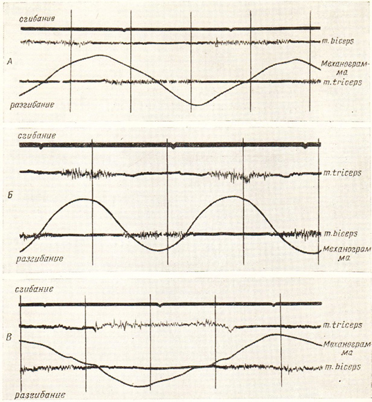
3 The activity of the antagonist muscles is strongly dependent on the speed of movement. During slow movement, the activity of the antagonist muscles corresponds to the phase of the movement for which they are responsible. Specifically: During flexion, the muscles responsible for flexion are active, while during extension, the extensor muscles are active. By increasing the speed of movement, the extensor muscle can become active towards the end of the flexion phase. In this case, the extensor muscle (antagonist) acts as a brake. During fast movements there are also phases with simultaneous activity of the antagonist muscles (AV Samsonova, 1998).
Read more:- How muscles and bones are related.
- Tibialis posterior muscle.
- The muscles that move the foot.
- Long flexor muscle.
- tibial fasciitis.
- lower leg muscles.
- abdominal muscle.
- lower leg flexion.