There are three main elements to abnormal alignment of the bones of the foot: foot adduction, valgus alignment, and flattening of the longitudinal arch. Anteroposterior radiographs can show signs of foot adduction by measuring the talonavicular angle and the angle between the first metatarsal and the talus. A talonavicular angle greater than 7 degrees indicates lateral talar subluxation. With normal arches, the downward angle between the first metatarsal bone and the talus (Meary's angle) should be no more than 4 degrees, an angle of 15-30 degrees is considered moderate flatfoot, while an angle of more than 30 degrees is considered pronoventral flatfoot applies. An upward angle of more than 4 degrees is called pes cavus.

- X-ray of the foot
- What an x-ray of the foot shows
- ICD-10
- causes
- Materials and Methods.
- Results and discussion
- Results and discussion
- Diagnosis of clubfoot in MRI and CT scan of the feet
- methods of choice
- What a club foot x-ray will show
- Typical manifestations or signs of clubfoot in children
- Causes of Clubfoot
- Where does the pain come from?
- Treatment methods for flat feet
- Indications and contraindications
- Carrying out the diagnostic procedure
- Drugs used in therapeutic blockades
- 'Novocaine'.
- 'Lidocaine.
- 'Bupivacaine'.
- Possible complications and measures to avoid them
X-ray of the foot
The foot is a flexible joint made up of bones, muscle joints and soft tissues. Only when all its structural parts are preserved and located in the right places can a person move freely and lead a familiar life.
An x-ray is an image of shadows that vary in density, size and structure. They are arranged in a certain order or overlap one another.
X-rays can pass through tissue, bones and muscles and emerge on the opposite side. Anatomical structures absorb the rays unevenly, so that they are heterogeneous when they emerge. Bones and soft tissues have different levels of radioactivity, and this is how the image of the organs, the order in which they are arranged, is formed.
During medical X-ray examinations, the beam emerges from the camera tube and the image is recorded on an image receiver: fluorescent screen, plate, sensor cassette.
In the image, all points that have the same radius appear to be in the same plane and colored the same - even if they are at different depths of the foot. Therefore, an image captured in a projection does not provide complete and reliable information.
X-ray images are meaningful if they are taken in two or three projections: straight, lateral, oblique.
What an x-ray of the foot shows

X-ray images clearly show the bones and joint spaces. Not only can the integrity of the bones be assessed, but also sprains or tumors. By analyzing the relative alignment and structure, the doctor identifies joint diseases, inflammation or tumors.
- bone and soft tissue cysts;
- Bone and soft tissue tumors
- metastases;
- softening of cartilage tissue;
- Bone softening in osteoporosis, rickets;
- Osteoporosis – diffuse thickening of the bones of the skeleton and bone fragility;
- Bone growth disorders;
- Gout;
- Arthritis and arthrosis (Arth;)
- osteoarthritis and cartilage inflammation;
- bone fractures and fractures;
- osteoarthritis (osteomyelitis);
- Reiter's syndrome;
- Sprains, tears of ligaments, muscles and tendons;
- Changes in the height of the arch of the foot.
ICD-10
Longitudinal flatfoot is a common pathology that occurs in approximately 20 % of patients with flat feet. The remaining cases are transverse or combined flat feet (a combination of longitudinal and transverse flat feet). It is usually acquired; Congenital forms are rare and are usually associated with more pronounced anatomical abnormalities and clinical signs.
Longitudinal flatfoot can occur at any age, but is particularly common in children. All children are born with obvious flat feet, and the arches of the feet do not form until the age of 3, so it is not possible to diagnose this pathology before this age. Most adult patients suffering from this condition are those who spend long periods of time on their feet due to their occupation. Slow flatfoot is treated by podiatrists.
causes
In about 3 % of cases, longitudinal flatfoot is congenital, which is due to intrauterine anomalies in the formation of the bones and ligaments of the foot. In addition, trauma such as fractures of the tarsal and metatarsal bones or ankle fractures can also lead to the development of this condition. A distinction is also made between paralytic flatfoot, which is caused by paralysis or paresis of the muscles of the foot and lower limbs, and wobbly flatfoot, which arises from bone deformity due to excessive softness.
However, the most common condition is static longitudinal flatfoot, which is caused by weakness of the ligamentous and muscular apparatus of the distal parts of the lower limbs. Factors that favor the development of static flat feet include obesity, pregnancy, excessive physical activity, jobs that require long periods of standing (salespeople, receptionists, wood turners, etc.), wearing uncomfortable, poor-quality footwear and weakening of the ligaments and Muscles of the foot due to aging or insufficient physical activity.
Materials and Methods.
Between 2008 and 2018, 13 patients with heel foot deformity aged 1.5 to 15 years were treated in the Center for Pediatric Orthopedics of the N. Priorov Scientific and Research Institute of Traumatology. In 8 children the deformity affected both feet, in 5 cases the deformity was unilateral. A total of 21 feet were treated surgically. In 8 children, heel deformity was due to a congenital central nervous system defect, spinal hernia (SMH), which was operated on in all patients in infancy; in 2 cases, the heel deformity was secondary and developed after treatment of equinovarietal foot deformity on the background of SMH; in 1 case, the heel deformity was secondary and developed after treatment for clubfoot; 1 child was diagnosed with caudal regression syndrome with severe malformations of the caudal spine and spinal cord and severe neurological deficits; In 1 child, the heel deformity was the result of a genetic pathology, a congenital structural myopathy and connective tissue dysplasia with generalized hypotonia and the development of a syndrome of symptoms of the so-called 'sluggish child'. 'Sluggish child' syndrome. We differentiated according to the type of foot deformity: heel deformity (without valgus or varus component) - 4 feet, heel-ankle deformity - 13 feet and heel-ankle deformity - 4 feet.
Surgical treatment tactics depended on the stiffness of the deformity, the age of the child, and the degree of muscle dysfunction in the lower leg. Marked trophic disorders of the soft tissues of the foot and the lower third of the tibia were contraindications to surgical correction. A single-stage surgical correction without prior use of a brace was performed when passive correction of the extensor contracture of the ankle to midfoot position without obvious soft tissue tension in the absence of circulatory disorders (skin pallor on the hindfoot surface and capillary reaction) and without the formation of rigid valgus deformity of the hallux valgus due to the tension the shortened extensor tendons was possible. For rigid deformities in which skin deficits on the dorsal surface with signs of circulatory disorders and/or the formation of a rigid toe rolling deformity were noted when attempting to lengthen the foot, a premedial correction was performed with the foot in the equine position to gradually tighten the tendons and soft tissues to stretch. In children under 12 years of age, heel foot deformity was corrected by releasing the soft tissues of the ankle joints (11 feet), taking into account the function of the growth plates. The release resulted in dissection of the joint capsules of the ankle, subtalar, scaphoid, and cuneiform joints. Foot unloading made it possible to correct the heel deformity of the foot with the following radiometric indices: heel-shin angle 30-50°, Meary angle 20-35°, talocrural sagittal angle 30-50°.
Results and discussion
The results of treatment were examined in 11 patients. The follow-up period ranged from 1 to 9 years. All treated patients demonstrated improved foot support by reducing deformity. Clinically, the feet of all patients were in a functionally favorable position, the tibia-foot relationship was restored, the ankle was mobile, the range of motion was 15-20°, and the gait improved, with the sole of the foot supported.
Clinical example (Illustration 1). Patient N., 8 years old. Diagnosis: 'CMH. Neurogenic deformity of the heels on both feet. The patient complained of difficulty walking and being able to stand only on the heels of both feet. For stability, the child propped himself up on his toes, leaning on the dorsal surface of the phalanges on which corns had formed. Clinical: Feet in upright position up to 40°, up to middle position with difficulty, no flexion. Muscle strength: right and left anterior tibialis muscles - 5 points, fibula muscles - 5 points, toe flexors - 3 points, toe extensors - 3 points, right and left posterior tibialis muscles - 0 points. Computed tomography of the feet: heel deformity of both feet, heel-shin angle right - 30°, left - 25°; Heel support angle right – 40°, left – 50°. Podography – pathological redistribution of the load, support only on the heel of both feet. The operation was carried out on the right and left foot in stages: relief of the ankle joints, transfer of the long fibula to the calcaneal tuberosity, lengthening of the tibialis anterior tendon. Ankle fixation with mild hypercorrection – 2 months. After removing the metal structures, the foot is fixed in the tunica. Nine years after treatment, the patient is 17 years old. Clinically, the feet are in moderate alignment, gait is satisfactory, and the foot is plantarflexed. Active foot flexion: The amplitude of active flexion of the left foot is 35°, that of the right foot is 10°. Subgraphy: Comparison of the dynamics shows significant improvement, even distribution of forefoot and rearfoot. X-ray image: feet in corrected position, osteoarticular conditions satisfactory: metacarpophalangeal angle right 70°, left 75°; Heel support angle right 25°, left 27°. Analysis of gait biomechanics using digital processing of the support response showed that an asymmetric butterfly curve with a high posterior thrust parameter was observed before treatment, indicating posterior region overload. After treatment, an even distribution of forefoot and hindfoot dynamics was observed in the right and left feet, and the butterfly curve became more symmetrical, consistent with a normal bipedal curve.
Results and discussion
The results of treatment were examined in 11 patients. The follow-up period ranged from 1 to 9 years. All treated patients demonstrated improvement in foot support by reducing deformity. Clinically, the feet of all patients were in a functionally favorable position, the relationship between the lower extremity and the foot was restored, the ankle was mobile, the range of motion was 15-20°, the gait was improved, and the support was over the sole of the foot given.
Clinical example. (Fig. 1). Patient N., 8 years old. Diagnosis: 'CMH. Neurogenic deformity of the heels on both feet. The patient complains of difficulty walking and the ability to stand only on the heels of both feet. To stabilize himself, the child pressed his toes together, leaning on the dorsal surface of the phalanges on which corns had formed. Clinically: The feet can be extended up to 40° in an upright position, but with difficulty in a medial position and flexion is not possible. Muscle strength: right and left tibialis anterior - 5 pts, fibulae - 5 pts, toe flexors - 3 pts, toe extensors - 3 pts, right and left posterior tibial muscle group - 0 pts Computed tomography of the feet: heel deformity of both feet, heel-shin -Angle right – 30°, left – 25°; Heel support angle right – 40°, left – 50°. Podography – pathological redistribution of the load, support only on the heel of both feet. The operation was performed on the right and left foot in stages: unloading the ankle joints, transferring the long fibula to the calcaneus tuberosus and lengthening the tibialis anterior tendon. Spinal fixation with mild hypercorrection – 2 months. After removing the metal structures, the foot is fixed in the tunica. Nine years after treatment, the patient is 17 years old. Clinically, the feet are moderately aligned, the gait is satisfactory, and the sole of the foot is supportive. Active foot flexion: Amplitude of active flexion of the left foot: 35°, right foot: 10°. Subgraphy: Comparison in dynamics shows significant improvement, even distribution of forefoot and rearfoot. X-ray image: feet in corrected position, osteoarticular conditions satisfactory: angle of the metacarpal bone on the right: 70°, left: 75°; Heel support angle right: 25°, left: 27°. Analysis of gait biomechanics using digital processing of the support response showed that an asymmetric butterfly curve with a high posterior thrust parameter was observed before treatment, suggesting overloading of the posterior region. After treatment, the forefoot and hindfoot dynamic parameters on the right and left feet were evenly distributed, and the butterfly curve became more symmetrical, consistent with a normal bipedal curve.
Diagnosis of clubfoot in MRI and CT scan of the feet
Classification: congenital clubfoot, clubfoot position, congenital clubfoot equinovarus, neurogenic clubfoot and clubfoot arthrogrypsis.
methods of choice
- Radiological examination in two projections (dorsal and lateral)
- The X-ray examination is not carried out at birth, but only 4 months after splinting in order to determine possible indications for treatment or to plan an operation
- To obtain an image of the foot in maximum dorsiflexion (assessing Achilles tendon shortening).
What a club foot x-ray will show
- Image of the foot in corrected position
- Pelvic-back angle less than 15° (normal 15-40°) and side angle less than 25° (normal 25-45°)
- Subluxation of the navicular bone (dorsal view)
- Ossification nuclei in the talus, heel, elbow and metatarsal bones are always visible at birth
- Ossification of the nucleus pulposus is not evident in the navicular bone (ossified until 3 years of age), requiring accurate assessment of navicular subluxation.
- The position of the ankle joint can be assessed by determining the angle between the axis of the ankle bone and the first metatarsal bone.
- Tibial carpal angle greater than 90° (normal 60-90°)
- Back-to-front inclination of the heel bone
- Adduction deformity: In the distal projection, the talar axis is lateral to the base of the first metatarsal.
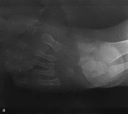
a, b crookedness at 5 months of age: a) Dorsal foot radiograph shows shortening of the Achilles tendon, lack of posterior to anterior tilt of the calcaneus, and horizontal involvement of the calcaneus, resulting in a reduced hip angle;
Typical manifestations or signs of clubfoot in children
- A palpable anterior edge of the heel bone
- Thin skin with fine wrinkles in this area
- Shortened Achilles tendon, palpable like a tight Achilles tendon
- Laterally displaced ankle joint
- Low heel height
- Thin calves
Four stages of clubfoot treatment in children:
– Phase I: chiropractic manipulation and application of a plaster splint (immediately after birth).
– Stage II: surgical correction of periarticular contracture (at 4-6 months of age).
– Stage III: Splinting (if the adducted position of the foot continues).
– Phase IV: Correction of recurrent deformities and late bone displacements.
Treatment of clubfoot involves manual manipulation and plaster splints (Stage I).
An uncomplicated clubfoot can be adequately treated with treatment levels I and II.
An important component of all stages of clubfoot treatment in children is a comprehensive massage and the right choice of footwear. The selection of corrective shoes for clubfoot is carried out in consultation with a podiatrist, after an examination and appropriate podometric and plantographic tests.
Causes of Clubfoot
General weakness of the tendon-muscular apparatus of the lower limbs as well as dysplastic changes in the foot skeleton are considered one of the causes of flat feet and clubfoot at this age.
There are a number of theories that explain the etiopathogenetic mechanisms in the development of flat feet:
- static-mechanical theory;
- vestibular theory;
- Anatomical theory;
- Theory of constitutional connective tissue weakness;
- Theory of inherited muscle weakness.
Where does the pain come from?
Etiologically, five types of flat feet are distinguished:
Congenital flatfoot occurs in varying degrees of severity (mild, moderate and severe). The most severe degree of congenital flat foot, called rocker foot, occurs in 2.8-11.9 % of cases and is diagnosed immediately after birth. The etiopathogenesis of this deformity is not yet fully understood. The most likely cause is a malformation of the embryo, a developmental delay at a certain stage of embryonic development. This malformation is considered a congenital defect.
In recent years, the history of static flatfoot has changed and is now being interpreted more broadly. In 78 % children with static flat feet, dysplastic changes in the foot skeleton were found in combination with neurological symptoms or metabolic abnormalities in the connective tissue.
A paralyzed flat foot occurs due to paralysis of the muscles that form and support the arch of the foot. Traumatic flatfoot is caused by the consequences of ankle and foot injuries, as well as soft tissue and tendon-ligament injuries.
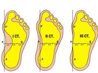
A distinction is made between mild, moderate and severe flat feet. Normally, the angle formed by the lines along the lower contour of the heel bone and the first metatarsal bone with the apex on the heel bone is 125 °, the height of the longitudinal arch is 39-40 mm, the angle of the heel bone to the support plane is 20-25 ° and the hindfoot is inclined 5-7°. In preschool children, the normal height of the longitudinal arch of the foot varies between 19 and 24 mm.
With a mild degree of flatfoot, it is necessary to reduce the height of the longitudinal arch to 15-20 mm, the angle of the arch height to 140°, the angle of the heel bone to 15°, the valgus position of the hindfoot to 10° and the anterior abduction of the foot to 8-10°.
Treatment methods for flat feet
Complete healing of flat feet is possible only in childhood, since the skeletal and musculoskeletal system in children is still being formed and by eliminating the pathology, subsequent fixation of the normal shape of the foot can be achieved. For adults, it's just a matter of improving the situation somewhat and stopping further deformities of the foot.
Treatment for flat feet in adults is primarily aimed at relieving pain and strengthening the muscles and ligaments of the foot.
It is very important to wear insoles as they help to properly distribute pressure on the surface of the foot. Orthoses for the spine restore the correct position of the foot and at the same time act as a shock absorber.
Shoes must be comfortable, not too tight, with a wide toe box and a low heel.
The orthopedist at the family doctor makes the diagnosis (degree of flat foot) and prescribes individual treatment. The sooner you see a doctor, the less deformed your foot will be. First degree flat feet can still be corrected, later the deformation can only be slowed down.
The goal of drug treatment for flat feet is to relieve pain.
Indications and contraindications
Ultrasound examination of the hip joint in newborns is indicated in all children up to 6 months of age to detect normal values or abnormalities. It is often performed within a few days after birth, especially if the woman has experienced adverse external or internal factors during pregnancy. Here are the most important indications for an ultrasound examination in a newborn:
- Premature babies in whom the likelihood of developing dysplasia is quite high. Babies from multiple pregnancies also belong to this risk group;
- breech or pelvic birth in which joint displacement occurs;
- A difficult pregnancy, especially if complicated by severe toxemia or vitamin and micronutrient deficiencies;
- if during pregnancy the woman takes drugs from various clinical and pharmacological groups - antibiotics, diuretics, cytostatics, immunomodulators, antiviral drugs;
- Acute viral, bacterial or fungal infections that the woman has contracted during pregnancy. Diseases of the intestines or respiratory tract are particularly dangerous in the second and third trimesters, when the fetal hip joints begin to form and the ossification nuclei of the femoral heads are formed.
Any of these factors can cause the development of dysplasia. An ultrasound scan is performed either immediately after birth or a few days after the mother and child are discharged.
After birth, the pediatrician examines the child once or twice a month to check his development. He or she may recommend an ultrasound scan if any of the signs of joint abnormalities are detected: asymmetric skin folds in the groin or buttocks, shortening of the leg, a specific clicking sound when the child lifts the hip.
Carrying out the diagnostic procedure
For the ultrasound examination, the child is placed on its side and the joint being examined is bent upwards at an angle of 20°. A gel is applied to the hip joint to help the probe slide and improve the conduction of sound waves. During manipulation, all joint and periarticular structures are displayed on one screen. The doctor takes images in the positions necessary to assess the condition of the tissue. As a rule, there are five positions: starting position, flexion and extension of the leg, abduction and adduction of the leg.
In order not to disrupt the examination, the referring pediatrician recommends that the child should not be fed immediately before the examination. The child must be full so as not to become gluttonous, but regurgitation can interfere with the diagnosis.
Drugs used in therapeutic blockades
First, let's look at the most commonly used local anesthetics.
'Novocaine'.
Can be injected into nerves and tissue, has an anesthetic effect, has an antispasmodic effect. It exerts strong irritation in the pathological focus and switches off peripheral innervation. The drug improves tissue trophism:
- reduces the permeability of blood vessel walls;
- has antiseptic and bacteriostatic effects
- increases resistance to allergens;
- balances vascular tone;
- Improves nerve trophism.
It is considered the safest analgesic and has minimal side effects. It has a certain level of toxicity.
'Lidocaine.
A local anesthetic that has a more intense and longer-lasting effect compared to Novocaine. It is used in the form of hydrochloride. Blocks potential-dependent sodium channels, thereby blocking the generation of impulses in nerve endings and the conduction of impulses along the nerves. It not only blocks pain impulses, but also other impulses. It dilates blood vessels and has no local irritant effects. A few minutes after injecting lidocaine, the pain subsides and the muscles relax. Toxicity is lower than Novocaine.
'Bupivacaine'.
A drug from the amide series. Its effect develops slowly but lasts a long time. It is about 16 times stronger than novocaine. The entire group of amide anesthetics is more stable compared to the ester anesthetics (Novocaine). Their prolonged effect can be explained by the fact that they have a longer half-life. This is because these drugs are processed in the liver and not in plasma. 'Bupivacaine prevents the pain impulse from spreading along the nerves by blocking the sodium channels of the neuron membrane. The blockade occurs sequentially, depending on the size of the nerve fibers (small fibers are more sensitive than large ones), the degree of activation (a higher pulse frequency opens more channels to be blocked) and the degree of myelination (fibers with a myelin sheath are better blocked). The effect of the treatment occurs after 5-10 minutes.
Possible complications and measures to avoid them
The complication rate for surgical blocks is extremely low at 0.5 %. Possible complications include reactions to the drug, symptoms of intoxication (vomiting, dizziness, palpitations), vascular damage at the injection sites. To prevent complications, additional medications are administered and the patient is advised to lie down for 1-2 hours after the procedure. Physical exercise is not recommended immediately, but is often the case when patients want to get rid of the pain. If no attention is paid to physical activity, the symptoms will return and worsen.
A spinal embolism can cause bleeding, infection at the puncture site, and damage to internal membranes and soft tissues. The latter is usually due to the doctor's inexperience. Anaphylactic shock can also occur. Therefore, the procedure is carried out only in a medical facility. To avoid complications, a trial injection is given.
When blocking a heel spur, complications occur in 15-20 % of people. These are in most cases due to inappropriate administration of corticosteroids and the patient's response to them. These may include soft tissue necrosis, abscesses, and tears in the heel fascia. It is necessary to examine the heel daily after surgery in order to detect an undesirable reaction in time. The first symptoms are pain, redness and/or flushing of the skin and numbness. Complications may be delayed and may not occur until several weeks after the procedure. This is due to the prolonged action of glucocorticosteroids.

Neurologist, neurophysiologist, homeopath, nutritionist and hirudotherapist with over 27 years of experience. Graduated from the St. Petersburg State Medical Academy. Highest qualification category. Member of the Association of Neurologists of Russia.
Read more:- Alignment of heel to toe.
- Why does a child develop clubfoot?.
- baby splashing.
- The bones of the human heel.
- Clubfoot in children therapeutic exercises 7 years old.
- What is clubfoot?.
- Photo of scraped feet.
- The right footwear for hiking.