A rarer disorder characterized by a shortened neck is Klippel-Feil syndrome. This is an inherited disorder caused by shortening or fusion of the cervical vertebrae. Klippel-Feil syndrome rarely occurs in isolation. More often, accompanying congenital anomalies affecting the musculoskeletal system, internal organs and systems are found.

- One child's leg is shorter than the other
- Diagnosis of asymmetry in children
- What risks are associated with short neck syndrome?
- Treatment of short neck syndrome
- What the symptoms of dysplasia can be
- What risks are associated with dysplasia?
- Causes of hip dysplasia.
- Treatment of dysplasia in newborns.
- Environmental factors
- diagnosis
- Causes of hip dysplasia in children
- Symptoms of hip dysplasia in children
- The child has one leg shorter than the other.
- Scoliosis and poor posture in children and adolescents
- Lameness after a fracture
- Main symptoms of intermittent claudication
- Types of hand deformities in children
- treatment and prognosis
One child's leg is shorter than the other
Short leg syndrome is an anomaly in child development that should be diagnosed and corrected as early as possible. Undiagnosed and untreated short legs can lead to lameness, permanent joint changes, or curvature of the spine.
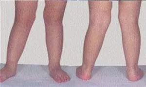
Pathological asymmetry of the lower limbs occurs when one leg is 0.5 cm or more shorter than the other. The shortening of one leg in relation to the other is usually divided into true shortening and false shortening. True shortening is a shortening of one leg relative to the other, with the different leg lengths due to different bone lengths. It usually develops in the fetus in the womb and is clearly visible on ultrasound.
Incorrect or dislocated shortening is caused by an imbalance between the segments of the lower limbs, resulting in one of the baby's legs being shorter than the other. The most common cause of incorrect shortening of one of the lower limbs in young children is hip dysplasia. Dysplasia is a dislocation of the hip joint in newborns with varying degrees of severity. When the head of the femur partially protrudes from the hip socket, doctors call it a subluxation. If the head of the femur protrudes completely from the hip socket and fat and connective tissue begins to grow in the hip socket itself, this is a hip dislocation. X-ray examination is the only way to detect the pathology and make an accurate diagnosis.
Another cause of false asymmetry in an infant, in which one leg becomes shorter than the other, may be muscle hypertension, that is, excessive muscle tension. If the baby has one leg shorter than the other due to hypertension, the excessive muscle tension causes the asymmetry. This condition may not be due to the infant's natural muscle tone, but rather due to neuronal dysregulation or brain dysfunction. Therefore, a child with asymmetry of the lower limbs due to hypertension should always consult a neurologist.
Diagnosis of asymmetry in children

Doctors can diagnose limb asymmetry as early as fetal development. In this case, doctors will clarify the diagnosis after the child is born and then prescribe treatment for the child.
If the orthopedist detects abnormalities in the child's development during a routine examination in the first months of life, he observes it and recommends preventive treatment. If hip dysplasia is suspected in the third month of life, an X-ray examination is carried out, which, together with the results of ultrasound examination, provides information about the presence or absence of this pathology in the child. If the diagnosis of dysplasia is confirmed, the doctor will determine a treatment regimen for the child depending on the severity of the disease.
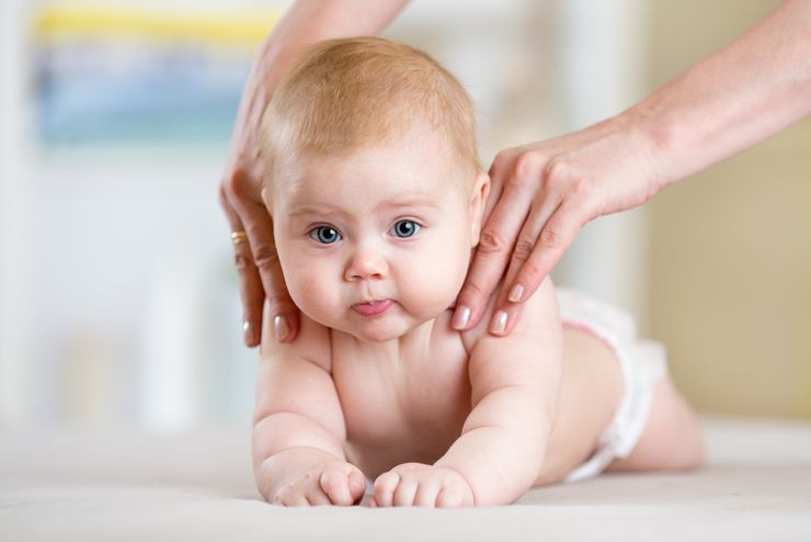
A simple examination for asymmetry of the baby's legs can be done by the mother herself. To do this, the child should be placed on his stomach on a changing table, with the hip and groin folds symmetrical and of the same depth. The child should then be rolled onto his back and the depth and symmetry of the skin folds should be reassessed. The child should be evaluated for limitation of hip abduction and for clicking or jerking during abduction. Asymmetric skin folds with varying depth and resistance or clicking during hip abduction may indicate hip dysplasia.
Another test for dysplasia is to bend the baby's legs at the knees while lying down. If one knee is lower than the other when you bend your legs, dysplasia is very likely.
Another test is the hypertonicity test. In this test, the child is placed on their back and asked to grasp an adult's fingers. If the child, having grabbed the adult's hands, begins to actively, symmetrically and fully move his legs, trying to pull himself up on his hands, everything is fine. If the child's legs are constantly crossed when moving, hypertension is suspected. Frequent crying, shaking the chin, excessive vomiting, stiffness while awake, and constantly clenched arms and legs while sleeping can also indicate hypertension. These symptoms should be reported to the pediatrician, who will refer the child to a neurologist.
What risks are associated with short neck syndrome?
The likelihood of complications depends on the severity of the pathology. The most common consequence of short neck syndrome is pinching of the nerve roots, of which there are many in the cervical spine. This leads to:
In addition to the nerve roots, there are also large blood vessels in the cervical spine that supply the brain. Shortening of the neck can cause them to become twisted or pinched. This can lead to dizziness and headaches.
Treatment of short neck syndrome
The cervical spine is corrected using conservative or surgical methods. The most common treatment is surgery:
These methods improve blood circulation in the clavicle zone, strengthen the muscle corset and restore the correct position of the vertebrae.
Surgical intervention is indicated when conservative treatment methods are ineffective or complications arise that threaten the life or health of the child.
The main symptom of pathology is a visible shortening of the neck and retraction of the neck into the shoulders.
If appropriate therapy is applied and all medical recommendations are followed, short neck syndrome can be easily corrected. This is confirmed by the many reports from parents who have been confronted with this unpleasant diagnosis. Since it is a genetic disease, the consequences are difficult to predict. Even if the pathology is successfully corrected, there is a risk of complications in other organs and systems.
What the symptoms of dysplasia can be
During the fetal period, the following may be affected or delayed:
- the formation and development of the acetabulum;
- the development of the upper parts (head and neck) of the femur;
- the formation of the correct relationship between the hip socket and the head.
These abnormalities result in the femoral head not being able to be fixed in the correct position, causing it to move outward and upward.
Pathological joint mobility develops, which can manifest itself in the following symptoms, among others
- Instability of the hip joint: The anatomy is preserved, but there is evidence of underdevelopment of the joint, allowing the femoral head to move freely up and down in the socket.
IMPORTANT
Normally such a movement should not occur. This leads to pre-dislocation, subluxation and dislocation.
- PredislocationDislocation: The head of the femur is displaced towards the upper edge of the joint socket, but does not protrude beyond the joint socket.
- Subluxation: Only part of the femoral head is in the socket. The other part is shifted upwards. The ligaments are overstretched and deformed.
- Contortion: The head of the femur is located outside the joint cavity. It is shifted upwards in relation to the joint socket. The hip socket lies below the femoral head.
What risks are associated with dysplasia?
If the dysplasia is not treated, the shortening of a limb is already advanced by the time the child can stand and walk. As a result, there is a compensatory curvature of the spine and disruption of the internal organs, which ultimately leads to severe disability.
- children whose parents had DTS at birth;
- children born prematurely;
- Children whose mothers had bad habits during pregnancy (tobacco, alcohol, drug abuse), resulting in chronic fetal toxicity;
- infants in non-cerebral position;
- children of mothers who suffered from early or late gestosis;
- children of mothers who have undergone an intrauterine infection;
- Children of mothers whose pregnancy occurred in polluted areas (near large factories, major highways, etc.);
- Children of mothers with serious somatic diseases, infectious diseases and those taking medications during pregnancy;
- Children of mothers with anemia, endocrine disorders and avitaminosis during pregnancy.
Causes of hip dysplasia.
There are several factors that contribute to the development of hip dysplasia:
- Genetic – the occurrence of the disease is due to a possible hereditary predisposition.
- Hormonal – the effect of hormones on the body during pregnancy. This leads to a reduction in muscle tone, resulting in instability of the hip joint.
- Bone and cartilage disorders – during fetal development: vitamin and micronutrient deficiencies; fixation in the womb; limited fetal mobility in the uterine cavity.
Diagnosing grade 1 and 2 hip dysplasia is not easy because there are no visible symptoms, no pain and the baby does not complain of anything. Therefore, parents should pay attention to the appearance and behavior of the newborn.
The following symptoms are characteristic of dysplasia:
- Asymmetrical positioning and additional folds on the buttocks and tendons;
- The child expresses discomfort when trying to bend his legs at the knees;
- One leg is slightly shorter than the other;
- The hip joint is excessively mobile;
- The amount of movement in the joints varies;
- A wobbly gait, walking on tiptoes, or limping.
The combination of several symptoms is the basis for a comprehensive examination by an orthopedist/traumatologist. The earlier the pathology is detected, the easier it is to treat.
Further examination methods in a child with suspected dysplasia.
- Ultrasound examination is the method of choice for diagnosing hip dysplasia in the first months of life. It is safe and provides sufficient information to confirm the diagnosis.
- The radiological examination shows the late ossification of the femoral head, the shape of the acetabulum and the femoral head and their differences in size.
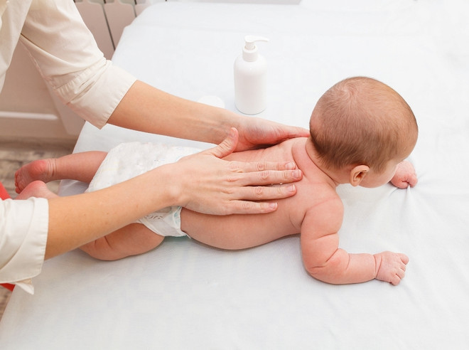
Treatment of dysplasia in newborns.
If hip dysplasia is diagnosed in the maternity ward, treatment begins before the diagnosis is confirmed by ultrasound.
The most common treatment regimen: extensive rocking for infants in the first four months, the use of a Frekeh pillow or Pawlik stirrup until the end of the first six months, and then various diverticular splints. The duration of treatment and the choice of orthoses depend on the severity of the dysplasia. From the first week of life, therapeutic massage is recommended to prevent muscle atrophy and improve blood circulation in the affected limbs, as this will help eliminate the pathology as quickly as possible.
Parents should remember that any treatment must be recommended by an orthopedic traumatologist. The treatment is lengthy and requires a lot of patience from parents and child as well as regular treatment.
Environmental factors
Various substances with estrogenic effects pollute the environment and accumulate through the food chain. They come from e.g. B. from insecticides, natural plant estrogens and the plastics industry. This negatively affects the formation of the genitals in boys and can lead to the development of hypospadias.
The characteristics of hypospadias concern the urethra: it is incomplete and its opening to the outside is in an unnatural position. Consequently, the urethra is not located at the tip of the glans, as is normally the case, but elsewhere along the ventral fascia of the penis (on the inside).
Main symptoms of penile hypospadias:
The disease does not cause any symptoms. The distal form without deviation is not associated with functional limitations and is only a cosmetic problem. In proximal hypospadias, the urine flow may be less controlled. A higher degree anomaly must definitely be corrected surgically. In addition to the aesthetic aspect, the undisturbed development of sexual function and normal bladder emptying play an important role in the therapeutic decision.
Table – symptoms of hypospadias in children depending on the form of the disease
The external urinary outlet is narrowed and deeply located. In the cephalic form of hypospadias, urination is impaired. Penile curvature increases with the onset of sexual activity.
It is characterized by a pronounced curvature of the body of the penis and impaired urination. In venous hypospadias, urine is released in a thin stream. The penis must be elevated so that the child does not urinate on the legs.
The metus can be located at different levels of the posterior surface of the penis. Urinating while lying on your stomach is difficult. With truncal hypospadias, a painful erection is noted. If sexual intercourse is possible, fertilization of the egg does not occur because semen does not enter the vagina.
diagnosis
Hypospadias is most commonly diagnosed at birth. Not only is the opening in the wrong place, but the foreskin is often not fully developed on the underside. The tip of the penis remains open. The diagnosis of hypospadias is made based on a physical examination of the penis. Only in this way can the urologist assess the location of the urethral meatus and the presence of accompanying anomalies such as a congenitally curved penis or cryptorchidism.
Although hypospadias is visible from birth, it can also go unnoticed. To avoid this, a thorough examination of the newborn is enough. Early diagnosis is important to avoid physical and psychological consequences.

The appearance of the foreskin often draws attention to the problem. About 8 out of 100 boys with hypospadias also have cryptorchidism (testicular weakness).
Anomalies associated with hypospadias:
- cryptorchidism (7 %);
- inguinal hernias (12 %);
- mental retardation (6%);
- cardiac abnormalities (5%);
- Musculoskeletal abnormalities (3%).
The most severe forms of hypospadias are often associated with other anatomical abnormalities of the penis or urinary tract. The most common is congenital penile curvature. Cryptorchidism, hydrocephalus, inguinal hernia, and renal malformations may also occur.
These developmental anomalies should be taken into account because they may be associated with an intersexual condition, in which a person has both male and female sexual characteristics. The causes of intersexuality include changes in sex chromosomes and hormones.
An ultrasound scan of the bladder and kidneys will be ordered to rule out any co-existing abnormalities. MRI of the pelvis, uroflowmetry, urethrography and urethroscopy are mandatory.
Causes of hip dysplasia in children
There are several factors that can contribute to the development of hip dysplasia in a child
- Inheritance. This pathology is more likely to occur in children whose father and mother suffered from congenital hip dysplasia;
- Severe toxicity;
- taking medications during pregnancy;
- Large fetus;
- abdominal presentation;
- lack of water;
- gynecological problems.
Symptoms of hip dysplasia in children
- instability of the hip joint;
- Displacement and return of the femoral head to its original position;
- Limited abduction of the affected hip joint;
- Asymmetrical folds in the back of the thighs;
- visible shortening of the affected limbs.
The first symptom in newborns is hip instability, but this resolves spontaneously in 80 % cases.
The child has one leg shorter than the other.
Ministry of Health
Main Department of Health Care of the Executive Committee of the Brest Region
225903 Brest Region Malorita 96 Sovetskaya Street, Malorita
Registration office: Tel. 8(01651) 20312, 20313, 20314
Email: [email protected]
- Home page
- Info
- Arthroplasty waiting list
- Algorithm for the patient's path through medical control
- Going through a medical check-up
- Algorithm for passing a driving test
- Algorithm for passing periodic and preliminary medical examinations
- Schedule for the admission of citizens and legal entities to the Health Department of the Executive Committee of the Brest Region
- POLICY FOR THE PROCESSING OF PERSONAL DATA IN THE HEALTH INSTITUTION 'CENTRAL DISTRICT HOSPITAL MALORITA
- Healthy lifestyle
- Electronic Prescribing Questionnaire
- COVID-19 information material:
- corruption
- News
- Order your ticket
- Timetable
- Administration
- Departments
- Health center departments
- Parent organizations
- Therapeutic areas
- Subordinate facilities
- Telephone directory
- Photo gallery
- Personal visit
- addresses
- Parent organizations
- PROCEDURE FOR PROCESSING INQUIRIES
- Subsequent diagnosis
- Services for RB citizens
- Services for foreigners
- Payment for services via the ERIP system
- Medical examination
- List of administrative procedures
- Hotline
- Senior executives
- ORDER FORM FOR MEDICAL DOCUMENTS
Scoliosis and poor posture in children and adolescents
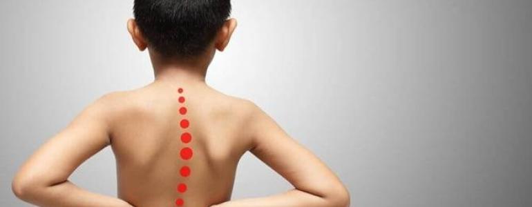
The above image is for illustrative purposes only.
It is not without reason that the 21st - and also the 20th - century is called the age of the sedentary person or even the sedentary civilization. The development of modern means of transport, the emergence of large urban agglomerations, the emergence of more and more machines to facilitate work at work, at home and at school have led to a radical change in our way of life. It is estimated that we sit for up to 15 hours a day and consciously 'switch off' one of the most important systems in our body - the musculoskeletal system. This inactivity inevitably affects the fitness and physical health of the skeleton and muscles. They become weaker day after day and suffer from a variety of diseases that are known to orthopedists, therapists and, unfortunately, pediatricians.
Causes of Postural Damage
Hypodynamia (decreased muscle mobility and contractility) occurs in both adults and children who prefer to walk to school rather than use public transportation. A few hours in the classroom, then extracurricular activities that often have nothing to do with sports - that doesn't leave much time for running. If homework, computers and television are added to the mix, there is no time left for active leisure activities.
The results are disappointing: gymnastics lessons (2 hours per week), sometimes swimming pool, rhythmic gymnastics or the popular martial arts school for boys - that's it. In the hectic everyday life of a school child there is no room for walks or outdoor games.
Today's children are growing and maturing faster, but their physical health is declining. Although a sedentary lifestyle does not contribute to flexibility and mobility, it does lead to hypoxia and a weakened immune system.About 70 % of children have poor posture and 10 % have scoliosis, a sideways curvature of the spine.
Lameness after a fracture
The main causes of lameness after a fracture are.
- the repositioning (reattachment of the bone fragments resulting from the fracture) was not carried out correctly and the injured leg was shortened;
- the patient put weight on the lower extremity before the doctor's appointment, which may have led to displacement of the bone fragments;
- Mobilization (immobilization of the lower limb, e.g. by applying a plaster cast) leads to soft tissue atrophy;
- the patient is afraid of putting strain on the limb.
Treatment for limping after a fracture depends on the cause and location of the fracture. Often a second repositioning procedure must be performed (the doctor must reattach the bone fragments). However, most often non-invasive (that is, without penetrating the tissue) treatment methods help to eliminate the condition:
- massage the injured limb;
- Physical therapy;
- Discussions with a doctor (not necessarily a psychotherapist) who will explain the processes of consolidation (fusion) of the bone fragments and convince you that you can put weight on your leg again after the fracture.
Main symptoms of intermittent claudication
In intermittent claudication (intermittent claudication), the gait 'fails' after a certain load on the lower limbs - for example, after a person has walked a certain distance.
Intermittent claudication has the following main causes:
- Diseases of the vessels supplying blood to the lower limbs (atherosclerosis, obliterative arteritis, thrombosis, etc.).
- Diseases of the musculoskeletal system. This disorder is most often caused by a narrowing of the spinal canal. It is also known as neurogenic claudication.
The difference in clinical symptoms is that in claudication caused by vascular diseases, pain in the legs is more common, while the symptoms of neurogenic claudication are mainly weakness of the lower limbs and paresthesia - a disorder of soft tissue sensitivity.
Treatment of intermittent claudication should begin with smoking and alcohol cessation. Drug treatment depends on the cause of claudication:
- drugs to improve blood flow to soft tissues;
- drugs to improve nerve conductivity;
- Physiotherapy (including hydrogen sulfide and radon baths).
A highly qualified specialist in our clinic will explain more about intermittent claudication treatment. You can make an appointment at https://www.dobrobut.com/.
Types of hand deformities in children
The following hand development abnormalities are most common in children
- Syndactyly. This malformation is considered one of the most common. In this anomaly, the fingers are fused together. There are different forms of this deformity. The fingers are partially or completely fused together, and the anomaly may affect only the skin or the bones may also be fused together.
- Polydactyly. The presence of an additional sixth finger is characteristic. It can occur on one or both hands, less commonly on the feet. In most cases, the sixth finger is underdeveloped and therefore looks more like an additional extension. In rare cases, the finger has bones, ligaments and tendons and is fully functional.
- Brachydactyly, ectrodactyly. These defects are characterized by the absence of phalanges, underdevelopment and shortening of the fingers. Brachydactyly has nail plates. This anomaly is often accompanied by fusion of the fingers.
- Camptodactyly. This is the name given to a malformation in which a flexion deformity is observed. The fingers cannot be fully extended either in a passive state or during active activities. Due to the lack of a hand fold, the skin is greatly stretched. The anomaly most commonly affects the little finger.
- Hypoplasia. This is a specific malformation of the thumb. Depending on development, it can manifest itself in different ways. The thumb is often shortened or emaciated. Sometimes it is only connected to the body through the skin or is missing completely.
- Concretion. This anomaly is also called a fusion defect. The main feature of this anomaly is that two fingers sit on one metacarpal bone, causing dysfunction of the hand. The space between the fingers is very narrow. The hand may be shorter than normal.
- split hand. Also called 'crab claw' because the hand is split in two. Can occur on one or both hands. Often occurs on the legs as well.
- Brachymetacarpy. With this malformation, the metacarpal bone is very short. The fingers are also short due to the 'collapsed' metacarpal bone. This anomaly is especially noticeable when the child clenches his fist.
- gigantism. Enlarged fingers or single phalanges. More rarely, there is an enlargement of all parts of the hand.
- Congenital amputation of fingers or phalanges. Very rare. The limbs end in a stump, which may worsen as the child grows. The stumps are often deformed. This is caused by uneven growth of bones and soft tissues.
- Macrodactyly and megalodactyly. What is characteristic is that one or more fingers are longer and thicker. In most cases, the finger is still functional but has a significant cosmetic defect.
treatment and prognosis
Finger deformities in children are treated surgically in most cases. Depending on the type of abnormality, tissue dissection or anastomosis may be performed and additional parts may be removed. Underdeveloped (shortened) toes or missing toes are more difficult to treat. In this case, reconstructive surgery with prostheses can be performed.
In each case, the treatment is determined individually by the doctor, depending on the type and severity of the malformation and the patient's condition.
Surgical removal of toe deformities in children is recommended before the age of one year, with the timing of surgery being chosen on a case-by-case basis. Modern medicine gives a favorable prognosis in most cases.
Conservative treatment is occasionally used when the deformities are minor, such as: B. in polydactyly, when the growths consist of skin and contain no bones or ligaments. Various physiotherapy treatments and therapeutic exercises are also used to restore functionality to the fingers and hands.
- Legs of different lengths in a child.
- shortening of the hip.
- One leg is shorter than the other.
- Leg splint for newborns.
- ectrodactyly.
- Feeling that one leg is longer than the other.
- One foot is bigger than the other.
- How to determine leg shortening.