Certain types of medicinal plants have anti-inflammatory, pain-relieving or pain-inhibiting effects and stimulate blood circulation. Here are examples of their use in practice:
- HEALING AND RECOVERY MUSCLE-SKELETAL SYSTEM
- Why you should come to our clinic:
- PRINCIPLE
- GRIGORENKO Andrei Alekseevich
- Tereshkin Vitaly Vladimirovich
- where to find him
- Functions of the ankle
- Pain from a spinal fracture
- Pain in the leg with a fracture
- How long do you have to walk on crutches after a fracture?
- How to go to elbow rests (sticks)?
- treatment of dislocations
- prevention
- Types of carpal bone injuries
- signs
- Where can swelling occur?
- Important to know!
- How do I notice swelling?
- Rehabilitation after bypass surgery
- Pain after bypass surgery / complications after bypass surgery
- How to prevent change
- Can a Tattoo Hide Stretch Marks?
HEALING AND RECOVERY
MUSCLE-SKELETAL SYSTEM
The constant stress our feet are subjected to is made worse by excessive weight, heavy lifting, or constant walking in heels.
Pain in the ankle when walking can be a result of uncomfortable footwear or a serious illness, but the problem isn't always in the lower limbs.
Ankle pain is often the result of trauma (fractures of the ankle or heel and foot, sprained ligaments, torn muscles), osteoarthritis, gout and Achilles tendinitis.
In order to ensure effective treatment, the actual cause of the pain must be determined. If your ankle is constantly swollen and painful when you walk, you should see a doctor as soon as possible. Prolonged misalignment of the ankle can contribute to the disease progressing into a chronic condition that is extremely destructive and more difficult to treat.
Why you should come to our clinic:

Leg pain is caused by a number of symptoms resulting from various diseases of the lower limbs. They are expressed in acute or long-lasting pain sensations. Most often they occur in connection with joint, muscular or vascular diseases, but they are also the result of trauma or neurological abnormalities.
Inflammatory processes often occur in the periarticular capsules. This condition is called synovitis. If the joint capsule of the Achilles tendon is affected, it is referred to as Achilles tendon bursitis, which is characterized by a build-up of inflammatory fluid in the joint, leading to thickening of the tissue and a partial loss of joint mobility.
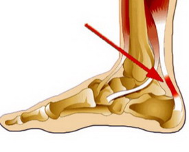
The Achilles tendon is the strongest tendon in the human body. It connects the calf muscle to the heel bone. The Achilles tendon supports the entire weight of your body when you walk, run, or exercise.
PRINCIPLE
GRIGORENKO
Andrei Alekseevich
Founder and director of the clinic.
Specialist in neurology, Doctor of Osteopathy Europe (DOE), rehabilitator, orthopaedist.
Tereshkin
Vitaly Vladimirovich
Osteopath, manual therapist, rehabilitation therapist.
Specialist in restorative medicine, negative pressure gradient therapy, medical stretching and remedial gymnastics.
where to find him
For example, a person's legs are highly extended, and the ankle, another name for the ankle, acts as a transmitter of force from the contracting muscles to the legs. Many people have heard these terms before, but not everyone is aware of them,

.
This part of the body has a special feature: it is neither a muscle nor an independent joint, it is not an independent part of the lower extremity, but a complex joint based on the connection with the ankle joint. When walking, this part of the body experiences the mechanical pressure of the weight and absorbs it.


Functions of the ankle

- It supports the weight of the human body;
- Even distribution of the total weight of the body on the foot;
- Functions related to upright posture;
- Active function that allows jumping, running, and more.
- rotation function. Allows the person to rotate around their own axis, keeping the legs in the same position during the rotation, ie not moving;
- damping function. Allows humans to cushion the force of impact while walking or running.

Because the ankle is a highly stressed area of the human anatomy, all types of ankle injuries are very common, especially if you are an active person who plays sports or moves around a lot. Let's take a look at the types of ankle injuries.
Pain from a spinal fracture
A spinal fracture and its characteristic pain occurs when a person falls from a great height or hits his head while diving; when there is a strong impact on the back (e.g. car accidents, falls from rocks); when a person becomes trapped under debris. The pain from spinal fractures is felt in the back and is very severe, especially when the person tries to move. The most dangerous thing about these types of fractures is that they can damage the spinal cord, which is located in the spinal canal. Spinal cord injuries can occur when vertebrae are broken off, when they are fractured, and also when they are dislocated. A spinal cord injury causes paralysis of the arms and legs, sometimes the entire body, with the affected person losing all feeling and the ability to move freely.
Pain from hand fractures occurs along the bone line. The limb takes on an unnatural shape, there may be stiffness where there are no joints, and the limb may be swollen.
If a fracture or dislocation of the carpal bones is suspected, the wrist is strapped into a wide splint that begins in the middle of the forearm and ends at the fingertips. Beforehand, cotton wool, gauze or something similar is put in the hand to bend the fingers. Cold is applied to the injured area.
Pain in the leg with a fracture
Pain in lower limb fractures occurs along the bones, the limb swells, becomes unnaturally shaped and stiff where it should not be, and the joints are not localized. As first aid for lower limb fractures, a splint (such as a plywood board, stick, cardboard, or similar) is placed over the injured limb. The length of the splint should be such that it can be applied from above the edge of the pool (possibly up to the armpits) to the heels. In this way, the injured extremity can be fully relieved. When applying the splint, do not lift the injured leg, but keep it in the position it is in, and then place the splint under the lower back, knee and heel, paying attention to the leg not to move. Put a cold object on the injured area.
Rib fractures result from falls from a height, chest compressions, direct impacts, etc. The pain from a rib fracture is sharp and occurs when the person breathes, coughs, or changes body position. When the ribs are injured, the victim usually does not take a deep breath, resulting in shallow breathing. The main danger with broken ribs is that the pleura and lungs can be injured by the sharp edges of the bone fragments. When the lungs are injured, subcutaneous emphysema can develop when air enters the subcutaneous tissue. This causes flattening of the intercostal spaces, which looks like edema.
You can feel the area to find out if it's a swelling - if it is, you'll feel a cracking sound that's like small blisters bursting on your fingers.
How long do you have to walk on crutches after a fracture?
If you have a hip fracture With a hip fracture, you must first undergo skeletal traction. This takes between 1.5 and 2 months. A plaster cast will then be applied and you will not be allowed to bear weight on your leg again for 3 to 4 months.
femoral neck fracture can be treated in several ways:
- Immobilization – a plaster cast is applied and the leg is rested. It is not recommended to treat a femoral neck fracture in this way as muscle wasting may occur.
- Osteosynthesis - Bone fragments are reduced with screws or pins. This method is slightly better, but scarring may occur.
- Endoprosthesis – a metal prosthesis is inserted. The best option. However, walking on crutches is also required here. 3-6 months.
If ankle fracture treated without a cast, you only need to use crutches for 1.5 to 2 months. With a cast and surgical treatment, this time can easily increase to 2 to 2.5 months. In the worst case of a bone strain, the treatment period can be extended to 4 to 6 months.
How to go to elbow rests (sticks)?
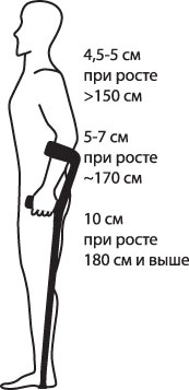
Walking on elbow crutches is not much different from walking on armpit crutches. If you have weak arms, it is better to use armpit crutches. This makes walking easier.
Elbow crutches have the advantage that they are lighter and take up less space.
You can read about walking techniques at the beginning of this article. It is important to choose the right size crutches. When walking with crutches, the weight of the load is not on the armpits, but on the hands.
How to find the right elbow crutches:
- Stand up straight with the tip of the crutch about six inches from your foot.
- Bend your arms 15-20 degrees at the elbows.
- The cuff that wraps around your forearm should be 5-10 centimeters from the elbow.
- The cuff should be neither too tight nor too loose.
treatment of dislocations
Before treating a sprain at home, the doctor will examine the injured leg and take an X-ray. The specialist feels the foot and checks its mobility. This is to ensure that there are no broken bones or dislocations. If necessary, an ultrasound examination and a computed tomography are ordered. Only after a thorough examination can the doctor make an accurate diagnosis and prescribe the necessary treatment.
Once the specialist has confirmed that it is a sprain, the patient is advised to rest the injured leg completely. Ideally, the patient should remain in bed for the first few days to reduce the load on the injured leg as much as possible. During these days, an ice compress should be placed on the ankle periodically. The ice pack is applied for about 30 minutes, then a break of about an hour is taken. At least five such treatments per day should be carried out.

If there is swelling, the leg is wrapped with an elastic bandage from the toes to the middle of the lower leg, but not too tightly. When the swelling goes down, the bandage can be removed. In bed, place a bolster or pillow under your leg so that your ankle is above heart level. This allows the fluid to drain better from the injury site. For severe sprains, a plaster cast can be applied for 2 weeks.
Anti-inflammatory gels or ointments such as ibuprofen or fastum gel are applied to the injured area of the leg to heal the dislocation quickly.
After 5 days, when the main symptoms of the dislocation subside, the doctor prescribes physiotherapeutic treatments such as magnetotherapy, ultrasound, UHF.

prevention
To avoid an ankle sprain or prevent its recurrence, a few simple rules should be followed. Athletes should do a short jog before training or competing to warm up muscles. A special bandage can be used to support the joint.
Once recovered, you must be very careful; the ankle is still weak and the slightest bump can lead to re-injury. Wear comfortable shoes that fit well and have non-slip soles. High heels should be avoided temporarily.
Types of carpal bone injuries
Broken bones are one of the most common injuries due to their small size and fragility. Fractures usually result from a fall onto an outstretched hand. Wrist pain and swelling are the most common symptoms. Any pressure on the bone can cause severe pain. Sometimes a wrist bone fracture causes only mild discomfort and pain in the injured area. A fracture can be difficult to diagnose because it is often mistaken for a wrist dislocation. A fracture often involves a tear in the ligament that connects the bones.
The patient may also be diagnosed with a rare condition known as idiopathic avascular necrosis of bone. In this case, the bone dies without a fracture.
signs
If you experience sudden, severe, or increasing wrist pain, it's important to see a doctor right away. Medical help should also be sought if a wrist injury is accompanied by the following symptoms:
A wrist injury can be diagnosed in a number of ways. The first step is usually a medical history and an orthopedic examination. This can be followed by further investigations:
- X-rays of the wrist
- Ultrasound examination of the wrist
- CT scan of the wrist
- Magnetic resonance imaging of the wrist
- blood test
- Taking a sample of fluid or tissue from the joint.
Where can swelling occur?
Swelling during pregnancy is most common in the feet, ankles, and lower legs. The causes are clear: Excess liquid first collects at the bottom, where it is pulled down by gravity. It usually all starts with a parasitosis – a slight swelling with pallor and reduced elasticity of the skin. Another popular spot for parasitosis and swelling is the hands. Often the excess liquid also leaves traces on the face; in addition to the swelling, a blocked nose – the so-called 'pregnant rhinitis'3 – can also occur.
Important to know!
For most people, catarrh drops and sprays are among the most harmless medications. Not in pregnancy - many of them are dangerous for the fetus 4 ! Remember to consult your doctor before treating a runny nose.
Even in early pregnancy, the woman's body temperature rises to a level that is more comfortable for the fetus - just over 37 °C. This does not mean you have a cold or a virus. If you want to learn more about basal body temperature, read our article.
How do I notice swelling?
Severe swelling during pregnancy is hard to miss. If the swelling is less obvious, especially if the extra fluid is slow to build up, the following signs will help you spot it:
- A suspicious weight development is observed. If you're following your doctor's recommended diet, but your weekly weight gain is above the norm for this period of pregnancy, there's likely fluid retention somewhere in your body.
- Rings press into the fingers, shoes press. The signs of pasticcio in pregnancy are easiest to spot from things that fit on your hand or foot - they start to pinch. By the way, it's better to remove the rings while it's still possible.
Rehabilitation after bypass surgery

The rehabilitation period after bypass surgery is very important. In general, further recovery depends on how well this phase of treatment is managed. Rehabilitation after bypass surgery should be divided into three phases. The first phase begins in the hospital when the patient begins breathing and walking exercises under the guidance of a physical therapist. The second phase continues in the sanatorium, where, under the guidance of professionals, the patient gradually walks more and becomes accustomed to everyday life. If the bypass operation was planned and the postoperative period went smoothly, the patient's exercise capacity gradually increases and becomes better than before the operation. That is basically the aim of the operation. Although the sternum is often opened during the operation and then connected with metal staples, there is no need to fear that it will come loose. During this period, asymmetrical movements in the upper part of the shoulder girdle should be limited and the habit of holding hands behind the back or carrying something heavy in one hand or on one arm should be avoided. Patients who have undergone minimally invasive surgery are very fortunate not to face these problems. The third phase is the outpatient phase. You exercise at home under the close supervision of your treating cardiologist, who uses stress tests to assess whether your exercise program is right or not.
In normal cases, physical activity is not contraindicated but beneficial. It is important for the treating physician and the patient to verify safety. The basic method is to do a stress test (usually an echocardiographic stress test). This test should be carried out 3-4 weeks after the operation on the recommendation of the cardiologist. This test assesses the body's response to exercise and detects arrhythmias and signs of myocardial ischemia (poor blood flow to the heart). If the test is negative (that is, no ischemia is detected) and the doctor considers the changes in blood pressure and pulse during exercise to be appropriate, we recommend that the patient undergo regular cardio exercise.
Pain after bypass surgery / complications after bypass surgery
All patients experience pain in the early postoperative period after bypass surgery. Postoperative wound pain occurs. It is important to know that the heart is functioning almost 'normally' for a few days after coronary bypass surgery. In addition to pain, the patient's discomfort is also caused by a drop in hemoglobin, sometimes a brain response to the artificial circulation. This is important:
- If the pain is difficult to bear, take painkillers (usually all patients stop taking painkillers after 7-10 days)
- Increase your reduced hemoglobin. This often requires long-term use of iron supplements.
- Confirm that there are no signs of myocardial ischemia (using a stress test) and resume physical activity.
- Keep in touch with your cardiologist to ensure all your questions are answered in a timely manner.
How to prevent change
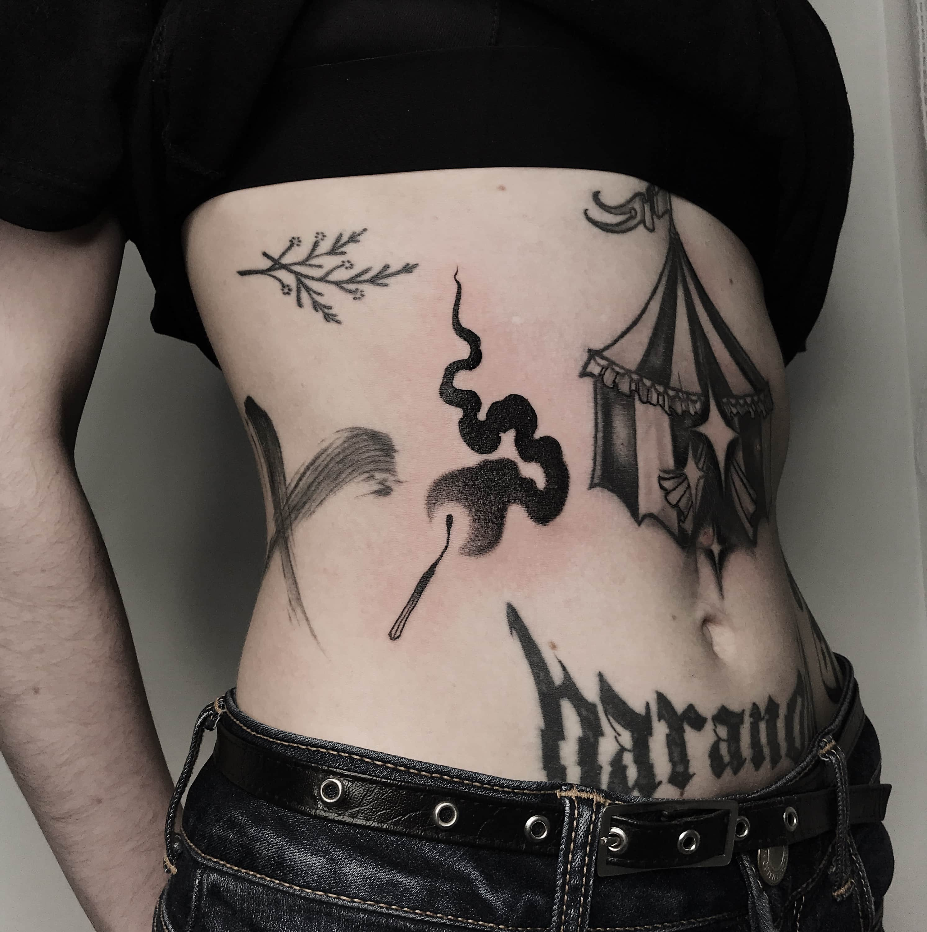
In order to reduce the risk of the tattoo stretching with weight gain or distorting after weight loss, it is important to choose the right body locations for the tattoo. It is advisable to place the sketch where the layer of fat is thinnest, based on the anatomical features of the person. Suitable for drawing:
It is better for girls to abandon the idea of sticking tattoos on the chest, stomach, buttocks or inner arm. These parts of a woman's body are more prone to age-related changes and become deformed during pregnancy and breastfeeding.
Small tattoos change less than large-scale compositions with weight gain or loss. If you shift the weight up or down smoothly and without big jumps, the subject will not be damaged.
Proper care of the tattoo during the healing phase is also important. This will help the pigment settle into the tissue well.
A good master in a proven salon should be consulted for a design. A professionally executed work with high-quality pigments will please the owner for a long time - it will not fade or lose its color, it will keep its shape even under the influence of changes in a person's weight and lifestyle.
Can a Tattoo Hide Stretch Marks?
Girls often worry that the image on the stomach may warp during pregnancy. In fact, during pregnancy, the skin is very sharp and greatly stretched. However, after childbirth, the abdominal muscles quickly return to their original appearance, especially if the woman is young and exercised before pregnancy. A lot depends on the elasticity of the skin. To improve them, it is necessary to apply special creams. When the mother's figure has recovered, the tattoo will also return to normal. Minor adjustments may be necessary.
Tattoo retouching is sometimes required due to stretch marks caused by a sudden stretching of the skin fibers. They initially have a bluish-purple hue, but then become lighter. Stretch mark scars disrupt the pattern and therefore need to be corrected.
Unsightly stretch marks can also be concealed with a suitable sketch. To do this, you need to contact an experienced handyman. Complex compositions with a plethora of additional details can help hide the flaw.
It is recommended to tattoo stretch marks at least a year after delivery. By this time, the woman's body and skin have fully recovered.
In summary, pregnancy, intense exercise, and weight changes throughout life are not barriers to getting a tattoo. A quality tattoo after losing weight or gaining muscle mass will preserve the original aesthetics. If the sketch is still distorted, it can always be corrected: retouch, improve the contours of the picture, add new details. In extreme cases, it is acceptable to completely cover the destroyed composition with a new one. The most important thing is that you contact a professional handyman who will suggest the best way to solve the problem.
- The hock in which the person is located.
- Torn ligament in leg how long will it take to heal.
- The hock in which the person is located.
- The pubic ligament where it is located Photo.
- The Ligamentum Treitz, where it is located.
- Hip Amputation Surgery Code.
- Take off the ankles.
- Pelvic ankle bones.