flexion and extension is painful due to nerve compression from the swollen tissue. A hematoma is bleeding into the soft tissues caused by rupturing of blood vessels and capillaries. The foot first turns purple, then blue and after some time yellow.

- What is ankle pain and what to do if it hurts
- causes of pain
- Possible diseases
- Where is she performing?
- Functions of the ankle
- Physiological function of the ankle
- Ligaments of the ankle:
- Osteoarthritis of the ankle
- Anatomy and function of the ankle
- Ankle injuries and treatment of ankle pain
- fracture
- Types of lateral malleolus injuries
- Lateral ankle sprain
- Treatment of a broken ankle
- Consequences of a broken ankle
- Treatment methods for ankle fractures
- Recovering from an ankle injury
- construction and location
- functions
- Important NOTE
- Conclusion
What is ankle pain and what to do if it hurts
The hock joint of the lower limbs has a complex structure. Walking, carrying heavy objects, or doing physical exercises puts stress on this part of the leg. In the musculoskeletal system, the ankle is the most important connection between the bones of the lower limbs and the foot. Its outer and inner parts act as a stabilizer.
We first define the ankle. The ankle joint is an important part of the hock joint, which is located at the lower part of the lower limbs. It is located where the calf meets the foot, on either side of the Achilles tendon. A distinction is made between the inner or medial part and the outer or lateral part.
reference. Many people wonder: how is the ankle different from the hock and what is the difference between the two? Both names for this part of the leg are correct and mean the same thing. The ankle is a slang, common name. In medicine and science, it is called the ankle.

The ankle is made up of two bones: the tibia and fibula. Accordingly, the outer (lateral) side of the ankle is formed by the tibia and the medial side by the fibula.
In between is the ankle bone (talus) with three joints. Together they form the ankle, which allows the foot to move freely in multiple directions.
The illustration shows where the ankle is located in humans.

The main function of the ankle is to connect the ligaments that hold the human foot together. There are four types of ligaments that start here:
causes of pain
The causes of external ankle pain are obvious: uncomfortable footwear (wrong size, high heels), constant stress on the ankle (during sports or carrying heavy loads), and skeletal stress from pregnancy.

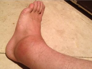
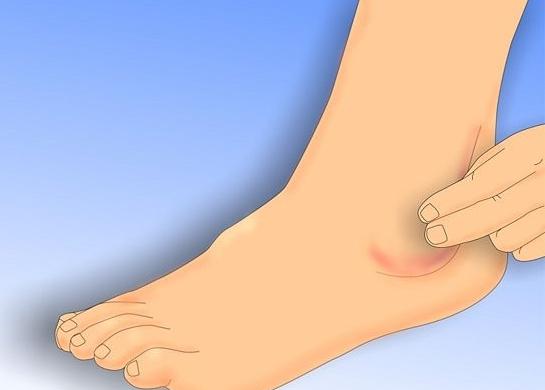
But internal processes in the body also have an influence. For example, overweight and obesity increase the pressure on the legs. In addition, the complex physiology of the ankle can lead to injury and inflammation, which in turn can damage adjacent joints, muscles, and bones.
The photo shows a patient with a painful ankle on the ankle side.
Possible diseases
Specialists distinguish a number of diseases that arise as a result of physiological disorders, age and the presence of other diseases:
- arthritis. It is caused by metabolic disorders or inflammation of various origins. The disease is divided into post-traumatic and rheumatoid diseases. The former is caused by trauma and repetitive injury, the latter is hereditary and often leads to joint destruction. Symptoms: pain on exertion and/or at rest, reddening of the skin around the joint and swelling.
- Osteoarthritis or arthrosis of the ankle. Denotes damage to the cartilaginous part of the joint. This disease often leads to deformity of the foot. The causes of osteoarthritis include wearing uncomfortable shoes, being overweight and old age. Symptoms: swelling, change in the appearance of the joint, crunching in the ankle, increase in pain with increased physical exertion, change in the weather, etc.
- Gout. This condition is caused by a malfunction of the kidneys. It is more common in older people, mainly in men. The cause is an accumulation of uric acid in the blood, which manifests itself as crystalline deposits in the joints. It is characterized by severe pain and the formation of strange growths on the hands or feet that eventually resolve.
Where is she performing?
The legs are elongated, a part of the body called the ankle is responsible for transmitting the force of the contracting muscles. Many have heard these words, but not everyone is aware that

.
This part of the body has a peculiarity: it is neither a muscle nor a separate joint, it is not a separate part of the lower limbs, and it is a complex connection that relies on the connection with the ankle joint. When walking, this part of the body experiences the mechanical pressure of the weight and absorbs it.


Functions of the ankle

- It supports the weight of the human body;
- Even distribution of the entire body weight on the foot;
- upright posture function;
- The active function allows jumping, running and more.
- rotation function. Allows the person to rotate around their own axis, keeping the legs in the same position during the rotation, i.e. not moving;
- cushioning function. Allows a person to absorb the force of impact while walking or running.

Because the ankle is a highly stressed part of the human anatomy, ankle injuries of all kinds are very common, especially if you're an active person who plays sports or just moves around a lot. Let's look at the types of ankle injuries.
Physiological function of the ankle

The ankle is the surface where many ligaments are attached that hold the foot together.
Ligaments of the ankle:
- The lower tendon that holds the extensor muscles together. This band lies just above the ankle joint and wraps around it like a band. It's just over an inch wide, so it's very strong. Most often, it is injured by a direct hit with a blunt or sharp object. Local swelling and bleeding occur at the site of inflammation. Function of the foot is not affected, but straightening the toes can cause pain in the injured area.
- Heel bone-fibula ligament of the heel bone (Ligamentum calcanei fibulae). It is a short and wide band that lies on the lateral side of the hock. It begins at the lower pole of the fibula and attaches to the heel bone. It is commonly injured when walking in high-heeled sandals. This is due to the openness of the foot and its loose position in the last of the shoe. Most ankle ligaments can withstand a load of over 450 kg. Therefore, with a sudden load, there is sometimes a tear in the bone fragments to which the ligament is attached.
- The anterior intertrochanteric ligament (intertrochanteric syndesmosis) – is a tendon that runs between the fibula and tibia. It is very rarely injured, but often tears when the ankle is externalized.
- Medial (deltoid) The ligament is located between the tibia and the anterior articular surface of the thigh. The band holds the foot in the sagittal plane and prevents the ankle from dislocating in that direction.
The muscles that attach around the ankle:
- Long Fibula. This is the pronator, which allows the foot to move sideways. It originates on the side of the knee joint just below the hip.
- The short fibula muscle is medial to the long fibula and has the same function.
- The anterior tibialis muscle is located just in front of the tibia near the periosteum. It has the task of stretching the foot and the big toe.
Osteoarthritis of the ankle
Occasionally, an injury to either ankle will result in a breach of the integrity of the skin on the ankle. This can lead to infection of the soft tissue area with infectious agents (as seen in the photo).
The ligaments are notoriously poorly perfused, which slows the development of the infectious process. Within 10-15 days, inflammation develops at the site of injury, affecting the synovial membrane of the talus. Gradually, the inflammation leads to destruction of the joint surface, impairment of joint movement and ossification.
Interesting fact!!! Small nodules sometimes appear around the inflamed joint, indicating reactive rheumatoid arthritis. This symptom makes it necessary to urgently consult a specialist, since delaying treatment can undermine its effectiveness.
Anatomy and function of the ankle
To understand where the ankle is located and what role it plays in humans, we need to take a look at human anatomy. The human tibia consists of two bones, the tibia and fibula, to which the kneecap is attached.
Distal end of the fibula The outer (lateral) hock and inner (medial) hock arise from the distal epiphysis of the tibia. The ankle joint or hock joint provides our mobility.
The inner malleolus allows inward rotation without loading the foot and the outer malleolus allows outward rotation.
The ligaments of the lateral malleolus, medial malleolus, and syndesmosis ligaments firmly hold the bony elements that make up the joint in place.
Ankle injuries and treatment of ankle pain
fracture
ankle fractures (ankle fractures) are a common occurrence. These can be pronation fractures, where the ankle is rotated excessively outward (pronation), or supination fractures, where the foot is rotated inward.
complaints of patients is pain at the site of injury, an inability to support the foot, and an inability to walk independently.
What to do in these cases?
- First: to raise the injured leg.
- Secondly: Apply ice wrapped in a cloth or towel and leave on for at least 30 minutes.
If the joint is severely swollen and the swelling does not go awayYou need to go to a trauma center as soon as you move it and if the slightest movement causes severe pain.
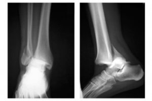
The trauma surgeon will examine you and take x-rays.
For isolated lateral malleolus fractures, a plaster cast is applied from the top third of the tibia to the fingertips.
You will be sedated for about three weeks. A follow-up examination is then carried out and the plaster cast removed. Your traumatologist will refer you to physical therapy, which includes massage, baths, and physical therapy.
If you've broken two ankles without displacement of a bone fragment, a plaster cast is applied to the middle third of the thigh, including the knee joint. In this case, the patient is sedated for six weeks. If the fractures are displaced, they are reconnected under local anesthesia.
A plaster cast is then applied for about six weeks. Sometimes it is not possible to put the pieces back together. Surgical intervention is then required to repair the fractures with plates and screws.
The injured leg is immobilized in a cast for six weeks, followed by exercise and physical therapy.
Types of lateral malleolus injuries
Since the lateral ankle is exposed to constant stress, fractures and dislocations can occur. In the first case there is a complete, in the second case a partial loss of mobility of the affected person. Loss of connection between the ankle and foot occurs when the anterior and posterior ligaments of the ankle and heel bone are damaged.
The following types of ankle injuries are distinguished:
- pronation In this case, an external force tries to rotate the fork outwards.When this occurs, an external force attempts to rotate the fork outward, resulting in a ruptured ligament and possible fracture. The most common type of injury accounts for about 75 % of all injuries.
- supination. It is characterized by the opposite direction of external action. However, the effects are the same.
- broken bones. A common cause is impact, a sudden pressure surge to the area of the foot.
Treatment regimens have been developed for each case described. Knowing where the ankle is can help to determine the nature of the injury based on the external symptoms, to provide first aid and to be in a difficult situation.
If corrective action is not taken in time, there is a high probability of post-traumatic exacerbations. This includes ankle osteoarthritis, which occurs when the ankle is not treated properly.
Lateral ankle sprain
Most frequently The most common injury is a sprained ligament, not a tear in the bony structure. It is caused by a sudden twisting of the foot without detaching it from its base, or by a strong one mechanical violent impact. As a result, cracks form in the connective tissue structure, and fluid such as intercellular exudate and blood begins to accumulate.
Possible consequences are the loss of mobility and flexibility of the ligaments.
- a fall from a height, especially if the landing puts pressure on the heel area;
- falling from a height, especially if the foot is pinched in the heel area while running or walking;
- spontaneous twisting of the foot when walking on uneven ground.
The first symptom of such an injury is a sharp pain in the ankle area. Swelling appears 10-15 minutes later, and small hematomas may also form. If the load on the lower extremity persists, the symptoms worsen.
There may also be a slight grinding sensation when walking or changing foot position.
- There may be slight pain without anything changing on the outside. These will subside within a few days.
- Significant pain and swelling can occur.
- The pain is constant and the swelling covers much of the lower leg.
If these symptoms appear, you should stop putting weight on the ankle and rest. The pain and swelling are relieved with cold compresses and painkillers.
Danger! If the pain worsens within 1-2 hours, an ambulance must be called. The cause can be not only a sprain, but also a broken bone or fracture.
Treatment of a broken ankle
After a comprehensive diagnosis and confirmation of the diagnosis of a broken ankle, the trauma surgeon must prescribe treatment for the patient. If the fracture did not cause a displacement of bone fragments, a plaster cast is applied to the injury site.
If the x-ray shows a displaced fracture, the trauma surgeon will treat the patient. This injury is usually treated with a one-stage closed reduction. No deep anesthesia is required for this surgical procedure; a local anesthetic is sufficient. After surgical treatment of an ankle fracture, the patient is prescribed painkillers, anti-inflammatory drugs, edema inhibitors, etc.
After the operation, the patient is given a special plaster shoe that immobilizes the ankle in an overcorrection position. The specialist doctor sends the patient for a follow-up X-ray to ensure that the bone and joint are in the correct position. To relieve the pain that accompanies this type of injury, specialists continuously administer painkillers. In order to speed up the healing of the bone, patients are advised to visit a physiotherapy office.
In order for the ankle to heal in the correct position, the patient must wear a cast for 4-12 weeks:
After the cast is removed, the patient should undergo rehabilitation to exercise the joint and restore mobility in the lower extremity. For this purpose, visits to a massage therapist and physical exercises are recommended. To avoid damaging the injured ankle, physical activity should be increased gradually.
Consequences of a broken ankle
Some patients treated for a broken ankle may experience unpleasant consequences:
- pain that occurs with any physical activity on the injured lower limb;
- pain in the ankle area when the weather changes;
- Intermittent swelling of the leg;
- lameness;
- changes in gait;
- Osteoarthritis causing deformation of the joint, etc.
If such complications arise, the patient should visit a medical center and receive physical therapy.
Treatment methods for ankle fractures

Once the patient arrives at the hospital, doctors take an X-ray to determine the nature of the injury and recommend treatment.
For open or complicated fractures with dislocations, surgical treatment is indicated, during which the bone fragments are repositioned, fixed with special plates (osteosynthesis) and covered with a plaster cast.
If the dislocation is not present or is not very severe, conservative treatment is recommended: immobilisation of the limb, application of a cast or splint (for 4 to 12 weeks), pain medication.
After this time, the doctor removes the cast and conducts an X-ray diagnosis. After this period, the patient must undergo rehabilitation treatment.
Recovering from an ankle injury
Methods of recovery after an ankle fracture:
- therapeutic exerciseswhich can be performed a few days after cast and repositioning;
- massagemassage to accelerate callus formation and restore blood supply to the injured limb;
- physical therapyPhysiotherapy: electro-magnetophoresis, hydromassage, ultrasound and shock wave therapy. In shock wave therapy, acoustic waves of a certain frequency are applied to the injured area of the lower limb, which accelerates the consolidation and recovery of muscle tissue and improves blood circulation. UWT also reduces the risk of scarring. Shock wave therapy can be offered at the Health+ Medical Center not only for rehabilitation after broken bones, but also for the treatment of diseases such as osteochondrosis or herniated discs.
The best prevention of re-fractures is to be careful and avoid traumatic situations.
Oleg Petrovich Tatarinov

Top doctor, neurologist, physiotherapist, UWT specialist, leading specialist of the Health Plus network
medical experience above 40 years
construction and location
The anatomy is divided into two ankles:

The articular surfaces of the ankles, together with the articular surfaces of the tibia, fibula and talus, form a very important anatomical structure of the lower limb – the ankle joint, through which:
- ensures the correct function of the human foot;
- supports the function of the foot;
- Allows vertical movement - walking, running, jumping;
- rotation of the body around the vertical axis without lifting the feet from the floor;
- Cushioning of the body during movement.
Both ankles are easy to feel on the outside of the skin as they are superficial.

Explanation of the location of the ankles: 1 - lateral ankle, 2 - medial ankle
The articular surfaces of the ankles are covered with smooth hyaline cartilage. The joint cavity contains a small amount of synovial fluid, which acts as a lubricant, protects the cartilage tissue, prevents premature wear, nourishes the cartilage, which does not have its own blood vessels, and provides cushioning in the joint during movement.
The inner malleolus attaches to the medial articular surface of the ankle, while the lateral malleolus attaches to the lateral articular surface.
The ligaments that strengthen the ankle are attached to both ankles. The tendons of some muscles of the lower limbs also attach to this anatomical formation.

functions
The main function of the ankles is to provide a firm connection between the bones of the lower limbs and the bones of the foot and to provide good cushioning during movement.
Another important function of the ankle is to limit high amplitude movement and injury to the ankle. So, this anatomical formation protects the lower limbs from possible injuries.
In addition, the ankles allow the ankle to move in a well-defined direction. Dorsiflexion is usually 40-50º and plantar flexion is 20-30º. Of course, all these values are completely individual and the anteroposterior movement of the foot can vary between 60 and 140º.
In the sagittal axis, 2 types of movements are possible with the following amplitudes:

Normal range of motion of the ankle
Important NOTE
It must be said that the ankle is about the second most common injury site among athletes and of course among hikers. The knee comes first because it suffers the most injuries. Sprains, fractures and dislocations occur at the ankle. This is by no means a complete list of the possible complications that can arise with this part of the body. This is due to the primary function of the ankle. And that is very important in the human body.
Where is this body part? And what functions does it fulfill? We will now enumerate everything in detail. Because Homo sapiens is an upright walking species, the load on our spine and legs is distributed differently than other mammals. In particular, after a person has learned to walk, most of their weight is not on their shoulders and back, but on their legs. They are under tremendous pressure throughout the day (the bones alone can withstand 1,600 kilograms of pressure). The ankle is the place where the weight of the body is transferred from foot to foot. And that part of the body is kind of a shock absorber.
Anatomically, it is an extension of the tibia. More specifically, there are two ankles - the lateral malleolus and the inner malleolus. The first is the distal end of the fibula, which forms the tibia. So where is the medial ankle located? Where the shin connects to the foot. They run parallel to each other and basically form an angle that is at the junction of these parts. Externally, the ankle is a bony protuberance that is visible from the outside and inside of the foot. In some people it is pronounced, in others it is barely noticeable, but everyone has it.
Conclusion
The ankle, on the other hand, is not a medical term. It usually refers to the narrowest part of the ankle. Most often it is located where the calf meets the foot. This is the area where the calf muscle contracts and the foot has the least volume. It is this area that is most often referred to by those who speak of narrow ankles in women. Hence the idiom mentioned above.
However, for those who know where the ankle actually is, this term will seem absurd because when speaking of it it is difficult to use the adjective 'narrow'. Rather, it can mean that it protrudes too much or, on the contrary, is not noticeable at all.
Read more:- Ligament damage in the right ankle.
- ligaments in the ankle.
- The lateral ankle is.
- The hock in which the person is located.
- Broken ankle.
- Treatment of torn ligaments in the ankle.
- Anterior fibula ligament tear in the ankle.
- ligaments in the ankle.