X-ray of the foot in side projection in a patient with severe flatfoot. This patient underwent metatarsal arthrodesis and tendon transfer and calcaneal osteotomy.
- Anterior shin muscle (tibialis anterior)
- Construction
- nerve supply
- variant
- Material and methods.
- Results
- Non-steroidal anti-inflammatory drugs
- orthoses
- Symptoms of tibial nerve neuropathy
- Diagnosis of tibial neuropathy
- treatment and recovery.
- Types of injuries: tendinitis, dislocations and tears
- Symptoms of an injury to the triceps muscle
- causes
- symptoms
- causes
- symptoms
- Causes of tendinitis
- Symptoms of tendonitis
- Non-surgical treatment
- AdobeStock_208633988-1.jpg
Anterior shin muscle (tibialis anterior)
The tibialis anterior (tibialis anterior) muscle is a muscle that begins along the upper two-thirds of the lateral (outer) surface of the tibia in humans and inserts on the medial side of the sphenoid bone and first metatarsal bone. It acts on dorsiflexion and inversion of the foot. This muscle is mainly located near the shinbone.
It is located on the lateral side of the tibia; it is thick and fleshy on top and tendinous on the underside. The tibialis anterior muscle overlaps with the anterior tibial vessels and the deep peroneal nerve at the superior end of the tibia.
Construction
The fibers of this undulatory muscle run relatively parallel to the plantar surface and terminate in a tendon visible on the antero-posterior dorsal side of the foot near the ankle.
The fibers of this undulatory muscle run relatively parallel to the plantar surface and terminate in a tendon visible on the antero-medial dorsal side of the foot near the ankle.
The muscle traverses the medialmost portions of the transverse and cruciate ligaments of the tibia and inserts at the medial and inferior surfaces of the medial malleolus and at the base of the first metatarsal.
nerve supply
Deep oculomotor nerve (peroneus), branch of the common oculomotor nerve (peroneus) (L4, L5, S1).
variant
The deep part of the muscle rarely enters the talus, alternatively the tendon may pull to the head of the first metatarsal or to the base of the first phalangeal phalanx.
tibiofascialis anterior, a small muscle from the lower part of the tibia to the transverse or cruciate ligament of the tibia or deep fascia.
Material and methods.
Between 2005 and 2017, 37 patients with long-standing Achilles tendon rupture were treated in the No. 1 Trauma and Orthopedic Department of Samara State Medical University Hospital. The diastasis between the ends of the Achilles tendon in the neutral position of the ankle was more than 10 cm. Due to retraction of the triceps muscle, the tendon cannot be closed through, requiring repair of the diastasis between the distal and proximal ends.
Group 1 (control) consisted of 21 patients who underwent inverted flap autoplasty (Chernavsky, Krasnov) and VY-plasty. Group 2 consisted of 16 patients who underwent our proposed new surgical treatment [4]. All subjects in the study and control groups had a significant difference (more than 4 cm) in the circumference of the symmetrical areas of the upper third of the tibia, indicating pronounced hypotrophy of the triceps muscle. Both groups were comparable in terms of age, sex, mechanism of injury and time of referral to the Department of Traumatology and Orthopedics No. 1 of Samara State Medical University, where surgical treatment was performed.
Before the operation, the patients were examined in the biomechanics laboratory of the SamSMU clinic (electromyography, podometry, measurement of foot flexion strength) and ultrasound diagnosis of the injured area was performed.
The team of authors from the Department of Traumatology, Orthopedics and Extremity Surgery of the Acad. RAS AF Patent No. 2537888 of the Russian Federation of 11/13/14 for the invention of a new method of surgical treatment of patients with long-lasting rupture of the Achilles tendon with pronounced diastasis of its ends (more than 10 cm) consists in transposition of the tendons of the short fibula and of the posterior tibialis muscle to the distal end of the Achilles tendon in optimal tension [4].

The proposed method is presented graphically. Figure 1 shows the proximal (1) and distal (2) ends of the Achilles tendon. The tendon (3) of the short fibula (4) is selected and held in place. The tendon (5) of the posterior tibialis muscle (6) was isolated and prepared for fixation.
Results
Treatment outcomes in control patients (n=21) who underwent plastic surgery of the Achilles tendon defect (according to Chernavsky, Krasnov, VY plastic), we found that the muscle strength of the plantar flexor muscles (Lovett scale) on the injured side averaged 3.43 points, compared with 4.63 in the control group. The muscle strength 5 in the main group was 62.5 % of the patients and 14.3 % in the control group.
Analysis of podometric indices examined features of step timing such as double step duration, foot transfer time, single and double support duration, and the order of foot part contact with the support. Limb-to-limb comparisons were made using the subjects' coefficient of gait asymmetry, which was defined as the ratio of longer to shorter rest periods minus one, multiplied by 100 %. Based on this ratio, gait asymmetry of up to 5 % was considered normal, 5 to 10 % was latent claudication, and greater than 10 % was overt claudication.
Gait asymmetry of up to 5° was 81.25 % in the main group and 14.3 % in the control group. Gait asymmetry of 5-10 % was 18.75 % in the main group and 71.43 % in the control group. There was no asymmetry over 10% in the main group and 14,27% in the control group.
The results show that the main target group had the best walking efficiency.
As can be seen from the functional indices obtained, the patients in the control group had a significant decrease in the strength of the soleus flexor muscles on the injured side and persistent gait asymmetry in the late postoperative period. This shows that even a high-quality replacement (plication) of the Achilles tendon defect with significant hypotrophy of the triceps muscle cannot fully compensate for the function of normal active flexion at the ankle and was the reason for the development of the new method.
Non-steroidal anti-inflammatory drugs
Medications like ibuprofen and naproxen can help reduce pain and inflammation. If you take them half an hour before training, you can reduce the development of inflammatory changes in the tendon area. Tendon thickening is usually the result of degenerative changes in the tendon. Taking medication has no effect on this. If you need to take medication for more than 1 month, discuss the issue with your doctor.
The ankle can be immobilized for 6-8 weeks with a short ankle splint or an orthopedic boot. This relieves the functional strain on the tendon and reduces the swelling. However, it should be remembered that immobilization leads to hypotrophy of the lower leg muscles (reduced muscle strength) and should therefore only be used when other conservative measures have failed.
orthoses
Orthotics and harnesses are effective for most patients. In this case, orthoses are insoles. This is the most common conservative treatment for flat feet.
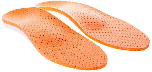
For patients with minimal changes in foot shape, simple factory-made insoles may suffice. Custom-made insoles are usually required for moderate to severe flat feet. The latter, of course, are more expensive than factory insoles, but allow better control of foot alignment.
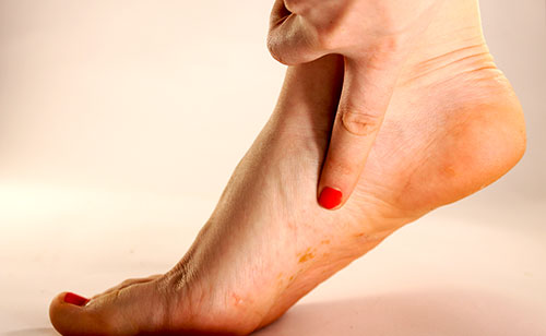
Symptoms of tibial nerve neuropathy
Depending on the location of the lesion, the clinical picture of tibial neuropathy can be divided into several syndromes.
Tibial neuropathy in the popliteal fossa is manifested by limited downward flexion of the foot and limited toe movement. The patient is unable to stand on their toes. Walking from heel to toe without rolling the foot on the toes is typical. There is atrophy of the posterior calf muscle group and foot muscles. The muscular atrophy of the foot makes it look like a claw foot. The Achilles tendon reflex is reduced. Sensory disorders include abnormalities of touch and pain sensation on the entire tibia on the back and outer edge of the lower third, on the sole, overall (dorsal and plantar surfaces) on the skin of the first 3.5 toes and on the back of the remaining 1.5 toes. Neuropathy of the tibial nerve of traumatic origin is characterized by a pronounced causal syndrome with hyperpathia (peroneal hypersensitivity), edema, trophic changes and autonomic dysfunction.
Tarsal syndrome is sometimes triggered by prolonged walking or running. It is characterized by burning pain in the sole of the foot, which often radiates into the calf muscle. Patients describe the pain as deep and note that it increases in intensity with standing and walking. There is hypoesthesia of both the inner and outer edges of the foot, some flattening of the foot, and slight clawing of the toes. The motor function of the ankle is fully intact and the Achilles reflex is intact. The nerve entrapment at the point between the inside of the ankle and the Achilles tendon is painful and results in a positive Tinel's sign.
Neuropathy at the level of the median soleus nerve is typical of long-distance runners and marathon runners. It manifests itself as pain and paresthesia on the inner edge of the sole of the foot and in the first 2-3 toes of the foot. Pathognomonic is the presence of a point in the scaphoid, percussion of which causes burning pain in the big toe.
Diagnosis of tibial neuropathy
When making a diagnosis, it is important to take a medical history. In order to determine the nature of damage to the tibial nerve, it is necessary to learn about trauma or overload, joint diseases, metabolic and endocrine disorders, orthopedic diseases, etc. A thorough examination by a neurologist of the strength of each muscle group of the lower leg and foot, the sensory sphere of this area, identification of trigger points and Tinel's sign allows diagnosis of the degree of damage.
Electromyography and electroneurography are of secondary importance. An ultrasound examination can determine the type of nerve damage. If indicated, ankle x-rays, foot x-rays, or CT scans of the ankle are done. In disputed cases, a diagnostic trigger point blockade is performed, the positive effect of which confirms the compressive nature of the neuropathy.
treatment and recovery.
Depending on the severity of the tendon injury, the attending physician recommends conservative (possible in the case of a partial rupture and in the early stages of tendonitis) or surgical treatment (in the case of a complete rupture).
- Rest and immobilisation of the injured limb
- Anti-inflammatory drug therapy
- Immobilization (splint or cast)
- Physiotherapeutic treatments (laser therapy, ultrasound, magnetic therapy, electrophoresis, etc.).
- Remedial exercises
- massage
In the trauma department, the torn tendon is repaired by suturing. Further rehabilitation is analogous to conservative treatment.
The length of rehabilitation depends on the severity of the injury and averages four to six months.
For qualified medical care in Moscow, you can contact NSC No. 2 (Central Clinical Hospital of the Russian Academy of Sciences).
Types of injuries: tendinitis, dislocations and tears
Tendon ruptures can occur at the point where the tendon attaches to the bone, at the junction between the muscle and the tendon, or in the tendon itself. When small pieces of bone detach along with the tendon, a burst fracture is diagnosed.
The main causes of triceps brachii tendon injury include:
- strenuous physical activity
- Local inflammatory processes (deep scratches, abrasions) cause tendinitis.
- Age over 40 years
- Chronic diseases (arthritis, bunions, etc.).
- Taking corticosteroid medications
- Mechanical injuries
Symptoms of an injury to the triceps muscle
- Pain and swelling on the back of the elbow (characteristic of tendinitis)
- When stretching or flexing the joint, the patient tightens the muscle, causing the tendon to stretch and causing pain
- Characteristic clicking sound
- muscle cramps (when stretched)
- nodules under the skin
- swelling and redness of the skin
- Painful touch of the tendon
- Decreased strength in the limb
Inflammation and sprains of the tendon of the triceps muscle are treated conservatively:
- Immobilization of the injured tendon (stopping all activities that cause pain)
- Application of cold to the painful area
- Use of nonsteroidal anti-inflammatory drugs.
- Use of orthoses, splints to relieve the tendon and promote its regeneration.
- Physiotherapy (electrophoresis, ultrasound, cold therapy, etc.).
- Exercise therapy (recommended once inflammation and pain have subsided).
Surgical treatment is recommended for tendon rupture. During rehabilitation, heavy physical work and sports should be avoided. The patient is recommended to wear an immobilizing bandage, undergo physiotherapy and physical therapy under the supervision of a doctor.
Timely and qualified treatment is the key to a speedy recovery. Otherwise, permanent changes in the tendon tissue can occur.
Qualified medical assistance can be obtained in Moscow at NCC No. 2 (Central Clinical Hospital of the Russian Academy of Sciences).
causes
The tibial nerve is a continuation of the sciatic nerve. Its damage can occur in the following cases:
- Injuries to the lower limbs of various etiologies (fracture, ankle dislocation or sprain, tendon rupture, ankle ligament sprain). Traumatic factors and the resulting swelling lead to direct damage or compression of the tibial nerve, which impairs the transmission of nerve impulses.
- Deforming changes in the foot (flatfoot, valgus).
- Pressure on the feet or uncomfortable positioning of the feet for a long period of time, e.g. B. by pushing the lower limbs with a heavy object.
- Diseases of the knee and ankle joint (arthritis, arthrosis).
- Endocrine disorders, thyroid disorders and diabetes.
- Tumors of the tibial nerve of benign and malignant origin.
- Poor circulation in the lower limbs and feet (vasculitis).
- Poisoning of the body with toxic and chemical compounds, including products containing alcohol.
- Inflammatory pathologies of infectious etiology in various sites, including those affecting multiple nerve fibers.
- Prolonged treatment with drugs that negatively affect CNS function.
- Weakening of the body's defenses.
- Unhealthy working conditions.
- Inadequate nutrition of the tissues of the lower limbs and foot.
- Prolonged stay of the lower limbs at low temperatures.
- Inadequate nutrition, which reduces the absorption of B vitamins.
- Pathological processes affecting the spine.
- people who practice various sports professionally;
- persons with working conditions that require constant standing, including walking;
- Overweight or obese individuals - high stress on the lower limbs increases the risk of damage to the tibial nerve;
- the choice of inappropriate footwear: high heels, thin soles.
symptoms
Neuropathy is accompanied by a clinical picture, the severity of which depends on the extent of nerve fiber damage.
Nerve dysfunction can be recognized by abnormalities in foot flexion and toe mobility. When walking, the foot is misaligned - the center of gravity is on the heel. The muscular apparatus of the lower limbs and foot shows clear signs of atrophy and deformity.
With traumatic neuropathy, the ankle swells, blood circulation is disturbed, tissue sensitivity is increased, and severe pain occurs, which intensifies when touching the damaged area.
When the nerve neuropathy is caused by endocrine disorders or infectious lesions, the patient loses feeling in the lower leg and foot. The pain syndrome persists and can vary in type and severity. The pain intensifies when walking and during physical exertion. The patient may feel involuntary contractions of individual muscles or spasms in the lower limbs.
In addition, patients may experience neurotrophic disorders such as dryness of the epidermis of the lower legs and feet, keratosis of the dermis, brittleness of the nails, pallor of the skin, and reduced local temperature and excessive sweating.
causes
Most modern doctors agree that shin splints are caused by excessive stress on muscle tissue, leading to muscle damage. Injuries are limited to the anterior and posterior tibial muscles. Sometimes shin splints spread to the calf and camel muscles as well.
Shin splints are most common in athletes who run and jump, but can also occur in dancers and military personnel engaged in general physical training. Occasionally, tibial stress syndrome is diagnosed in people who engage in physical activity, when they engage in intense exercise or simply change their rhythm. Muscle strains while running (both uphill and downhill) and on rough terrain are considered the most traumatic. Uncomfortable or poorly worn shoes can also lead to injuries.
The cause of pain is sudden exertion, especially if it is accompanied by acceleration. Untrained people and those who start training without a sufficient warm-up phase are particularly at risk. As a result of a muscular imbalance, the muscles are micro-traumatized and their attachment points are overstretched.
symptoms
The main symptom of a 'cleft tibia' is pain, which can occur in both the front and the back of the tibia. The symptoms appear at the beginning of physical activity, but usually subside over time. If a person ignores the symptoms of the ailment, the pain can become permanent.
The pain in tibial stress syndrome can be sharp or dull. They increase in intensity as they are scanned. The symptoms gradually subside with rest.
Causes of tendinitis
- sports injuries
- excessive physical activity
- Muscle imbalance, muscle weakness in the lower limbs
- intensive training without proper preparation
- metabolic disorder
- rheumatism
- weakened immune system
- infectious diseases
- Flat feet, different leg lengths and other anatomical abnormalities
In addition, tendinitis can develop against the background of taking quinolone and fluoroquinolone antibiotics.
Symptoms of tendonitis
The thigh muscle group includes the five adductors, which stabilize the pelvis and move the hip toward the midline. These muscles 'work' when walking, balancing while standing and crossing your legs. When tendonitis develops in the quadriceps tendon area, there is pain in the groin, which increases with hip flexion. They can appear immediately or gradually.
Patients complain of stiffness and pain in the hip joint, redness, local temperature increase, swelling or thickening of the affected area. Sometimes a characteristic cracking sound is heard. In particularly severe cases, complete immobilization of the limb is possible.
Non-surgical treatment
If doctors find an incomplete supraspinatus tear in the shoulder, treatment is immobilization (shoulder immobilization), prescription drugs, or physical therapy.
Immobilization involves placing the shoulder in a position that allows the tendon to heal properly without affecting its length. Anti-inflammatory drugs, vitamins and cartilage-protecting agents are then prescribed as required. Another therapeutic measure is physiotherapy, which also includes magnetotherapy and electrophoresis.
Conservative therapy is a relatively lengthy process and can be done at home.
AdobeStock_208633988-1.jpg

AdobeStock_208633988-1.jpg
If examination reveals a complete tear of the supraspinatus muscle, surgical treatment is required. This is done in the hospital through an open approach (the more traumatic option) or by arthroscopy. Arthroscopy is the method of choice because access to the tendon is less traumatic and the postoperative and rehabilitation period is shorter.
Read more:- supporting muscle.
- Anterior shin muscle (tibialis anterior).
- Tibialis posterior muscle.
- Syndrome of the tibial nerve.
- Exercises for the triceps tibialis muscle.
- Long fibula muscle.
- The long section of the big toe.
- Innervation of the lower leg muscles.