To relieve symptoms, orthopedists prescribe exercises to stretch and strengthen the muscles of the foot. Some patients are helped by special devices that relieve pressure on the plantar fascia. Physiotherapy methods generally include:
- Heel pain in athletes: diagnosis, treatment, prevention.
- Part 1 Clinical manifestations and diagnosis.
- Plantar fasciitis, heel bone periosteopathy and heel spurs.
- How is plantar fasciitis treated?
- Was this page useful?
- Test your knowledge
- Diagnosis of plantar fasciitis
- Treatment of longitudinal fibromatosis
- Prevention of plantar fasciitis.
- Heel spurs (plantar fasciitis). Kind of treatment.
- surgical treatment
- Related preventive methods
- symptoms of the disease
- diagnosis
- Where does plantar fasciitis come from?
- What Are the Symptoms of Plantar Fasciitis?
- ICD-10
- causes
- Treatment of necrotizing fasciitis
- prognosis and prevention
Heel pain in athletes: diagnosis, treatment, prevention.
Nechaev Vladimir. Graduated in sports medicine from the University of Tartu, Estonia in 1977. From 1979 to 2000 he was a member of the national teams in marathon, walking training and triathlon.National teams in the sports of walking and triathlon. He is currently the President of the Russian Podiatry League (www.podiatr.ru, www.ploscostopie.ru), Chief Physician of the Clinic for Foot – Back – Posture (Chernoglovka, Moscow Region); Sports champion in marathon running.
- plantar fasciitis and heel spurs;
- Pressure bruise/fracture of heel bone;
- Partial tear of the plantar fascia;
- Periosteopathy of medial heel convexity;
- heel tunnel neuropathy;
- Myofascial pain syndrome of the heel.
- orthopedic insoles;
- less strain on the feet, more rest;
- stretching of the foot muscles;
- foot massage;
- Exercises to develop and maintain foot function;
- pain in the heel area;
- the nature of the pain varies from a sharp, disabling ache to a dull, constant ache;
- The pain increases in the morning with the first steps;
- the pain becomes more unbearable when the person changes position from sitting to standing for long periods;
- climbing stairs is difficult because of the pain.
- Age – the condition is most common in people between the ages of 40 and 60.
- A preference for certain physical activities. People who do a lot of long-distance running and jumping can develop plantar fasciitis much earlier. People who love to dance and dance ballet also belong to this group. All of these types of physical activity put stress on the plantar fascia.
- Obesity – Being overweight also puts strain on the plantar fascia.
- footwork. Factory workers, teachers, shop assistants, and others who spend most of their time standing (especially on hard surfaces) can damage the plantar fascia.
- pain in the lower part of the foot, which increases month by month,
- A feeling of discomfort in the arch of the foot,
- pain in the foot that gets worse when standing,
- swelling in the heel.
- flat feet (found in 90 % of patients); high or low arch of foot;
- overweight;
- Diseases of the large joints (arthritis) and the spine;
- neurodystrophic and vascular diseases (obliterative arteritis, atherosclerosis of the lower limbs);
- prolonged standing on your feet due to professional conditions (salespeople, hairdressers);
- uncomfortable footwear;
- Endocrine and metabolic diseases (diabetes, gout);
- Long-term foot overload in athletes who run.
- surgical intervention. The necrectomy is performed as soon as possible after admission to the surgical department. Areas of necrosis are excised to the unchanged tissue and the wound is left open. Wound recovery occurs within 24 hours. If the pathological process is advanced, amputation may be required.
- antibiotic therapy. Antibiotic administration begins upon admission. Broad-spectrum antibiotics are used initially, and the prescription is adjusted once pathogen susceptibility is established.
- systemic therapy. Infusion therapy to correct acid-base and water-salt balance is continued during surgery and hospitalization. Vitamins and trace elements are prescribed. Donor plasma is administered to stimulate the immune system. Hyperbaric oxygenation is used to accelerate wound healing, neutralize endotoxins and eliminate tissue hypoxia.
With these injuries, the instrument-based diagnostic methods are often powerless and often lead the treating physician away from the actual cause of the pain and draw his attention to secondary lesions that are not relevant at this time. However, knowledge of the specific clinical signs of each possible heel pathology, as well as a set of simple functional and manual diagnostic tests, allow not only to differentiate these injuries, but also to identify precursor symptoms. 'Heel pain' in athletes can therefore often present a diagnostic problem for the clinician. Despite the numerous scientific studies published over the last 30 years on the etiology, pathogenesis and treatment of heel pain, doctors are still dissatisfied with the effectiveness of the proposed therapies. For example, according to statistics, the median length of treatment for plantar heel pain in the United States is 13.3 months. This length of rehabilitation is completely unacceptable for competitive sports. However, new therapeutic technologies such as eccentric exercise, custom insoles, special taping, shock wave therapy, platelet-rich autografts, various reparative drugs and innovative therapeutic strategies can drastically reduce the treatment time of heel plantar pain in athletes.
Part 1 Clinical manifestations and diagnosis.
Plantar fasciitis, heel bone periosteopathy and heel spurs.
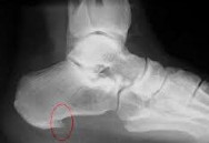
Fig. 1 Injury to the plantar part of the heel
1 - plantar fasciitis;
2 – heel spur;
3 - periosteopathy;
4 - stress fracture;
5 – rupture of plantar fascia;
6 – Periosteopathy of the medial convexity of the heel bone
The above pathologies are so closely interrelated that it makes sense to treat them together.
plantar fasciitis – is one of the most common causes of heel pain. However, the diagnosis of plantar fasciitis itself has limitations, since this pathology usually manifests itself as a syndrome of anatomical and functional anomalies in the heel area. For this reason, in recent years, the diagnosis of plantar fasciitis has often been replaced by chronic plantar heel pain syndrome (CPHP) in the foreign literature.
Most authors agree that plantar fasciitis (Fig. 1) is a low-grade inflammation of the plantar fascia (plantar fascia) resulting from repeated chronic microtrauma and degeneration of its fibers (similar to Achilles tendinosis). The fascia becomes thick and dense with the typical symptoms of chronic inflammation (degeneration of fibers, perivascular dilatation and swelling, collagen proliferation). The presence of plantar fasciitis is best confirmed by an ultrasound diagnosis.
A review study found that subjects with CPHP syndrome had an average soleus aponeurosis thickness of 4.0 mm. This is more than double the norm [AM McMillan et al 2009]. These injuries are referred to as 'fatigue' or 'overuse injuries'.
The first and most frequent complaint of the patients is the so-called 'starting pain': pain when taking the first steps after sleeping or when getting up after a rest phase as well as pain at the beginning of the warm-up training. As a rule, one of the heels hurts. At the beginning of the disease, the initial pain in the heel subsides after 5-10 steps or after a 2-3 minute warm-up. However, as the lesion expands, the initial pain initially subsides in intensity, but with exercise duration, heel pain worsens and begins to impair performance during exercise loads.
How is plantar fasciitis treated?
To reduce foot pain and stress, you should:
Use of ultrasound in the heel area to improve blood circulation and relieve pain (extracorporeal shock wave therapy);
Copyright © 2023 Merck & Co, Inc, Rahway, NJ, USA and its subsidiaries. All rights reserved.
Was this page useful?
Test your knowledge
Provided by Merck & Co, Inc, of Rahway, NJ, USA (known as MSD outside of the US and Canada), which uses advanced science to save and improve lives around the world. Learn more about MSD textbooks and our commitment to the Global Medical Knowledge Initiative.
Copyright © 2023 Merck & Co, Inc, Rahway, NJ, USA and its subsidiaries. All rights reserved.
Diagnosis of plantar fasciitis
The diagnosis of plantar fasciitis is confirmed by intense pressure of the thumb on the heel bone when the foot is dorsiflexed, causing pain. Pain along the medial border of the plantar fascia is also possible. If clinical findings are inconclusive, the presence of a heel spur on x-ray can confirm the diagnosis; however, the absence of lesions on the radiograph does not preclude the diagnosis, and visible spurs do not cause the symptoms of plantar fasciitis. In addition, in some cases, heel spurs are difficult to see on x-ray and appear as friable foci of cancellous bone suggestive of spondyloarthropathies (e.g., ankylosing spondylitis or reactive arthritis). An MRI examination is indicated if there is a suspicion of an acute fascial tear.
Other conditions that cause heel pain can mimic plantar fasciitis
Acute severe heel pain with redness and warmth may indicate gout. Podagra is a condition caused by hyperuricemia (blood uric acid levels >6.8 mg/dl [>0.4 mmol/l]) and the precipitation of sodium mononitrate crystals in and around guides the joints. Read more .
Treatment of longitudinal fibromatosis
To reduce stress and pain in the fascia, shorten your stride and avoid going barefoot. Activities that put stress on the foot from impact, such as B. running, should be avoided. Some of the most effective treatments for calf fasciitis include stretching exercises for the calf and plantar fascia muscles, and the use of orthotics to stretch the lower leg and plantar fascia muscles while you sleep. Orthotics can also reduce fascial tension and pain. In addition, motion correction, weight reduction in obese patients, nonsteroidal anti-inflammatory drugs (NSAIDs), cooling massage, and sometimes glucocorticoid injections are used. However, corticosteroid administration can exacerbate degenerative changes in the plantar fascia, leading many clinicians to limit their use (see 'Corticosteroid use in obese patients'). Recommendations for the use of corticosteroid injections Use of glucocorticoid injections ).
In refractory cases, physical therapy, oral glucocorticoids, and immobilization may be recommended before surgical treatment is considered. The newest form of treatment for unresponsive plantar fasciitis is extracorporeal shock wave therapy (ECWT), which uses a hand-held applicator to deliver low-frequency pulsed waves to the painful area. Pulsed shock wave therapy is a safe, non-invasive method that increases metabolism and blood flow, repairing damaged tissue and speeding healing. EHT is used in major medical centers (1 Treatment Note Plantar fasciitis is characterized by pain at the insertion of the longitudinal fascia at the heel bone (calcaneal enthesopathy) that may radiate along the medial border of the longitudinal bone. Read more).
Prevention of plantar fasciitis.
Plantar fasciitis is treatable, but as everyone knows, prevention is better than cure. If you are at risk, you should take regular preventive measures. In fact:
Everyone has done stretching exercises at least once in their life to stretch specific muscle groups. It is important to note that heel fascia stretching exercises should only be performed under medical supervision until you learn how to do them properly. This is very important to avoid further injury. Based on the results of the diagnosis, your doctor will help you choose the appropriate prophylaxis.
In our clinic, we only practice a comprehensive approach to the treatment of fasciitis. Why do things by halves when you can also treat? In the ZARTA orthopedic and rehabilitation center you will experience a really effective treatment that will meet your expectations!
Eliminating pain and restoring the full functionality of your foot - that's our job!
Heel spurs (plantar fasciitis). Kind of treatment.


– Vascular diseases of the lower limbs (rheumatoid arthritis, deforming osteoarthritis, diabetic polyneuropathy)
There are different methods of treating this disease and the choice depends on the severity of the pain and the timing of treatment, among other things.
The most harmless treatment method is the medicinal one.
Heel spur therapy consists of prescribed pain medication, special exercises, therapeutic massage and some physical treatments:

The drug blockade method.
If drug treatment does not give a positive result, then therapeutic blockade is performed. This procedure relieves the patient from severe piercing pain, eliminates the inflammation, thereby improving the patient's well-being. The main difference from other injections is that when there is a therapeutic blockage, the drug is injected directly into the pain site. This procedure is more complicated than regular injections and can only be performed by a doctor!
In addition, hormonal drugs such as Diprospan or hydrocortisone are used to relieve pain. These belong to the group of glucocorticoids, which have an anti-inflammatory and anti-oedematous effect.
Non-steroidal anti-inflammatory drugs are also used.
The injections relieve pain and allow the patient to move independently.
surgical treatment
When treatment does not produce the desired results, it usually occurs late and at an advanced stage of the disease. Then a surgical procedure is performed, which is usually performed under local anesthesia. During the procedure, the surgeon shrinks the bony prominence in the heel, restoring the foot to its natural state.
This treatment method is very effective if done correctly. The pain and inflammation disappear very quickly. With successful treatment and regular follow-up care, the patient can 'forget' the problem for several years.
Related preventive methods
Heel spurs can be treated as a complex condition. Even if the pain subsides, you should follow your doctor's advice to prevent it from coming back.
– Regularly perform special exercises,
- Take medication if necessary.
Regular check-ups with your orthopedist are essential for prevention. This is necessary in order to monitor your condition and well-being and to prevent future pain syndromes.
Copyright © 2019 Center for Traumatology and Orthopedics
Southern Medical District of Leningrad Region
Not the official website of the SCMB in Gatchina
Medicine, traumatology, fracture treatment, foot surgery, arthroscopy, arthroplasty.
symptoms of the disease
Plantar fasciitis has characteristic symptoms. These symptoms occur when the foot's cushioning ability is severely compromised. The most common symptoms are.
Pain syndrome does not have to come on suddenly. Sometimes the patient walks for a long time with a limp in the affected limb. Since he is forced to put more weight on the good leg, plantar fasciitis soon begins there as well. At this time, when both legs are already hurting, the disease is particularly active.
diagnosis
In most cases, a clinical examination by a specialist doctor is sufficient to make a diagnosis. The typical clinical symptoms point directly to plantar fasciitis. In cases of doubt, a differential diagnosis can be made:
An X-ray can already show a bone spur. The ultrasound examination complements the examination, since some small bone outgrowths are not always visible in the X-ray image. Ultrasound can also detect osteoporosis. An MRI scan is only performed in extreme cases, but it is the most informative because it has the highest resolution. A computed tomography can also be used to determine the condition of the plantar fascia itself.
Where does plantar fasciitis come from?
Under normal conditions, the plantar fascia acts as a shock-absorbing tendon, absorbing stress and supporting the arch of the foot. When too much pressure is applied to the fascia, micro damage or small tears form. The repeated stretching and micro tears cause inflammation of the foot fascia.
Another possible cause of inflammation is a biomechanical defect in the foot itself. People with flat feet or excessive foot deviation are prone to this condition.
There are several additional factors that increase the risk of developing plantar fasciitis:
What Are the Symptoms of Plantar Fasciitis?
The pain intensifies in the morning and after long periods of sitting. After walking for a few minutes, the fascia stretches and the pain in the foot subsides slightly. In some people, the pain goes away for a while, but returns after standing for a long time.
With fasciitis, the area of the foot near the heel bone is painful and the pain is described as stabbing. If the person concerned does sport, the pain often does not increase during but after the sport. By the way: Another possible cause of the symptoms is the formation of a heel spur, which you will read about in this article.
ICD-10
Heel spurs (plantar fasciitis, plantar fasciitis) is a very common disease that accounts for about 10 percent of all diseases of the musculoskeletal system. It usually occurs in patients over 40 years of age; Statistics show that about 20% of people in this age group suffer from %. Women are affected more often than men. The incidence decreases with age because the patient is not as physically active as usual. This disease is very rare in children.
causes
The disease is an enthesopathy, ie inflammatory and degenerative changes in the tendons where they attach to the bone. The pathological process is triggered by overuse and occurs at the point of attachment of the soleus aponeurosis to the calcaneus tubercle, less often at the point of attachment of the Achilles tendon to the posterior surface of the calcaneus. Predisposing factors include:
Certain rheumatic diseases, including psoriasis and rheumatoid arthritis, increase the likelihood of heel spurs. One cause of fasciitis in these disorders is repeated corticosteroid blockade, which causes the fibers of the soleus aponeurosis to degenerate.
Treatment of necrotizing fasciitis
This disease is treated by specialists in the field of abscess surgery. If symptoms of this pathology are detected, an emergency hospital admission and resuscitation is indicated. Infusion therapy is initiated during the transport phase. Water and saline solutions are transfused and hormonal drugs are administered. Shortness of breath requires urgent tracheal intubation with artificial ventilation. The treatment plan includes:
prognosis and prevention
The prognosis of necrotizing fasciitis is always difficult. According to various sources, between 20 and 47 % cases of this disease are fatal. In the other cases, functional disorders of various organs as a result of sepsis and acute multi-organ failure in the course of the disease can occur. After removal of the necrotic lesions, extensive wound areas remain, which must be closed by plastic surgery. Scarring with severe cosmetic defects and limb dysfunction may occur. Prevention includes measures to avoid immune disorders and to exclude or minimize other risk factors. If necrotizing fasciitis is suspected, urgent transport to a surgical hospital and immediate treatment upon admission are required.
1. Necrotizing fasciitis – an urgent medical condition / Nikolov VV // Intensive Care – 2015 – №2.
2. Necrotizing fasciitis / Grinev MV, Budko OA, Grinev KM // Bulletin of Surgery named after II Grekov – 2005- no. III Grekov – 2005- ;1.
4. Necrotizing fasciitis: early diagnosis and surgical treatment / Shaginyan GG, Chekanov MN, Shtofin SG // Siberian Medical Review. – 2011.
- Treatment of plantar fasciitis.
- fasciitis.
- tibial fasciitis.
- Treatment of plantar fasciitis at home.
- How to cure plantar fasciitis forum.
- Structure of the human heel.
- Longitudinal soleus muscle.
- Treatment of plantar fasciitis on the sole of the foot.