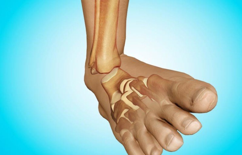
In this type of foot injury, the ankle bone stays in place while the adjacent heel bone, scaphoid bone, and ulna bone appear to detach. The soft tissues of the joint are severely traumatized and the blood vessels are damaged. The articular cavity and periarticular tissues are filled with a large hematoma. This leads to significant swelling, pain and, most importantly, reduced blood supply to the limb. The latter can be a trigger for the development of foot gangrene.

hip dislocation
Hip dislocations account for about 10 % of all dislocations. Hip dislocations are divided into anterior and posterior dislocations based on the position of the femoral head in front of or behind the hip socket (acetabulum). Both types of hip dislocations are divided into superior and inferior dislocations depending on whether the head is high or low.
Hip dislocations are divided according to their origin into:
Of the hardware methods for diagnosing hip dislocation, the following diagnostic procedures can be recommended and performed by the doctor:
Posterior hip dislocations
Posterior hip dislocations are the most common (up to 80 % of all dislocations) hip dislocations. With rapid flexion, adduction, and internal rotation of the leg within physiological limits, the femoral neck settles on the anteroinferior rim of the acetabular fossa. The formation of a fulcrum at this point creates a two-armed lever, the shorter end of which – the joint head – heavily loads and tears the rear, weakly reinforced part of the joint capsule. If the limb bends inward at less than a right angle with increased flexion and rotation, a dislocation occurs with a tear in the joint capsule over the tendon. If the hip is bent more than at right angles, a dislocation occurs with a tear in the joint capsule and the joint head emerging below the tendon. This head position can change in the form of an inward rotation (pivotization) of the hips if the force continues. In general, the lower the condyle, the greater the initial flexion and the deeper the posterior capsule tear.
MRI of the hip (frontal view) for hip dislocation helps assess the condition of the ligament, acetabulum, and articular cartilage..
About the disease
Hip dislocations account for about 2 % of all dislocations. When the left or right foot is dislocated, the ankle bone (talus) is usually displaced and the function of the entire foot is disrupted (biomechanical disruption). This injury is always characterized by damage to the attachment ligaments. Multitraumatic dislocations can be combined with fractures of the posterior or anterior bones of the foot.
Bone dislocations in the joints of the foot are accompanied by severe pain, swelling and changes in the normal shape of the organ. Anatomical abnormalities lead to functional limitations - passive and active movements of the foot are no longer possible, as is supporting the foot in general.
How is a foot sprain diagnosed? The diagnosis is made based on the patient's complaints, evidence of the injury, and the results of objective examination (examination and palpation of the foot). A definitive diagnosis is made with an X-ray examination. In most cases, the x-ray can rule out/confirm a possible bone fracture.
Treatment for a dislocated foot is usually conservative. Under anesthesia, the doctor returns the dislocated bone to its physiological position and applies a cast (which is later replaced with a cast). Treatment ends with an appropriate rehabilitation program.
species
Dislocations of the foot can occur in the following forms
- robe
- subtalar;
- Lateral tarsal and metatarsal bones – the dislocated bone swings outward;
- Metatarsal and metatarsal bones – the dislocated bone swings inward;
- Interphalangeal Dislocations.
A distinction is also made between complete dislocations, in which the bones are not in contact with each other, and incomplete dislocations, in which the bones are partially in contact with each other.
Traumatologists in St. Petersburg – opinions
I had an appointment with my child and the specialist did a check-up. Viktor Dmitrievich is kind, finds contact with the child, is positive, optimistic and has a pleasant energy. I chose the orthopedist based on his professional experience, I saw his books and I liked them.
The doctor approached my problem responsibly. He immediately did what needed to be done and told me what to do next. I would go back to this doctor if needed. I would highly recommend this doctor.
He is a professional doctor and I liked him. He communicated openly with me. The doctor listened to my complaints, examined me and immediately made a diagnosis. I started my treatment.
Andrew Juriewicz is a very kind, good-natured doctor and an expert in his field. During the visit, the doctor did everything necessary for my problem. The visit lasted about half an hour, the specialist examined me, advised me, prescribed treatment and made recommendations. My problem was completely solved. I chose a traumatologist with a free appointment. I recommend the doctor.
The doctor examined me, sent me for an X-ray and brought a special device to immobilize my hand. The doctor also gave me recommendations. Aleksey is very friendly and attentive, you can feel that he is a competent doctor. I would definitely recommend this trauma surgeon.
The doctor communicated politely. He did the research carefully and looked at the results. I got what I came for - recommendations. I went to this specialist because he has a lot of good references. I will go again if need be.
Good clinic and highly qualified specialists. I had an ultrasound scan of my joints (hip) and a consultation with an orthopedist. The visit lasted more than 40 minutes. That was enough. I felt very comfortable during the consultation and have full confidence in the doctor. I will return for another consultation after the MRI scan.
I had problems with my right knee. I didn't play any sports and had no injuries. Still, I was in pain. I have seen several doctors within a year. But only Dmitry Yurievich and her massage therapist Murat Zhumazhanovich solved my pain problem.
They made a diagnosis and prescribed treatment. I could have had a joint massage at their clinic or any other place I wanted. I chose Ligaymed for treatment and I have no regrets. My knee no longer bothers me and my soul is happy!
Treatment
One of the most dangerous injuries is dislocation of the foot, which you cannot determine yourself at home without specialist knowledge. Therefore, if you experience severe pain in the foot after physical manipulation, you should go to a medical institution, where you will receive adequate treatment. Regardless of the type of injury, the following treatment measures await you in the medical institution the following treatments:
- – The area from the fingertips to the hip third must be treated with a plaster cast. The duration of this treatment depends on the type of injury: ankle sprain - at least 21 days, plantar sprain - more than a month, metatarsal regeneration - about eight weeks;
- – After the plaster is removed by the specialist, the patient must perform a list of exercises for training the joints and additional orthopedic aids, massage, physiotherapy. Full recovery takes about two months.

Any dislocation of the foot requires a cast
Surgical intervention is sometimes required, which in most cases involves damage to the metatarsal bone. The patient is then prescribed a cast and orthopedic shoes for up to 12 months. It is not necessary to spend large sums of money on expensive medicines, since folk remedies do with a small amount of harm; the most effective recipes include:
- – Mugwort. Painful sensations are relieved by balm made from grated St. John's wort, which is applied to the damaged joint area and additionally covered with a damp, cold cloth.
- – Lavender oil. This is a great painkiller, it is recommended to prepare and infuse it at home, but this process is quite time-consuming and time-consuming, so you can give preference to ready-made options.
- – Tansy tincture. Compressions performed with this active ingredient shorten the time it takes for the joint to regain full mobility.
- – Tincture of cornflower twist. Used as an internal remedy, this remedy can relieve the pain of sprains.
- – Sugar and grated onion. A mix of these ingredients helps joints and ligaments recover quickly after a sprain. Simply apply the mixture to the injured area.
First aid measures for sprains
First aid for a sprained foot consists of the following algorithm of actions:
- The injured person should be placed on a comfortable, flat surface.
- The injured limb should then be elevated (the foot should be above the knee and hip joints) by placing a pillow, jacket or other suitable improvised device underneath it.
- To reduce post-traumatic swelling, the injury site should be cooled. Ice cream or frozen foods (e.g. a packet of dumplings) work well.
- If the skin is injured, an aseptic dressing should be placed on the wound.
- After the above steps, see a doctor with an X-ray machine and a trauma surgeon as soon as possible.
treatment of a dislocation
Treatment of a dislocation involves surgery to straighten the leg and return it to its natural position. Reduction can be closed - without surgery - or open, ie through a surgical incision.
It is not possible to give specific advice on how to treat a dislocated foot at home, since the help of an experienced traumatologist is essential. Once the dislocation is fixed, he can give you some advice on what to do about a dislocated foot to get you back on your feet as quickly as possible.
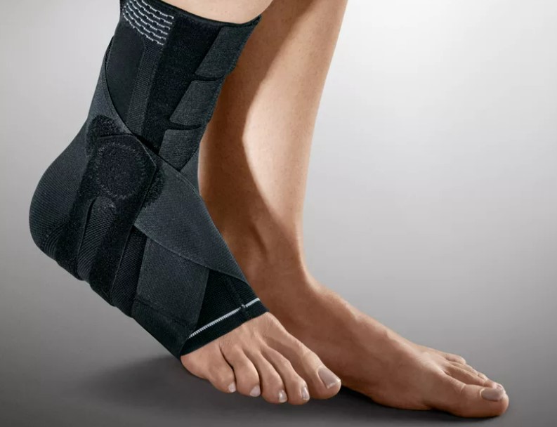
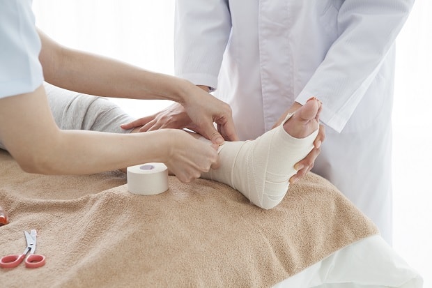
After the reduction, a fixation bandage is applied for a period of four weeks to two months. It should come as no surprise that when immobilizing the lower limbs, a bandage is applied to the lower third of the thigh - when immobilizing the knee joint. This is necessary because walking with an immobile ankle is very dangerous for the knee joint.

Moscow traumatologists – opinions
Very friendly and attentive doctor who is easy to talk to. This was my second visit and I came with the MRI that I had done on my first visit and he looked at it and prescribed the necessary treatment.
It was my first visit to this clinic and I didn't have to wait in line to see the orthopedic and trauma surgeon. The doctor was very thorough, attentive and conscientious. After the examination, he explained the treatment to me in detail. The treatment went very well.
He makes a good impression of a knowledgeable and attentive doctor. During the appointment he listened to me, explained and clarified everything and prescribed the treatment.
The doctor gave me good advice. I heard everything I wanted to hear from him. I had no reservations about him.
The doctor examined me, took X-rays and made recommendations. He is a nice, kind and responsible doctor and I recommend him to everyone.
I was very happy with my doctor. I liked him very much. I already knew him beforehand. The doctor is responsible and cares about his patients.
This is the second time we have seen this doctor. That was six months ago. Last time we really liked the way he conducted the visit. He answered all the questions I had. The exam was thorough, lasting over 40 minutes. He examined the child from head to toe. He was very kind to my daughter. The child liked the doctor very much. He recommended that we come back in six months, which we did. So this visit was planned, not that long because there were no problems, it was just a routine check-up. It was the same as last time. Ilya is very attentive and very knowledgeable. We will continue to go to him in the future.
He is a professional and very attentive doctor. He looked at the MRI result, explained everything clearly and prescribed a treatment.
He had a calm, high-quality and confident reception. Ibrahim Muradovic questioned me about my problem, questioned me, examined me and suggested exercises. He also suggested a diagnosis, recommended an ultrasound, and his opinion was confirmed. He also gave me recommendations on what to do next. I'm very satisfied.
prevention and rehabilitation

Once the cast is off, physical therapy follows, and prescribing physical therapy exercises is very useful. The exercises help train the injured joint.
It is advisable to break dislocated leg prevention into three equally important phases:
- In the first phase Prevention is very important to avoid injury, especially in the knee and hip joints of the injured limb. Regular exercise significantly improves blood circulation in the limb and changes the tension of all muscles in the leg. If, in the first phase of prevention, the specialist allows the injured person to move in a cast, care must be taken to ensure that the foot is positioned correctly. This is done to avoid foot deformities and possible later surgeries;
- second phase In the second phase of treatment (after removing the cast), the patient should pay special attention to strengthening the muscles that stabilize the arch of the foot. If the foot is dislocated, only insoles should be used to support it. These are called supinators. After the second stage and at the beginning of the third stage, the injured person can very carefully restore the spring function of the leg. For this it is necessary to perform not too large jumps and all kinds of hopping. It is advisable to do them on a soft surface (carpet or special mat);
- In the third phase The third phase is about restoring full range of motion to the leg. This can be achieved through special exercises and physical therapy. It is important not to deviate a millimeter from the recommendations of the specialist throughout the period of prevention. This will help the leg recover and return to normal as quickly as possible.
bruises and sprains
Foot bruises are associated with intense pain because the skin and muscle layer is very thin and the impact hits the periosteum, the connective tissue that covers the bone. This is quickly followed by swelling that increases over time. Because of this, you need to be treated before the hospital to reduce the swelling and pain. You can cool your foot with ice and try not to put any strain on it.
Sprains of the foot are quite rare. Appearance may depend on which joint the injury occurred. If the foot is dislocated in the subtalar joint, the following symptoms can occur: the foot is untypically displaced and the sole points inwards, the lateral malleolus is protruding and tight skin is visible over the ankle. The inner ankle, on the other hand, is pressed in and the skin is drawn in.
Sprains tend to swell very quickly. This makes reduction very difficult, so it is important that the injured person gets to a specialist clinic as soon as possible. It is not advisable to perform a dislocation on your own as it cannot be done without anesthesia.
When transporting the injured limb, it must be fixed with a splint or other suitable aid, placed on a roller and provided with something cool. Under no circumstances should the limb be stepped on or leaned against, as this can increase pain and worsen the dislocation.
Dislocations of the forefoot and midfoot cause swelling and deformity of the foot. The initial treatment should be the same as for this problem in other joints.
A direct blow to the foot, which causes severe pain and increasing swelling, can be either a bruise or a dislocation of the foot. These are difficult to tell apart from the outside. In such a case, the injured person must be taken to a hospital.
fractures
A common cause of a fracture is an unfortunate jump or fall on your foot. In posterior bone fractures, fragments can shift and compress tendons and skin, cutting off blood circulation. This leads to necrosis of the foot tissues. The first symptoms of such an injury are severe pain, swelling and bruising under the ankle. When the heel is pushed up, an increase in pain is felt.
This type of fracture requires urgent medical attention. It's important to get to the hospital before swelling develops and spreads. In this case, you must immobilize the leg with a splint, elevate it and cool it with ice.
When the heel bone is fractured, the tissues in the heel area swell, the foot flattens, and the tendon flattens out. At the same time, the heel widens optically. Even a light touch on the affected area can cause pain.
When the metatarsal bones that make up the forefoot are broken, pressure increases in pain and bleeding occurs.

If the toes are injured, the skin is more likely to be damaged. At the site of the injury, there is bleeding under the skin, severe toe pain occurs, and the axis and mobility of the toe are disturbed.
There are a few things to consider when treating a broken phalanx. A bandage must be applied to immobilize the finger. A wide bandage is used for this, which is wrapped around the finger several times. If several toes are affected, this should be done for each toe individually.
When the joint and ligaments are bruised, there is pain and swelling in the ankle area. All movements in the joint are restricted. However, the injured is able to step on the foot. First aid in this case consists of applying ice and elevating the limb. In addition, the joint should be protected with a tight bandage.
diagnosis
X-rays of the joints in two perpendicular projections are used to diagnose concomitant injuries to the bony structures. Make an appointment with a trauma surgeon or orthopedic traumatologist. The duration of the recovery process depends on qualified advice from the trauma surgeon and subsequent treatment.
A CT scan of the foot provides very accurate results. An MRI scan of the foot is also used to obtain the most accurate data possible.
Treatment
Self-reduction of the dislocation is contraindicated. First aid should consist of immobilizing the limb as much as possible: immobilization, fixation, ice wrapped in a plastic bag for at least 10 minutes. If the injury occurred in winter, shoes should not be removed as this can lead to further damage. If the pain is severe, painkillers should be given and the casualty taken to the nearest health center emergency room immediately.
The quality and timeliness of first aid determines the degree of recovery of normal foot function.
The choice of treatment depends on the type and severity of the injury and the presence of comorbidities.
In general, treatment can be conservative or surgical.
In the first case, immediate manual reduction is performed under local anesthesia (in a mechanism opposite to the injury that caused the problem). A plaster cast is then applied for two to twelve weeks (depending on the location and type of dislocation) and rest is provided. Pain medication may be prescribed.
The operation is performed under general anesthesia.
Rehabilitation after the operation is supervised by an orthopedic surgeon and consists of physical exercises to train and strengthen muscles, massage, gymnastics, physiotherapy and acupuncture. It is advisable not to lean on the injured leg for the first time.
Premature or inappropriate treatment leads to chronic pain as it becomes chronic and limits mobility of the limb.
A control x-ray is taken to check the quality and effectiveness of the treatment measures. The injured person has to wear orthopedic shoes for about a year until he fully recovers.
Read more:- dislocation of the foot.
- Dislocation of a bone in a joint.
- orthopedist, the.
- How do you tell if it's an ankle fracture or a dislocation?.
- The lateral dislocation is.
- How to make an appointment with a traumatologist.
- dislocation of the ankle.
- Consultation of an orthopedic traumatologist.
