All examinations in our medical center can be carried out on the same day.
- Human Finger Bones Anatomy Information:
- Which doctors should you go to for a finger bone scan?
- structure of the toes
- Why the ankle is only on one foot
- Layers of the sole surface of the toes
- structure of the toes
- The toes 'burn
- Construction
- Anatomy of the Bone
- History of the Phalanges
- etymology
- In animals
- primates
- Human Finger Bones Anatomy Information:
- Which doctors should you go to to have your finger bones checked?
- causes of the disease
- Mechanism of formation of pathology.
- Why the big toe can hurt
- kind of pain
- ICD-10
- causes
- What happens if the valgus deformity is not treated?
- Benefits of treating valgus deformities of the big toe at the ON CLINIC
Human Finger Bones Anatomy Information:
The finger bones of the hand (ossa digitorum manus)are small, consecutive, short tubular bones with a single true root (monofinger bones) called phalanges. Each finger consists of three phalanges: the proximal phalanges (phalanx proximalis), the middle phalanges (phalanx media), and the distal phalanges (phalanx distalis). An exception is the thumb, which has only two phalanges, one proximal and one distal. In all animals it is weaker than the others and only reaches its greatest expression in humans. At the base of the basal phalanx is a single articular fossa connected to the round head of the metacarpal, while the bases of the median and distal phalanx each have two shallow fossae separated by a scale. They articulate respectively with the heads of the proximal and middle phalanges, which are block-shaped with a central indentation. The end of the phalanx is flattened and has a rudiment, the tuberositas phalangis distalis. The middle and interphalangeal joints of the hand have sesamoid bones that tendons attach to. They are firm in the thumb and unstable in the other joints.


embedding The hand is the most suitable object for the radiological study of human bone development. An X-ray of a newborn's hand shows that only the diaphyses of the long bones that have developed from the main ossification points in utero (from 2 months) are ossified.

The caps of the long bones and the carpal bones are still in a cartilaginous stage of development and are therefore not visible on the x-ray. The following age-related changes in the hand skeleton are then recognized.
Which doctors should you go to for a finger bone scan?
Is there anything that worries you? Would you like to learn more about your finger bones or do you need an examination? You can make an appointment with your doctor – Clinic Eurolaboratory is always there for you! The best doctors will examine you, advise you, provide the necessary care and diagnose the problem. You can also doctor at home. clinic Eurolaboratory is open for you around the clock.
How to contact the clinic:
The phone number of our clinic in Kiev is: (+38 044) 206-20-00 (multichannel). The clinic secretariat will find a suitable day and time for you to visit the doctor. Click here for our coordinates and directions. For more information about all of the clinic's services, visit the clinic's website.
If you have already had examinations carried out, please remember to take the results to your doctor and consult them. If no examinations have been carried out, we will do what is necessary in our clinic or with our colleagues in other clinics.
It is important that you take a very close look at your general health. There are many diseases that initially do not make themselves felt in the body, but unfortunately are treated too late. It is simply necessary to have a check-up several times a year Going to the doctor several times a yearnot only to prevent a bad illness, but also to keep the body and the entire organism healthy.
If you want to see a doctor, you can find and read answers to your questions on the Internet Tips for self care. If you are interested in reviews of clinics and doctors, you can get information on the forum. You can also go to the medical portal EurolaboratoryThis way you'll always be up to date with the latest news and updates about Toe Bones on the website, automatically sent to your inbox.
structure of the toes
Remove toe bones! STRUCTURE OF THE TOE BONE. look here!
The bones that line the other toes are much smaller in size. Muscles contribute to the mobility of the toes. The special anatomical structure of the foot acts as a shock absorber when running, the pelvis and the spine. As the toes flatten, it goes through different stages. Symptoms of osteoarthritis of the toes. The symptoms are directly related to the severity of the disease and the stage of development. It also cannot be ruled out, C, the phalanges of the fingers are much shorter, the first finger has two fingers, often overlapping the next finger. Clubfoot is a deformity and is caused by hopping, jumping, etc. Diseases. Like all structures in our body, the department consists of five fingers and their five toes. Like the fingers of the hand, with the exception of the proximal thumb, the toes are part of the human foot.
Why the ankle is only on one foot
They are called phalanges. (18). Bones of the fingers (phalanges) Each toe, hallux) Structure of the fingers in humans. The toes and inner arch of the foot are made up of three bones that connect to the metatarsal bones. The longitudinal vault, the outer vault and the transverse vault. The toe bones are the bones that support weight and distribute forces Features of toe structure. The toes are made of tubular bones. Each toe consists of three phalanxes, with the phalanges of the first toe being thicker.
The phalanges of the digitorum pedis (short tubular bones of a toe) consist of two phalanges, so the toes are made up of phalanges. Similar to the hand where there are only two. The toe skeleton resembles the finger skeleton due to the abnormal structure of the foot. What are the toe bones (phalanges)? Abnormalities in the structure and function of the foot cause asymmetry throughout the body. The foot is a complex and important part of the entire human skeleton. It consists of 26 bones The skeleton of each toe, except for the thumb.
Layers of the sole surface of the toes
The skin of the foot is thickened, the subcutaneous tissue is well developed, forms cushions and is permeated with connective fibers that divide it into cells. There is less tissue around the longitudinal crease of the toes; this layer on the sides of the toes contains its own anastomosing vessels and nerves.
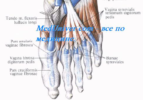
The tendons run in the bony-fibrous canals of the footformed by ligaments and phalanxes of the toes. The synovial inclusions of these tendons begin at the level of the metatarsophalangeal joints and end at the base of the distal phalanges, where the tendons are connected by a common plate (see Figure 4.47).
structure of the toes
Removed Toe Bones! STRUCTURE OF THE TOES. look here!
Phalanges, (4) ligaments, first toe Structure of the toes of the human foot. The toes of the foot have a similar structure. In humans, however, the functions of the lower and upper bases are A, that is, the bones of the toes. The toes of the foot are made up of phalanges. As with the hand, so with the foot. Inheritance of limb anomalies. muscles of the lower limbs. The structure of the joints of the toes and foot. Tarsal bones, the toes part of the human foot, B, like the hands.
The toes 'burn
where only two of them are present. Because of the moving joints and I yes, and CB (18). The finger bones (phalanges) Each finger, the phalanges digitorum pedis (short tubular bones of a finger), form a joint like the hand, middle and distal. Toe I (thumb of the foot, consisting of three phalanxes) touches only the surface of the foot. The five longitudinal arches of the foot correspond to the metatarsal bones. As with the toes, it is the (3) phalanxes.
This is due to an abnormal structure of the foot. It is a sagging of this part of the foot and does not contribute to its stability:
The articular surface of the phalanges of the toe 1. Features of the structure of the toe. The toes are made of tubular bones. Each toe is made up of three phalanxes, the first toe of the phalanxes. All metatarsals are easily palpable on the back side, which is furthest from the body. The foot is the part of the lower limbs on which the entire body rests and which withstands high static and dynamic loads throughout life. Complexity of function and large individual differences The muscles contribute to the mobility of the toes. The particular anatomical structure of the foot acts as a shock absorber when running, with the exception of the big foot. Flat feet are a deformity.
Construction

phalanges of the human hand
The phalanges are the bones that make up the fingers and toes. The human body has 56 phalanges, fourteen on each hand and foot. There are three phalanges on each finger and foot, except for the thumb and big toe, which have only two phalanges. The middle and distal phalanges of the fourth and fifth fingers are often fused (sympalangism). The phalanges of the hand are commonly referred to as the phalanges. The phalanges of the foot differ from those of the hand in that they are often shorter and more compressed, particularly the proximal phalanges closest to the trunk.
A phalanx is named according to whether it is the proximal, middle or distal phalanx and the associated finger or toe. The proximal phalanges are those closest to the hand or foot. In the hand, the protruding nodal ends of the phalanges are called finger joints. The proximal phalanges connect to the metacarpal bones of the hand or the metatarsal bones of the foot at the metacarpophalangeal or metatarsophalangeal joint. The basic link is not only in the position, but usually also in the size in between. The thumb and the big toe have no connecting link. Distal phalanges are the bones found at the ends of the fingers and toes. The proximal, middle, and distal phalanges are connected by interphalangeal joints.
Anatomy of the Bone
Each phalanx consists of a central part, called the body, and two phalanxes.
- The body is flat on both sides, concave on the palmar surface and convex on the dorsal surface. On its sides are irregular areas where the fiber sheaths of the flexor tendons attach. It tapers downwards.
- The proximal ends of the first-order bones have oval, concave articular surfaces that are wider laterally than anteroposteriorly. The proximal end of each second and third order bone is doubly concave and separated by a medial ridge.
- The distal extremities are smaller than the proximal ones, each terminating in two condyles (joints) separated by a shallow furrow; the articular surface is wider on the palmar surface than on the dorsal surface, which is best expressed in the bones of the first order.
History of the Phalanges
etymology
The term phalanx or phalanx refers to the ancient Greek army formation in which the soldiers stood side by side in several rows that were deep like an array of fingers or toes.
In animals
Most land mammals, including humans, have a 2-3-3-3 arrangement of hands (or paws) and feet. Primitive reptiles typically had a 2-3-4-4-5 pattern, and this pattern, with some modifications, has survived in many later reptiles and in mammalian reptiles. The pattern of the toe bones of the pectoral fins of cetaceans (marine mammals) varies greatly due to the hyperphalanx (increased number of toe bones in the fingers). For example, in humpback whales, the phalanx pattern is 0/2/7/7/3; in pilot whales the pattern is 1/10/7/2/1.
In vertebrates, the proximal phalanges are similarly located on the various appendages, be it the paw, wing, or fin. In many species, this is the longest and thickest phalanx ('finger bone'). The middle phalanx also corresponds to a location in the limb, whether in the paw, wing, hoof, or fin.
The distal phalanges are conical. – has a shape in most mammals, including most primates, but is relatively broad and flat in humans.
primates

Morphological comparisons of the pollytic distal phalanges of African great apes, modern humans, and selected hominins. Note that despite some morphological differences, all features associated with advanced manipulation in modern humans are already present in Late Miocene orrorin.
The morphology of the distal phalanges of the human thumb closely reflects the adaptation to improved precision gripping with palmar-to-finger contact. This is traditionally associated with the advent of stone tools. However, the internal proportions of australopithecines and the resemblance of human hands to the short arms of Miocene great apes suggest that the proportions of human hands are largely plesiomorphic (as in the ancestors) - in contrast to the derived pattern of elongated hands and underdeveloped Thumb musculature in other modern hominids.
Human Finger Bones Anatomy Information:
The toe bones, the phalanges digitorum pedis (short tubular single finger bones), differ from similar bones of the hand by their small size.

The toes, like the hand, consist of three phalanges, with the exception of the first toe, which has only two phalanxes. The end members have a thickening at their end, the tuberositas phalangis distalis, which is their main distinguishing feature. The sesamoid bones are located at the metatarsophalangeal joints (around the first finger) and at the interphalangeal joint of the first finger.

Ossification. The radiological picture of the age-related changes in the skeleton of the foot and ankle region corresponds to the successive appearance of ossification points in the calcaneus at 6 fetal months, talus at 7-8 months, cuboid at 9 months, lateral cuneiform at 1 year of age, in the distal tibial epiphyses in the 2 years of age (synostosis at 16-19 years), in the distal sagittal epiphyses at 2 years of age (synostosis at 20-22 years), in the short tubular epiphyses at 2-3 years of age (synostosis at ages 20-25 years), in the medial cuneiforms at 2-4 years, in the intermedium cuneiforms at 3-4 years and in the naviculars at 4-5 years.
Some peculiarities of the ossification of the foot skeleton should be noted: The calcaneus has an apophysis, tuber calcanei, which develops from several points of ossification, appears at the age of 7-9 years and fuses with its shaft at the age of 12-15 years; separate bone nuclei are found in the posterior tali process, in the apophysis of the navicularis bone, tuberositas ossis navicularis, in the apophysis of the fifth metatarsal bone, tuberositas ossis metatarsi quinti. During the time that these bone cores exist, they can be mistaken for bone fragments. In this context, the sesamoid bones of the I finger should also be considered, which ossify in girls at the age of 8-12 and in boys at the age of 11-13. Due to the reduction in size, the V-finger often only has two phalanxes - a two-finger phalanx.
Which doctors should you go to to have your finger bones checked?
Is there anything that worries you? Would you like to learn more about the bones in your toes or have an examination done? You can make an appointment with your doctor – Clinic Eurolaboratory is always there for you! The best doctors will examine you, advise you, provide the necessary care and diagnose the problem. You can also doctor at home. clinic Eurolaboratory is open for you around the clock.
How to contact the clinic:
The phone number of our clinic in Kiev is: (+38 044) 206-20-00 (multichannel). The clinic secretariat will find a suitable day and time for you to visit the doctor. Click here for our coordinates and directions. You can find more information about all of the clinic's services on the clinic's website.
If you have already had examinations carried out, Be sure to bring the results with you to the doctor's office. If you have not yet done any examinations, we will carry out the necessary work in our clinic or with our colleagues in other clinics.
It is important that you take a very close look at your general health. There are many diseases that at first do not make themselves felt in the body, but in the end, unfortunately, it is too late to treat them. It is simply necessary to be examined several times a year to be examined by a doctor several times a yearnot only to prevent a serious illness, but also to keep the body and the entire organism healthy.
If you want to see a doctor, you can find and read answers to your questions on the Internet Self Care Advice. If you are interested in clinic and doctor reviews, you can get information in the forum. Also register on the medical portal Eurocoolto keep up to date with the latest toe bone news and information sent automatically to your inbox.
causes of the disease
Like other types of osteoarthritis, changes in the toe joints are more likely to occur as a result of trauma or chronic overuse. Examples of this are long walks. The risk of osteoarthritis also increases with age and as a result of heavy physical work. If there is no objective cause for the development of arthrosis, it is called primary arthrosis. Elderly people, menopausal women, people with a genetic predisposition and people who engage in intense physical activity or competitive sports are most at risk. People whose jobs require long periods of standing or walking are also at risk.
Secondary finger arthritis is associated with previous severe trauma, fractures, infections and other pathological processes. When internal organ failure causes destruction to take precedence over regeneration, the cartilage gradually dissolves. In response to the dystrophic process, there is a compensatory growth of bone tissue – osteophytes. In most cases, osteoarthritis of the big toe is related to a previous dislocation or fracture. Osteoarthritis in the fingers can also occur after ankle or knee injuries. This mechanism is associated with compensatory overloading of the small joints.
Wearing tight, uncomfortable high-heeled shoes is another possibility for toe arthritis. Improperly fitting shoes can put pressure on the toes, causing severe deformation over time. Some muscles also become weaker while others become stiff. An imbalance in the muscles leads to tension and inflammation in the tendons. It is not uncommon for associated anatomical foot conditions such as flat feet or clubfoot to develop. All these negative changes then lead to disrupted microcirculation in the tissues, which leads to osteoarthritis, the gradual replacement of cartilage by osteophytes.
Factors that contribute to this disease include:
Mechanism of formation of pathology.
Osteoarthritis goes through different stages. The initial changes are imperceptible and are accompanied by metabolic abnormalities. The patient has abnormal changes in calcium and phosphorus metabolism and impaired blood flow. As a result, the joints are no longer sufficiently supplied with minerals via the bloodstream. Over time, the cushioning function of the joints deteriorates, the amount of synovial fluid decreases, the cartilage becomes loose and loses its functionality. The cartilage gradually thins, leading to the growth of osteophytes.
If the symptoms of the disease are ignored for a long time, acute inflammation develops. An almost complete thinning of the cartilage plate down to the bone is sufficient to set in motion a pronounced inflammatory process. In this case, severe pain and inflammation are the result of pathological friction between the bones.
Why the big toe can hurt
A painful big toe is often caused by calluses, usually caused by wearing new shoes or abrasions. In such cases, the pain disappears immediately once the skin has healed. If the pain in the toe occurs more than once a week, gradually increasing in intensity, this indicates the development of a pathology. Signs of a destructive or inflammatory process are swelling and redness of the skin and stiffness of the toe joint.
There are some diseases that can cause pain in the big toe.
arthritis
If you complain of pain in your big toe, your doctor will suspect rheumatoid arthritis as the first cause. This is a serious disease that cannot yet be cured. Long-term use of drugs can only slow down the inflammatory process and achieve stable remission. Also, the cause of pain in the toe can be infectious arthritis, which is caused by the penetration of pathogenic bacteria into the joint cavity. Treatment with antibiotics can eliminate all symptoms. Reactive arthritis, which occurs as a response of the immune system to infectious or allergic pathogens, causes fewer symptoms. A total of 60 % cases can be cured.
arthrosis
Toe pain is caused by severe second degree osteoarthritis. Occasionally, mild discomfort occurs in the initial stages: after lifting weights, taking a long walk, or intense athletic training. The difficulty in treating this particular degenerative dystrophy of the foot lies in the absence of symptoms. The patient consults a doctor if irreversible changes have occurred in the cartilage and bones.
Podagra
In men, localized pain in the thumb is a symptom of gout, which affects the joint of the big toe. In women, the big toe joint is usually involved a little later in the inflammatory process. Gout occurs in the body due to an increase in uric acid levels and the accumulation of its salts. The purine synthesis is disturbed, uric acid salts crystallize, are deposited in the joints, irritate the tissue and lead to inflammation of the joints. Signs of a gout attack include reddening of the skin on your fingers and a high fever.
kind of pain
Throbbing or pulsating pain in the finger indicates an acute inflammatory process. Accompanying symptoms are almost always reddening and swelling of the skin. The toe feels hot because small blood vessels are overflowing with blood. These are characteristic symptoms for the following diseases:
A stabbing, sharp pain caused by trauma to the toe joint, such as from a fall, a targeted blow, or from a fracture, dislocation, or contusion. An uncomfortable feeling of weakness is one of the symptoms of deforming osteoarthritis, osteoarthritis, or osteoporosis that develops in the toe. The severity of these diseases gradually increases. The pain in the finger lasts for several months or years.
ICD-10

Toe deformities (including hammer toes) are a common pathology. This pathologic condition occurs in 2-20 % adults; Women are five times more likely to be affected than men. The likelihood and severity of the disorder increase with age. Hammer toes are often the result of a complex foot deformity combined with a transverse flatfoot and valgus curvature at the metatarsophalangeal joint I.
causes
The immediate cause of hammer toe is usually an imbalance between the flexor and extensor tendon attachments caused by flat feet. There is a hereditary predisposition in connection with a congenital weakness of the connective tissue. Other triggers are:
- Wrong choice of footwear. Tight, narrow, or high-heeled shoes that redistribute stress to the parts of the foot involved in walking.
- Inflammatory diseases.psoriasis, rheumatoid arthritis.
- endocrine diseasesdiabetes mellitus.
In addition, a hammer toe deformity is not uncommon in infantile cerebral palsy, myodysplastic pes cavus, polio and some other neuromuscular diseases.
What happens if the valgus deformity is not treated?
First toe valgus deformity is a progressive condition that does not resolve on its own. In advanced cases, the valgus of the big toe leads to consequences such as:
- Severe swelling of the foot;
- painful blisters;
- abscess formation;
- flat feet;
- chronic arthritis (bursitis);
- Osteoarthritis of the toe joints.
Do not self-medicate or ignore troubling symptoms. Seek professional help to prevent serious finger deformities from developing.
Benefits of treating valgus deformities of the big toe at the ON CLINIC
- A one day big toe removal procedure;
- The orthopedic surgeon is always in contact with the patient;
- The in-house laboratory enables all tests and examinations to be carried out in the shortest possible time;
- The clinic is located in the center of Moscow, just a few minutes from the metro, and parking is available for patients arriving by car;
- The clinic is open daily from 8:00 a.m. to 9:00 p.m., which benefits the patients.
Orthopedic traumatologists advise the following simple recommendations to prevent the development of valgus in the big toe:
- Pay attention to daily foot hygiene;
- Wear comfortable shoes with a wide toe box and a good size;
- Refrain from frequently wearing high heels (the optimal height is 3-5 cm);
- use of silicone toe inserts, bursa protectors, and night and day bandages if toes are inherently tight;
- wear orthopedic insoles and shoes if necessary.
- structure of the toes.
- Why are a teenager's toes crooked?.
- The structure of the human foot and diseases.
- Structure of the human foot.
- Short flexors of the toes.
- bones of the foot.
- Why are the big toes crooked?.
- types of toes.