- deep veins. They ensure the outflow of blood from the lower limbs, draining the blood that has already been filtered.
- Superficial Veins. Supply the joints and muscle tissue with blood and supply them with important substances.
Structure of the veins of the lower limbs
- The human foot
- arch of the foot.
- structure of the foot
- When feet hurt
- General or diffuse, spreading to the whole foot
- Pain that is limited to a specific area (the size and extent of the area does not matter)
- unrolling of the foot
- Achilles tendon
- blood supply
- Consequences and prognosis of injuries
- Injury and disease prevention
- External pain in foot
- Causes of foot pain on the outer lateral side
- diagnosis
- The most important areas of the foot
- header area
- nerves of the lower limbs
- Examination of the bones and joints of the lower limbs
- General investigation
- goniometry
- Radiological diagnosis of the lower limbs
- Human Foot Anatomy Information:
- Which doctors should you see for a foot exam?
- Causes of foot pain
- diagnosis
The human foot
The foot is the part of the lower limbs on which the entire body rests and is subjected to high static and dynamic loads throughout life. The complexity of function and the large individual differences in the structure of the foot result from the large number of bones and their articulations, the architecture of the ligamentous apparatus and the number of muscles involved in maintaining the movement of the foot. The proper functioning of all structures gives the foot reliable functioning, stability and resistance to the weight of the whole body and the stresses encountered during movement.
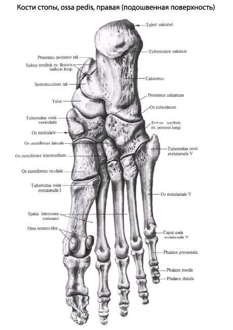
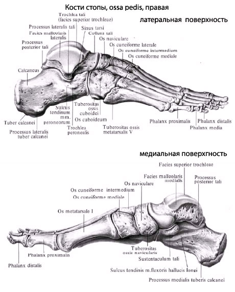
Each human foot is made up of 26 bones. The bones of the foot are broad and flat and connected by a large number of strong ligaments that limit movement but strengthen the foot for support. The strength of the foot as an overall structure is important for performing body movements and supporting body weight. Despite limited mobility. The foot can move easily on both smooth and uneven surfaces.
Each toe (there are five in total) has 3 phalanges, except for the big toe, which has 2 phalanges. The toe bones are connected to the metatarsal bones. The tarsal consists of 5 bones, each connecting to the corresponding phalanx on the distal side (farther from the body) and to the tarsal bones on the proximal side. The tarsal bones are made up of 7 bones: the heel bone, the ankle bone (talus), the tarsal bone (cubitus) and the three sphenoid bones – the outer (lateral), inner (medial) and middle. The talus and heel bones are the largest. The navicular bone connects the posterior talus to the three anterior sphenoid bones—the internal sphenoid, the external sphenoid, and the internal sphenoid. The navicular bone connects the calcaneus behind it with the 4th and 5th bones in front of it. The largest bone in the tarsus, the calcaneus, forms the heel. Attached to the heel is the Achilles tendon, a tendon of the calf muscle and cambioideus muscle at the back of the tibia. The tarsal bone in the form of the talus forms the ankle joint with the tibia and fibula. When standing, the ankle bone absorbs the entire body weight and distributes it between the forefoot and hindfoot.
arch of the foot.
The foot has five longitudinal arches and one transverse arch. The longitudinal arches of the foot begin at the heel and continue in convex lines to the metatarsal bones. The tallest and longest of the longitudinal vaults is the 2nd, and the lowest and shortest is the 4th. In summary, the longitudinal vaults can be summarized into two: the outer longitudinal vault and the inner longitudinal vault. In the forefoot, all longitudinal arches come together in an arch line pointing upwards and form the transverse arch of the foot.
The arches of the feet are formed by the bones of the foot, as well as tendons, ligaments, and muscles. The longitudinal muscles of the foot shorten and increase the longitudinal arch, while the oblique muscles narrow the foot and increase the transverse arch. In addition to the foot muscles, the muscles of the shin bone are also involved in the formation of the longitudinal arch of the foot. The strongest ligament that forms and maintains the longitudinal arch is the longitudinal ligament (tendon-muscle). The plantar fascia is important for maintaining the arch of the foot.
The human foot, lateral view (medial side).
structure of the foot
As those skilled in the art know, the foot has 3 points of contact with the ground, called the bony support: 2 in the forefoot and 1 in the heel.
The foot consists of 24 bones, a hundred muscles and many tendons and ligaments that have an elastic function in human movement.
The human toes are made up of the phalanges of the foot skeleton, the bones of which extend from the toes to the heel. The bones of the tarsal bones are similar to the metatarsals and phalanxes of the feet, but are less developed because they are less mobile. When walking on the ground, the heel, ie the lateral part of the foot, first encounters an obstacle, followed by the sole of the foot and then the toes.
The structure of the foot provides a soft load on the whole body (the longitudinal and transverse arches are responsible for softness).
Flat feet pose a risk for conscripts because the (longitudinal) arch is flattened, leading to deformation of the ligament and muscle structure. Unfortunately, flat feet are not the only foot problem that awaits people on their long journey through life.
When feet hurt
Who doesn't know foot pain at the end of a hard day's work? And who does not feel his feet hurt after a long walk? Every adult has experienced uncomfortable (and painful!) sensations at some point in their lives.
However, in science it is accepted to divide foot pain into different types:
General or diffuse, spreading to the whole foot
Diffuse foot pain is sometimes directly related to a specific strain or exertion (if the foot hurts when walking), but can also occur when the foot is at rest.
Pain as a result of excessive stress, but without any special additional clinical symptoms, is a clear and characteristic sign of calcium deficiency (osteopathy). Osteopathy can show up in those affected: rickets, osteoporosis in the elderly, osteomalacia.
An uncommon but notable symptom is that the bones themselves are painful to palpation or simply to pressure.
Diffuse pain is also caused by
- Long bed confinement in case of illness. Here the problems are not due to bone pain, but to a weakness in the muscular system.
- Weight gain with short periods of movement.
- Osteoporosis due to tissue damage (bone or soft tissue) or joint damage in the ankle group (or its condition).
- Organically caused vascular diseases that impair the function. These pains vary over time and are paroxysmal in nature.
Pain that is limited to a specific area (the size and extent of the area does not matter)
Pain in feet and toes And the point localization, that is, the localization of pain in strictly defined areas, which is caused by various factors. Touching and squeezing the toes is the basis of the diagnosis to identify the nature and cause of the pain - and eliminate it.
unrolling of the foot
When we stand still, 50 % of our weight is on the heel, 25 % on the forefoot, and the remaining 25 % on the metatarsal. When walking, the entire weight rests on the heel (1), then on the forefoot (2), then over the transverse arch (3) in the direction of the first metatarsophalangeal joint (4) to the tips of the toes.
The most important thing you can do is wear comfortable shoes. The three ingredients to keeping your feet healthy are cleanliness, neatly trimmed nails, and exercise.
Achilles tendon
The Achilles tendon is the largest tendon in the human body and strengthens the ankle at the rear end. It is formed by the union of the calf muscle and kidney muscle in the lower leg.
The powerful tendon stretched between the gastrocnemius muscle and the calcaneal tubercle plays an important role in the movement.

An important clinical issue is the potential for strain and overload of this structure. In this case, the trauma surgeon must perform a comprehensive treatment to restore function.
blood supply
The muscle function, the regeneration after stress and injury as well as the metabolism in the joint are made possible by the special anatomy of the circulatory network surrounding the joint. The arteries of the ankle are arranged similarly to the blood supply of the knee joint.
The anterior and posterior tibial artery and the brachial artery branch off at the lateral and medial malleolus and surround the joint on all sides. This arrangement of the arterial network allows for full function of this anatomical region.
The outflow of venous blood from this area occurs through the internal and external meshwork, which form important formations: the great saphenous vein and the internal tibial vein.
Consequences and prognosis of injuries
Untreated injuries and diseases of the Achilles tendon significantly increase the risk of complications. Untreated injuries can lead to permanent disability of the foot.
With timely treatment, the prognosis is favorable. In the event of an injury, the tendon can be successfully rehabilitated. Agility takes a long time to restore, but manages to fully restore it.
Injury and disease prevention
In order to prevent injuries and pathological processes in the area of \u200b\u200bthe Achilles tendon, the following rules should be observed:
- Only wear shoes that are comfortable, fit well, do not pinch and are made of natural materials.
- Avoid walking in high heels.
- Move, but don't put too much weight on your feet.
- Warm up before regular exercise.
- Only walk on a level surface.
- Make sure your feet don't get too cold.
So, the Achilles tendon is an important part of the lower limbs that is involved in the movement of the legs. If injury or inflammation occurs in this area, you should see your doctor as soon as possible and start treatment.
External pain in foot
The human foot has a complex structure. This allows humans to stand and move safely. The feet fulfill important functions every day: they help to maintain balance, push off the ground and cushion movements. This ensures that shocks resulting from contact with the ground are not transmitted to the spine and skull. This prevents damage to the brain and spinal cord. But even such a complex, perfect mechanism can fail. Overexertion often causes pain on the outside of the foot. This affects people of all ages.
The information in this section should not be used for self-diagnosis and self-medication. In case of pain or other aggravation of the condition, diagnostic tests should be ordered only by the attending physician. A specialist doctor should be consulted to diagnose and prescribe appropriate treatment.
Causes of foot pain on the outer lateral side
There are many known causes of foot problems, each requiring a specific therapeutic approach. Treatment should only be undertaken by a specialist after a series of investigations have been carried out to determine the actual cause of the external foot pain. The main causes of the symptom include the following abnormalities:
- Arthrosis;
- sprains;
- Osteoporosis;
- Achilles tendon injury;
- heel spur;
- Morton's neuroma;
- Valgus deformity of the foot.
However, it is important to remember that there are physiological causes of foot pain. This may include wearing uncomfortable footwear. Women are especially prone to this factor. High heels and narrow toes can cause pain in the outer part of the foot. As soon as the feet are freed from uncomfortable footwear, this symptom disappears. In such cases, no treatment is required. However, when the feet are systematically exposed to uncomfortable footwear, the situation worsens, leading to joint deformities. As a result, synovitis, arthritis, osteoarthritis, plantar fasciitis and other diseases can develop. Excessive physical exertion can also promote the development of these diseases.
diagnosis
Only a qualified doctor can diagnose a foot problem after conducting an examination and collecting all the necessary data. External diagnosis of foot pain includes the following methods:
| diagnostic technique | Time |
|---|---|
| X-ray of the foot | 15-30 minutes |
| Ultrasound examination of the foot | 30 minutes |
| CT scan of the foot | 15 minutes |
| MRI of the foot | 20 minutes |
Each of these methods provides information about the bones, muscles, tendons, blood vessels, and skin of the foot. The test can be performed at any medical facility that has the appropriate equipment. The cost of the diagnostic procedures varies. The most cost-effective examination is the ultrasound examination. Their price starts from 1110 rubles. Computed tomography and magnetic resonance imaging are considered the most expensive. The cost of these procedures starts from 4,000 rubles. But the accuracy of the data obtained fully justifies the cost.
The most important areas of the foot
Why cold and wet feet lead to cold or respiratory areas
Therefore, wet feet are the most typical cause of colds, which are accompanied by a runny nose and headache. And the left sinuses are projected onto the left fingertips and the right sinuses onto the right fingertips.
Even walking a little increases visual acuity and intraocular pressure normalizes.
- The inner ear, pharynx and bronchi are located on the front and sole of the foot.
- So when your feet get cold and wet, you can't avoid a cold with a cough, runny nose, and ringing in your ears.
- If you suffer frostbite. at the top of the front arch of the foot at the base of the second and third toes, the sore throat can spread to the lungs.
For adults, whose immune systems are usually already strong, frozen and wet feet don't always result in a cold. In children, on the other hand, there is a very strong connection between the foot and health.
header area
- Located in the middle of the big toe on the lateral side of the foot.
- When a child or adult has problems with the big toe, its curvature (big toe valgus), or persistent ingrown toenails, headaches and problems with memory, attention, and concentration are directly related to the big toe problem, for which the Brain and cerebellum are responsible.
- Wet and cold feet have an effect on headaches.
- The heart zone is located in the front part of the arch of the left foot.
- Older people have a slight limp in their left foot a day or two before a heart attack, which they often notice themselves ('Something got in their foot.') but doesn't pay much attention to it.
- this is a warning sign of an irregular heart rhythm and irregular nutrition of the heart muscle. It has already been established with absolute certainty that if the left foot is palpated nowadays, the heart area reacts with a sharp pain.
When a child has an outward valgus curvature of the foot (clubfoot), there is a direct link to the development of cardiovascular disease.
nerves of the lower limbs
The nervous system enables the brain to receive information from the different parts of the body and to set muscles in motion by causing them to contract or stretch. This ensures that all bodily functions are carried out. When the nervous system is damaged, the entire body suffers, even if the injury is localized.
There are two nerve plexuses for the innervation of the lower limbs:
The femoral nerve is one of the largest nerves in the lower limbs, making it the most important. This system is responsible for controlling the legs, direct movement, and other movement sequences.
Human nerves of the lower limbs
When the femoral nerve is paralyzed, the entire underlying system has no connection to the CNS (central nervous system), meaning control of the legs becomes impossible.
Therefore, it is important to keep the nerve plexuses intact, avoid damaging them, and keep the temperature in this area of the lower limbs constant and avoiding fluctuations.
Examination of the bones and joints of the lower limbs
When the first symptoms of a lower limb injury appear, a diagnosis should be made immediately so that the problem can be identified early.
The first symptoms can be:
- Drawing pains in the calf area;
- General weakness in the legs;
- nerve spasms;
- Constant hardening of various muscles.
However, if even slight pain occurs regularly, this also indicates a possible injury or illness.
General investigation
The doctor examines the lower limbs for any visible abnormalities (enlargement of the kneecap, swelling, bruising, blood clots, etc.). The specialist asks the patient to do various exercises and tells him if he feels any pain. In this way, the area in which the disease can appear is determined.
The patient's legs are then palpated to see if there are hardening or lumps that could indicate blood clots, neurological or muscular problems.
goniometry
Goniometry is an additional examination of the lower limbs using modern technology. It is used to diagnose deviations in the vibration amplitude of the joint and kneecap. If there are deviations from the norm, there is reason to think about and carry out further investigations.
goniometry
Radiological diagnosis of the lower limbs
There are several types of radiological diagnostics:
Human Foot Anatomy Information:
The foot is constructed like a flexible, moveable vault and works like that. The arched structure of the foot is not found in all animals, not even in the great apes, and is a peculiarity of humans due to their upright posture. This structure was developed due to the new functional demands on the human foot: increased load on the foot when standing upright, reduced support surface while saving building material and strength of the overall structure.
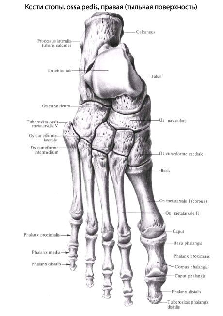
The bones of the foot, which are almost immovably connected to each other by fixed joints, form the so-called hard foot, which consists of 10 bones: os naviculare, ossa cuneiformia mediale, intermedium, laterale, os cuboideum, ossa metatarsalia I, II, III, IV, V. The long plantar ligament, or long plantar ligament, plays the most important role in strengthening the arch of the foot. It begins on the underside of the heel bone, runs forward and inserts with deep fibers at the tuberositas ossis cuboidei and with superficial fibers at the base of the metatarsal bone. The long plantar ligament traverses the bony sulcus (sulcus ossis cuboidei) and transforms this sulcus into a bony-fibrous canal through which the peronei longi tendon passes.
The general arch of the foot consists of five longitudinal arches and one transverse arch. The Longitudinal Vaults begin at a point on the heel bone and extend forward along the upwardly curved rays that correspond to the 5 rays of the foot. The sustentaculum of the talus plays an important role in the formation of the first (medial) arch. The longest and highest of the longitudinal arches is II. The longitudinal arches converge in a parabola shape at the front and form the transverse arch of the foot.
Which doctors should you see for a foot exam?
Is there anything that worries you? Want to learn more about the foot or need an investigation? You can make an appointment with your doctor – Clinic Eurolaboratory is always there for you! The best doctors will examine you, advise you, provide the necessary care and diagnose the problem. You can also doctor at home. clinic Eurolaboratory is open for you around the clock.
How to contact the clinic:
The phone number of our clinic in Kiev is: (+38 044) 206-20-00 (multichannel). The clinic secretariat will find a suitable day and time for you to visit the doctor. Click here for our coordinates and directions. Further details on all of the clinic's services can be found on the clinic's homepage.
If you have been examined before Be sure to take the results with you to your doctor's office. If the examinations have not been carried out, we will carry out the necessary work in our clinic or with our colleagues in other clinics.
You must be very careful about your general health. There are many diseases that do not initially make themselves felt in the body, but unfortunately it is too late to treat them in the end. You just have to get checked out several times a year visit a doctor several times a yearnot only to prevent a bad illness, but also to keep the body and the entire organism healthy.
If you want to see a doctor, you can find and read answers to your questions on the Internet Tips for self care. If you are interested in reviews of clinics and doctors, you can get information on the forum. You can also go to the medical portal Eurolaboratoryto keep up to date with foot pain news and information, automatically sent to your inbox.
Causes of foot pain
The complaints can be general, diffuse or limited to a specific area. Doctors do not always immediately recognize what is causing the symptoms. Foot pain usually has a variety of causes. The most common causes of complaints are:
- Injuries – fractures, bruises, hematomas;
- Excessive stress during physical activity;
- arthritis and arthrosis;
- inflammatory processes;
- benign and malignant tumors.
You can also perceive discomfort as a symptom of a disease. In osteoporosis, for example, the pain is severe, stabbing and long-lasting. In addition, the sufferer feels discomfort even at rest. In this case, pain in the foot caused by osteoporosis should be a reason to see a doctor. Otherwise, there can be serious complications that affect the joints and soft tissues. Then the person can remain disabled and only surgery can help.
diagnosis
The exact cause of the burning pain in the foot can be diagnosed by a specialist in the hospital. It is worth making an appointment with your family doctor. He or she will do an initial assessment and then likely refer you to a specialist for a diagnosis. The doctor may prescribe the following tests, among others:
| diagnostic technique | Time |
|---|---|
| Ultrasound examination of the foot | 30 minutes |
| CT scan of the foot | 15 minutes |
| X-ray of the foot bones | 20 minutes |
| MRI scan of the foot | 20 minutes |
Pain in the upper part of the foot can be temporary or chronic, depending on the general condition and health of the patient. However, if they occur frequently, a visit to the doctor should not be put off. Serious complications can be prevented by an appointment with an orthopaedist, traumatologist or rheumatologist. Some of these lead to bone and joint destruction that is extremely difficult to treat.
Read more:- The structure of the human foot and diseases.
- Determining the number of longitudinal arches in the human foot.
- Structure of the human lower limbs with captions.
- structure of the toes.
- structure of the toes.
- The tarsal bone hurts from above - what to do?.
- The largest bone in the skeleton of the human foot.
- The bones of the human heel.