Equally important are the rehabilitation measures. The disappearance of pain is not a sign of recovery. Curing a single symptom when treating a movable joint portends a favorable prognosis, but full recovery takes time. In this case, irradiation of the joint allows faster tissue healing and accelerates the process of complete healing of any of the above pathological conditions.

- ankle
- Construction
- Area
- ankle
- Causes of ankle pain and symptoms.
- arthritis of the ankle
- Osteoarthritis of the ankle
- diagnosis
- What is the structure and function of the lower ankle?
- Research Methods:
- Human Ankle Anatomy Information:
- Which doctors do you go to to have your ankle examined?
- First aid measures for a sprained ankle
- Diagnosis of an ankle injury
- Complications after the procedure
- Indications for surgery
- types of interventions
- arthroscopy
- Anatomy of the foot and ankle
- Indications for the examination
- X-ray diagnosis of neoplasms of the foot
- Diagnosis of fractures of the foot
- Type of pain in various pathologies, concomitant symptoms
- Injuries to the ankle
- ankle arthritis
- Osteoarthritis of the ankle
- Treatment of ankle pain - basic methods
- Symptomatic treatment
- Painkillers
- Physical therapy for ankle pain
- clinical picture of arthrosis
- How is osteoarthritis treated?
ankle
The ankle or ankle is the area where the foot and leg meet. The ankle consists of three joints: the actual ankle joint, the subtalar joint, and the inferior tibial plateau joint. The movements performed at this joint are dorsiflexion and plantar flexion of the foot. Typically, the term 'ankle' refers only to the area of the ankle. In medical terminology, the term 'ankle' may (without limitation) refer to the region in general or to the ankle specifically.
The main bones of the ankle region are the ankle bone (talus) and the shin and fibula (tibia) of the foot. The ankle is a synovial joint that connects the distal ends of the tibia and fibula of the lower extremity to the proximal end of the talus. The joint between the tibia and the ankle bone has a greater mass than the joint between the fibula and the ankle bone.
- 1 structure
- 1.1 Interface
- 1.2 hock
- 1.3 Bands
- 1.4 Retinaculum, tendons and their synovial membranes, vessels and nerves
- 1.5 Mechanoreceptors
- 3.1 Traumatic Injuries
- 3.1.1 Fractions
- 5.1 Development
Construction
Area
The ankle is the area at the transition between leg and foot. It extends downward (distal) from the narrowest part of the shinbone and encompasses the body-hugging parts of the foot (proximal) to the heel and top (dorsal) of the foot.
ankle
The hock is the only groove and spine in the human body, a term that identifies the bony structure with the joint wood of the same name. The bony structure of the ankle consists of three bones: the tibia, fibula, and talus. The articular surface of the tibia can be referred to as the plafond (French for 'ceiling'). The medial ossicles is a bony spur that branches off distally from the medial tibia. The most distal part of the fibula is called the lateral malleolus. Together, the ankles and their supporting ligaments stabilize the talus below the tibia.
Because the movement of the ankle is a major contributor to the alignment of the foot, some authors refer to it as the ankle and the ankle. Dorsiflexion and plantar flexion are movements that take place in the ankle. When the foot is flexed in the sole, the ankle also allows for some lateral, sliding, rotating, adducting, and abducting movements.
The bony arch formed by the tibial plateau and both ankles is called the 'insert' of the ankle (or talar insertion). The insole is a rectangular acetabular cup. The ankle joint consists of three joints: the ankle joint (also called the ankle joint, tibial joint, talar joint, talus joint), the subtalar joint (also called the talonavicular joint), and the tibiofibular (lower shin) joint. The articular surface of all bones of the ankle is covered with articular cartilage.
. The distances between the bones of the hock are as follows:
Causes of ankle pain and symptoms.

Injuries (dislocations, subluxations, tears, torn ligaments and fractures of the joint surfaces)
arthritis of the ankle
Ankle arthritis is inflammation of the joints. Arthritis can be acute and sudden or chronic and long-lasting. Arthritis is caused by autoimmune diseases and injuries. Autoimmune arthritis is arthritis in which immune cells damage the body's own tissues. Examples of this are rheumatoid arthritis, gouty arthritis or Bechterew's disease. Some autoimmune diseases develop as a result of infection. Autoimmune arthritis commonly affects small joints such as fingers, toes, and vertebral bodies. The ankle is rarely affected by immune cells. With any autoimmune disease, ankle arthritis is the exception rather than the rule. Nevertheless, the possibility of such an ankle pathology should not be overlooked. In trauma, unless accompanied by a dislocation or fracture, arthritis is the first and only symptom of the injury. Arthritis is characterized by the following symptoms in the joints: pain, swelling, limited mobility, local reddening of the skin and fever. Usually all of the above symptoms are present.
Osteoarthritis of the ankle
Osteoarthritis is a degenerative disease of the joints. It arises from a disruption in the trophic system. This means that the nutrition of the mobile joint is disturbed for some reason, which leads to a thinning of the joint surface and a decrease in the amount of synovial fluid. There is an opinion that degeneration occurs only in old age. It is true that the knee and hip joints are often affected in older people. However, the ankle is not affected. In contrast, osteoarthritis of the ankle is more common in young, healthy people with an active lifestyle. It is caused by excessive loads and frequent micro-injuries. Statistically, athletes involved in sports like volleyball, basketball, soccer, running, ice hockey, and figure skating are more likely to be affected by this condition. In these sports, the joint is subjected to maximum stress. During competition, athletes have high concentrations of adrenaline in their blood, which helps to alleviate the discomfort. As a result, they may not realize they are injured. After a few hours, the ankle begins to hurt, but athletes often ignore the pain or take painkillers. In the meantime, a slight inflammation disturbs the trophism of the tissue in the ankle. Frequent micro-injuries lead to a reduction in synovial fluid (the ankle's main shock absorber) and thinning of the cartilage.
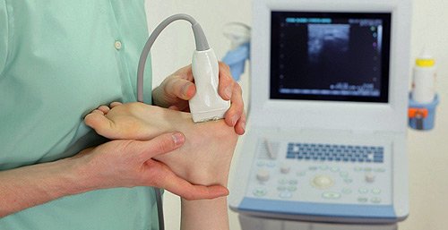
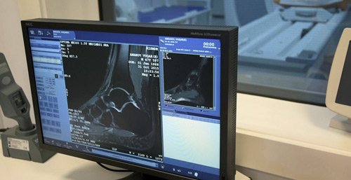
diagnosis
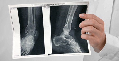
- An X-ray is an examination that shows the bony structures but not the soft tissues. X-rays are particularly useful for fractures and dislocations. With osteoarthritis, the stage of the process can be assessed using an X-ray. In the case of arthrosis, this examination is not very meaningful.
- Ultrasound can be used to diagnose ligamentous abnormalities and monitor the amount of synovial fluid.
- MRI is an exam that reveals both soft tissue and bone abnormalities. It is used in the diagnosis of arthritis and arthrosis.
Other diagnostic methods are also possible. The entire examination must be tailored to each individual patient by the doctor.
A consultation with our specialists helps to make the right diagnosis.
What is the structure and function of the lower ankle?
The lower ankle is much more complex than the upper ankle. In this multi-part joint, the ankle bone (talus), the heel bone (calcaneus), the cuboid bone (os cuboideum) and the heel bone (os naviculare) are connected to each other.
It works especially when walking uphill or downhill and on uneven terrain. Controlled by multiple ligaments, this joint is particularly vulnerable as its natural stability is reduced when the foot is lowered. This leads to a severe twisting of the ankle. The ligaments of the ankle can become stretched or torn. Broken bones, cartilage damage and trauma to the joint can also have consequences. The resulting deformation of the joint and uneven loading are the main causes of further damage to the ankle.
Ankle pain is often the result of accidents, overuse, congenital defects or long-term ligament injuries. This leads to cartilage damage, which only becomes noticeable in stage 3 or 4, when the cartilage has been damaged down to the bone.
Deformations and diseases in the legs lead to pain not only when walking, but also at rest. This has serious consequences for mobility and thus for the quality of life. We offer modern, functional therapy and surgery for the foot and ankle.
Early detection of possible causes through modern diagnostics and expertise is important for successful treatment. The aim is to prevent mechanical movement disorders of the foot or bone and soft tissue injuries in good time.
Computer-assisted examinations that precisely determine the pressure distribution in the foot and analyze the gait pattern help in selecting a suitable, individual treatment.
Research Methods:
There are various proven therapies from which the specialist selects the appropriate one. With conservative therapies such as active foot exercises, shock wave therapy and orthopedic techniques, an improvement can be achieved in the early stages; in advanced stages, a targeted operation can help.
General information and questions about the upper and lower ankle:
The ankle is the most stressed compared to other joints. During normal walking, it is loaded with seven times its own weight. The ankle is the guarantor for our upright gait.
Human Ankle Anatomy Information:
Ankle joint, art. talocruralisThe articular cuff is formed by the articular surfaces of the lower ends of the two tibias, which enclose the talus trochlea like a fork, with the inferior articular surface of the tibia attaching to the superior articular surface of the block and the articular surfaces of the hock bones attaching to the lateral surfaces of the block.
The articular sac attaches to the cartilaginous edge of the articular surfaces and encloses part of the anterior calcaneus neck. The collateral ligaments are located on the sides of the joint and run from the ankles to the adjacent tarsal bones. The medial ligament (Lig. mediale – deltoideum) is a lamella, resembling the Greek letter delta, which originates from the medial malleolus and fans out towards the three bones – talus, heel and scaphoid; the lateral ligament consists of three bundles that run from the lateral malleolus in three different directions: anterior – anterior talofibular ligament, inferior – calcaneofibular ligament and posterior – posterior tabofibular ligament.
The structure of the ankle is a block joint. Movement occurs about an anterior axis passing through the talus block, with the foot moving up (extension) and down (flexion) with the toes. The amplitude of these movements is 63-66°. Even in flexion, only very little sideways movement is possible because in this position the narrower posterior part of the talus block is not so tightly closed by the tibial foramen. During extension, on the other hand, these movements are completely impossible due to the strong compression of the block at the foramen ovale.

The ankle is nourished by the bony branches of the anterior tibial, post tibial and regopean arteries from the medial and lateral malleolar rete. regopea. The venous outflow occurs via the deep tibial veins – vv. tibiales anteriores, vv. tibiales posteriores, v. regopea. The lymphatic drainage takes place via the deep lymphatic vessels into the Nodi lymphatici poplitei. The joint capsule is supplied by the N. tibialis et. n. peroneus profundus innervated.
Which doctors do you go to to have your ankle examined?
Is there anything that worries you? Would you like to learn more about the ankle or would you like to be examined? You can make an appointment with a doctor. – Clinic Eurolaboratory is always at your service! The best doctors will examine you, advise you, provide the necessary care and diagnose the problem. You can also doctor at home. clinic Eurolaboratory is open for you around the clock.
How to contact the clinic:
The phone number of our clinic in Kiev is: (+38 044) 206-20-00 (multichannel). The clinic secretariat will find a suitable day and time for you to visit the doctor. Click here for our coordinates and directions. For more information about all the services of the clinic, please visit our personal page.If you have been examined before Be sure to bring the results with you to your doctor for a consultation. If you have not yet done any examinations, we will carry out the necessary work in our clinic or with our colleagues in other clinics.
It is important that you take a close look at your general health. There are many diseases that do not manifest themselves in our body at first, but, unfortunately, are treated too late. It is enough if you go to the doctor several times a year to be examined by a doctor several times a yearnot only to prevent a bad illness, but also to keep the body and the entire organism healthy.
If you want to see a doctor, you can find and read answers to your questions on the Internet Self Care Tips. If you are interested in opinions about clinics and doctors, you will find the necessary information in the forum. You can also go to the medical portal EurocoolSign up to keep up to date with the latest ankle news and information, automatically sent to your email address.
First aid measures for a sprained ankle
The following treatments are recommended for ankle ligaments:
- Limit exercise and movement. It is important to limit movement and loading of the ankle to prevent further damage.
- local cold. Ice application will help slow or reduce swelling and create a numbing sensation that reduces pain. It is useful to apply ice to the ankle injury site for the first 48 hours after injury. Never hold the ice for more than 20 minutes at a time to avoid frostbite. Pause for 1.5 hours before reapplying ice to allow tissue to regain normal temperature and trophism, and repeat as necessary. You can wrap the frozen product in a towel and put it on the injured area. This helps reduce pain and swelling. Ice should be applied as soon as possible after the injury. (The ice should not be applied directly to the skin. Also, do not leave the ice on while you sleep or keep it on for more than 30 minutes. This can cause frostbite).
- Elastic bandages. You will need to bandage your leg with an elastic bandage. But connect it properly, not too tight. If your toes get cold and numb, it means the bandage is too tight. An elastic bandage reduces swelling and limits movement of the joint. You can also sleep without a bandage. But remember to move around the leg with an elastic bandage.
- elevated position. Elevate the injured leg, e.g. B. on a sofa cushion or a bed. If you are sitting, you can place the leg on a chair to reduce swelling and pain.
Avoid: heating the injured area for the first week, alcohol and massage, which can increase swelling. For example, avoid hot baths and saunas. Heat has the opposite effect compared to ice. That means it stimulates blood flow.
Diagnosis of an ankle injury
First, the doctor will ask questions about how the injury occurred to determine the mechanism. This is important to diagnose various injuries. Physical exam of the ankle area can be painful because the doctor needs to determine where and what movement is causing the most pain in order to make a proper diagnosis.
The doctor may order an x-ray of the ankle to determine if there is a fracture.
Complications after the procedure
The procedure is non-invasive and completely non-traumatic and has no consequences if certain rules are observed, especially no more than once every six months. The permissible radiation exposure of the body must not exceed 5mSv. Sv stands for Sievert, which is the amount of energy absorbed by the body when exposed to radiation. It varies depending on the type of X-ray. More modern devices cause less harm to the patient's body.
The main complication after treatment is exceeding the permissible limit of radiation exposure.
Constant contraindications to the study are serious mental illnesses that prevent compliance with safety rules and the presence of metal prostheses in the study area.
Temporary contraindications are pregnancy (pregnant mothers are X-rayed only when absolutely necessary, shielding their abdomen with a lead apron) and a serious condition of the patient requiring resuscitation measures.
To further clarify the diagnosis, the patient can be prescribed other diagnostic procedures (ultrasound, MRI, CT).
[13], [14], [15]
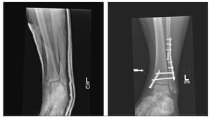
Indications for surgery
Not every fracture is suitable for surgical treatment. Simple injuries that are not associated with a dislocation can be treated conservatively and on an outpatient basis. If a dislocation has occurred, it can be repositioned by a one-stage reduction or traction.
Surgical treatment is always recommended for the following diseases:
- A fracture with a tear in the syndesmosis between the bones;
- Open fracture with extensive skin and soft tissue injuries;
- No effect after manual reduction of the fracture and traction.
There are no absolute contraindications to surgical intervention, but relative contraindications include shock, decompensated pathology, severe tissue trauma at the fracture site.
types of interventions
The most important joint operations are arthrodesis and endoprosthetics. Arthrodesis involves immobilization, while arthroplasty involves replacing the ankle joint with an artificial joint. In the first case, it is not possible to return to a normal, physiological gait, while a prosthesis gives a much better functional result, but requires a long rehabilitation and is more prone to complications.
In most cases, the specialist will insist on an operation on the ankle. After a fracture, the first step is to fuse the hock fragments and bone fragments together to allow the hock to heal faster. Only after the operation can the ankle fully recover and regain its function. If necessary, the orthopedist selects implants to immobilize the bones so that they do not shift and shift.
The operation should be performed by experienced doctors to minimize the risk of negative consequences. If the bone tissue cannot be fused, screws and metal plates are needed for the operation. Bone tissue becomes firmly anchored and the ankle remains in this state for 12 months or more. During this time, the affected leg must not be loaded. After a year, the plates are removed in another operation, a bandage is applied and the patient can only bear light weight on the ankle.
arthroscopy
This is a minimally invasive surgical procedure. Arthroscopy of the ankle is used for both treatment and diagnosis. The advantages of this method are that there is no need to make large incisions, there is no blood loss and the rehabilitation time is minimal.
The doctor performs the procedure through several small incisions no larger than 1 cm in diameter. If the surgery is diagnostic, a single incision is made through which an arthroscope is inserted and the joint cavity is examined. If resection of the joint cavity, removal of mice and joint debris, or ligament reconstruction is required, an additional incision is made for arthroscopic insertion of special surgical instruments.
Anatomy of the foot and ankle
The foot is a flexible structure of bones, joints, muscles, and soft tissue that allows for upright standing and activities such as walking, running, and jumping. There are five toes (phalanges) and five long bones in the forefoot.
The middle part of the foot consists of the pyramidal bones that form the arch of the foot. These include the three sphenoid bones, the cuboid bone, and the calcaneus.
The back part of the foot forms the heel and ankle. The ankle bone (talus) supports the shinbones (tibia and fibula) to form the ankle. The heel bone is the largest bone in the leg.
Muscles, tendons, and ligaments run along the surface of the feet, allowing for complex movements. The Achilles tendon connects the heel to the calf muscle and is important for running, jumping and standing on your toes.
The ankle allows the foot to move up and down. The hock is located below the ankle joint and allows lateral movement of the foot.
Numerous ligaments (made of rigid, flexible tissue) surround the ankle proper and subtalar joint and connect the bones of the foot.
Indications for the examination

Osteoarthritis is undoubtedly the most common foot disease, which is primarily caused by mechanical wear and tear of the articular cartilage. But inflammatory rheumatic diseases (rheumatoid arthritis, psoriatic arthritis, Bechterew's disease and Reiter's syndrome) often occur in the lower limbs or are first diagnosed there. In addition, the foot is commonly affected by cystic fibrosis and diabetic neuropathic osteoarthropathy.
X-ray diagnosis of neoplasms of the foot
Occasionally, isolated tumors can occur in the bone of the distal foot. Fortunately, most of these tumors are benign. An example is a solitary bone cyst, the enchondroma.
Some lesions have distinctive radiographic features. However, some are similar to others and may not be visible on x-ray alone.
In order to be able to assess these lesions, radiological features must be recognized. These can serve not only as diagnostic clues, but also to determine the growth rate or aggressiveness of the lesion.
A list of possible differential diagnoses can then be drawn up based on this data.
Diagnosis of fractures of the foot
In medical dictionaries, a fracture is simply defined as the collapse of a bone. However, the clinician must also know the anatomical location of the fracture, its direction, and whether it is a linear fracture or a comminuted fracture, and must be able to distinguish it from a dislocation.
The biomechanics of different fractures can vary, and accordingly the rate and type of fusion will vary.
An x-ray of the foot is the best way to screen and identify a fracture. X-rays reveal changes that describe the nature of the fracture and the location of the bone fragments.
Type of pain in various pathologies, concomitant symptoms
Injuries to the ankle
The most common form of traumatic ankle injury is a sprain or subluxation of the foot. When walking, running, or jumping, the foot can be placed awkwardly and twisted inward, overstretching the ligaments of the ankle. The more fibers are damaged, the more severe the injury and the more severe the pain symptoms. In addition to the pain, the leg reacts with severe swelling and bruising (bruising) is common.
Fractures are less common in this joint. However, a jump or fall from a height can cause a fracture of the heel bone or ankle. The symptoms are similar to those of a dislocation or sprain, but are much more intense. The foot can no longer bear weight, and palpating the injured joint causes a sharp, unbearable pain. The heel thickens and feels like it is turning inside out.
ankle arthritis
The common name for all inflammatory joint diseases is arthritis. Depending on the type of causative organism, arthritis can be viral or bacterial. Flu, chickenpox or gonorrhea can spread throughout the body and affect the joints. A characteristic rash appears around the affected joint, and the skin over the joint changes color. The patient often complains of a stabbing pain in the ankle, but can still move. The symptoms are similar to those of tuberculous arthritis.
However, if there is severe, throbbing pain in the joint, the doctor may suspect suppurative arthritis. When the disease is so reactive, your general well-being deteriorates with fever, chills, and weakness.
Osteoarthritis of the ankle
Professional athletes who continually subject their ankles to repeated sprains and strains eventually develop degenerative ankle disease. With repeated trauma to the surfaces of the ankle, fibula, and tibia, they lose their natural smoothness. The physiological sliding movement of the joint is disturbed, the movement becomes increasingly painful and the ankles swell, which even affects the choice of clothing and footwear.
Treatment of ankle pain - basic methods
Symptomatic treatment
The immediate treatment of ankle pain is what is known as symptomatic treatment, ie treating the symptom and not the cause. It is advisable to immobilize the joint or, in the event of an injury, to fix it. Applying cold to the joint works well. You can also apply an anti-edema ointment to the joint. Painkillers should be taken to relieve the pain.
Painkillers
There are a number of medications that can improve the condition of the ankle. The most effective include:
- Non-steroidal anti-inflammatory drugs (NSAIDs) – relieve pain and the acute phase of inflammation; they are used in the form of ointments, tablets and injectable solutions.
- Corticosteroids - used in the later stages of the degenerative process when 'heavy artillery' is needed.
- Chondroprotectors - are prescribed in remission, in the absence of pain symptoms, to prevent recurrence and to strengthen cartilage and ligaments. They are administered long-term and in the form of courses.
- Anti-edema agents – in the form of ointments applied to the affected joint.
Remember that medication should only be used with a doctor's prescription and under the supervision of a doctor.
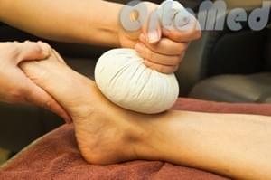
Physical therapy for ankle pain
The basis of physical therapy is the effect of natural or artificially reproduced natural substances. Not only is it relatively inexpensive, but there are practically no contraindications - it is suitable for everyone. The gentle and targeted effect of cold, ultrasonic waves and magnetic fields improves blood circulation and metabolism and enhances the effect of the drugs included in the treatment package.
clinical picture of arthrosis

As already mentioned, osteoarthritis of the ankle is a gradually progressive disease. Therefore, symptoms are minor in the initial stages but worsen over time.
The primary clinical symptom is pain in the ankle region. Initially, the pain is mild and occurs only with heavy physical exertion. Later, the pain syndrome occurs even with light exercise, and in later stages it can also occur at rest.
The characteristic feature is the appearance of pain. That is, the pain occurs at the beginning of the movement and becomes less pronounced as it 'spreads'. Some patients also report pain at night.
This pathological condition has periodic flare-ups in which symptoms worsen and then disappear.
In addition to the painful sensations, there is a complaint of a crunching and popping in the ankle when it is moved. There is also stiffness and restricted movement in the affected area.
With the progression of arthrosis, various deformations appear. Another common complication is synovitis, which leads to accumulation of excess fluid in the joint cavity.
How is osteoarthritis treated?
The treatment of osteoarthritis of the ankle must be comprehensive. At the initial stage, conservative treatment is prescribed.
For pain relief, the doctor prescribes non-steroidal anti-inflammatory drugs; if these are ineffective, intra-articular corticosteroid injections are given. Chondroprotective measures and measures to improve metabolic processes are also indicated.
Physiotherapeutic treatments such as electrophoresis or magnetotherapy are also mandatory.
Outside of an exacerbation, therapeutic exercise is indicated. In 2016, researchers from the BN Yeltsin-Ural Federal University conducted a study, the results of which confirm the importance of therapeutic exercises in the rehabilitation of patients with ankle osteoarthritis.
When conservative therapy is ineffective and the patient's quality of life decreases, surgical intervention such as ankle arthroplasty is performed.
- Structure of the human heel.
- Bones of the human ankle.
- dislocation of the ankle.
- Structure of the heel.
- The ankle bruise is where the picture is taken.
- Damaged ligaments of the ankle.
- Injury to the ankle.
- Treated subluxation of the ankle.