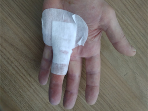
Patients with extensor tendon injuries can be treated in two ways: surgically or conservatively (i.e., splinting). The choice of treatment depends on the extent of the injury. Generally, open injuries and complete tears require surgical treatment. Closed injuries and partial tendon tears must be splinted. Both static and dynamic splinting are practiced. In dynamic splinting, the fingers are pulled back using elastic components to fix them in a specific position. Static splints, on the other hand, do not put any strain on the finger joints.
- Polyosteoarthritis of the fingers
- Causes of polyarticular finger disease
- Surgical treatment of congenital hand pathologies in children
- keywords
- MATERIAL AND METHODS
- Treatment
- Characteristics/clinical features
- Differential diagnosis.
- Assessment of clinical outcome
- Diagnosis of wrist ligament tears/injuries in the clinic
- Investigation methods
- Thumb amputation – nuances
- Disarticulation technique
- Needling technique
- injection of medication
- Indications for surgery
- Preparing for surgery
- Anatomy of the flexor tendon
- Causes of flexor tendon injuries
- Soft tissue diseases of the hand
Polyosteoarthritis of the fingers
Other names for this disease are polyarthritis of the finger and knuckle joints.
Polyosteoarthritis of the fingers (polyarticular arthritis of the fingers, 'knotted fingers') is a disease that occurs mainly in women. Although men sometimes suffer from it, they are affected about 10 times less often than women.
In women, the onset of the disease is usually directly related to the hormonal changes in the body - polysteoarthritis usually begins during menopause, between the ages of 40 and 45. The disease most often breaks out between the ages of 50 and 60 .
Causes of polyarticular finger disease
So far, researchers have not been able to identify a single cause for the development of the multi-joint disease. It is believed that the following causes include: Inherited predisposition – There are families in which polyosteoarthritis of the fingers extends over several generations of women. Apparently, certain unfavorable metabolic and cartilage patterns in the joints are inherited in these families. Due to this congenital weakening of the cartilage in people with a hereditary problem, the articular cartilage begins to degenerate during menopause, that is, it becomes damaged.

Cartilage degeneration means that the cartilage gradually loses its natural 'lubricant' and becomes dry and cracked. The friction of the 'dried out' cartilage causes inflammation in the joints and leads to the production of abnormal 'inflammatory synovial fluid'. This causes the joints to bulge from the inside, which can be very painful, and the joints themselves become deformed.
Although the risk of developing osteoarthritis is slightly increased in people whose parents or siblings have or were suffering from polyarthrosis, there are other circumstances in addition to hereditary predisposition that contribute to the development of the disease. It is obvious that polysteoarthritis occurs more often in people suffering from metabolic disorders, diabetes mellitus or diseases of the endocrine glands (thyroid, perihypothyroidism, etc.).
Surgical treatment of congenital hand pathologies in children
The article is based on an analysis of the results of surgical treatment of congenital hand pathologies in 335 children (592 operations on 408 hands). Patients had polydactyly, 125 had syndactyly, 26 had amniotomy, 25 had clinodactyly and 23 had other rare pathologies. The analysis showed that the Bilaut-Cloquet procedure was effective in symmetrical diverticulation of the first toe after 2 to 3 years. When doubling the first toe, if one toe is dysfunctional and the main toe is deformed, the axis of the main toe should be corrected at 1-2 years of age simultaneously with the removal of the additional toe. Finger separation for syndactyly should be done at 5-6 years of age by excising the triangular flaps and using a combined skin graft. At a younger age, fingers whose axis is curved during growth should be separated. A curvature of 15-20° is an indication for correction of the finger axis. Circumferential dissection of the amnion should be performed in one stage with skin grafting using offset triangular flaps. For complex hand malformations requiring multi-stage surgical interventions, treatment should be started at the earliest possible age.
keywords
According to the literature, the incidence of congenital hand malformations is between 0.1 and 1.94 per 1000 newborns. VL Andrianov and VI Sadofieva (1990) noted that the number of cases of this pathology doubled within 10 years and the percentage of severe combined defects increased from 18 to 64 % [1]. The problem of surgical treatment of congenital hand pathologies is far from solved, despite undoubted advances in development, including the introduction of microsurgical techniques [3~5]. There is still no consensus on the optimal timing (age limits) for surgical treatment, and the most effective treatment methods have not yet been identified. This requires in-depth analysis of the accumulated experience and further research.
MATERIAL AND METHODS
At TS Zatsepin Hospital No. 19, 335 patients with congenital hand pathology were treated surgically between 1984 and 1998. 592 operations were performed on 408 hands. Among these patients there were 185 boys and 150 girls.
As shown in Table 1, patients with hypoplastic hand defects (according to the classification of AM Volkov) predominated. The group of patients with polydactyly was the most numerous.
Table 1. Classification of patients according to the type of congenital hand pathology
Table 2. Age distribution of children with different types of congenital hand pathologies
The age of the children at the time of surgery ranged from 6 months to 15 years, determined primarily by the type of deformity (Table 2). The majority of children were operated on at preschool age.
The examination of the patients was comprehensive and included, in addition to clinical and radiological methods, determination of the peripheral blood circulation status: revascularization, Doppler ultrasound and vascular contrast examination (angiography was performed in seven children).
Treatment
Polydactyly In all cases, 136 patients had only one additional finger. Hexapalotomy was more common in the right (dominant) hand (76 patients), less common in the left hand (44), and even rarer in both hands (16 children). Only 2 patients had functionally active, normally shaped and normal sized additional middle fingers on both hands. Nine children had skin lumps that, for various reasons, were not removed at the age of 10-12 months, as is usual in our clinic. In the vast majority of cases, in 137 hands of 125 patients, the additional fingers were the result of branching of the main fingers, most commonly the thumb. Radial polydactyly occurred in 119 of 125 hands, while ulnar polydactyly (caused by branching of the fifth finger) occurred in 15 patients of 23 hands. Hand function was significantly less impaired in polydactyly than in other malformations, especially the pincer grip when the thumb was bifurcated.
Characteristics/clinical features
The following are the characteristics of tendon injuries according to the zone in which they occur:
- Zone I: Hammertoe;
- Zone II: Partial tendon injury without complete rupture;
- Zone III: Injury to the insertion site of the extensor tendon at the insertion of the middle phalanx, also called Weinstein contracture (characterized by flexion of the metatarsophalangeal joint, extensibility or hyperextensibility of the metacarpophalangeal joint);
- Zone IV: Injuries are often partial with/without loss of extension of the proximal interphalangeal joint;
- Zone V: open lacerations or blunt trauma (a possible consequence of these injuries may be a rupture of the sagittal bundle with tendon displacement, leading to difficulty in attempting to straighten the flexed interphalangeal joint);
- Zone VI: The metatarsophalangeal joint of the big toe can still be stretched by the tendon joints;
- Zone VII: Physical damage to extensor muscle retention.
Differential diagnosis.
- The hammertoe is characterized by a hanging finger joint. This occurs when the tendon has torn or separated from the bone. Often this separation occurs when the fingertip is hit with an object (such as a basketball) and is forcefully bent.
- Weinstein contracture describes the flexion of the middle joint of the fingers. The contracture can occur if the extensor tendon is torn or severed.
- Cuts on the back of the hand can damage the tendon. This can lead to difficulty extending the finger.
- Trigger finger syndrome (passive movement is not possible).
- Syndrome of the posterior intercostal nerve in the forearm: tenodesis present, no tear.
X-rays are recommended because there is often trauma to the surrounding tendon structures. For example, a bone fragment may have detached along with the tendon. The tendon is soft tissue, so the tear is not visible on the x-ray.
Assessment of clinical outcome
- Arm, Shoulder and Hand Impairment Questionnaire (DASH).
- The DASH rapid questionnaire is a shortened version of the Arm, Shoulder and Hand Disability Questionnaire. It is a shortened version of the DASH and is used to assess a patient's physical condition and symptoms.
- Gartland and Wearley scale. The most commonly used method in clinical practice to assess wrist and hand function.
Diagnosis of wrist ligament tears/injuries in the clinic
Diagnosing wrist ligament injuries begins with a simple examination and questioning of the patient. The doctor conducts a visual inspection. He determines the type of injury by palpation (feeling the hand). In addition, instrumental examination methods are recommended.
Our doctors have the necessary skills and knowledge to make a diagnosis quickly and efficiently. We are able to carry out examinations using modern, precise and high-quality equipment from the world's leading brands. The diagnosis is made in a very short time and your doctor can start treatment immediately.
Investigation methods
The main types of research include:
- ULTRA SOUND. The ultrasound examination assesses the condition of the wrist and determines the functionality of the tendons and ligaments. By examining the hand, the doctor detects nerve damage, pinching, and small bone fractures that are not visible on X-rays.
- X-ray examination in anterior and lateral projection. This examination serves to clarify the nature of the injury. It can also be used to confirm or refute a bone fracture. X-rays can also show the extent of injury to the hard tissue adjacent to the ligamentous apparatus.
- MRI (magnetic resonance imaging). This research is accurate and meaningful. It can be ordered to clarify the diagnosis.
Thumb amputation – nuances
The size of the amputation depends on the type of injury. After the operation, the stump must be mobile, it must be pain-free and there must be no swelling at the tip.
During the operation, the following details should be taken into account:
- During disarticulation, the length of the thumb and little finger is preserved as long as possible, and the other fingers even become short stumps.
- If it is not possible to maintain the size of the stump, the entire finger is removed.
- If there is a high risk of infection or gangrene, a total amputation is performed.
- When it comes to amputation, the person's occupation is also taken into account.
- The cosmetic result is important and sometimes plays a large role in the choice of type of surgery.
Disarticulation technique
Disarticulation is a surgical procedure that involves removing part of a limb. It is carried out in cases of acute necessity.
- Anesthesia is used.
- Healthy fingers are spared as much as possible, the injured finger is bent tightly and an incision is made on the inside.
- A vein or artery is tied off.
- The collateral ligaments are then severed and a phalanx is inserted through the incision.
- They are anesthetized and the remaining components are severed.
- The articular cartilage is removed.
- A flap of skin is placed over the wound. The seams are always placed on the inside. During amputations, tissue is always preserved as much as possible and flaps of skin are removed from the hand.
In the period after the operation, the wound should be cared for well and the function of the hand should be trained. Physiotherapy and exercises are prescribed to help the patient learn to manage the stump.
All instructions and prescriptions must be followed and pain medications taken so that recovery occurs as quickly as possible.

Needling technique
The fibula tendon can be crossed with a hypodermic needle through punctures in the skin and the finger can be stretched. The contracture usually resolves so that the procedure can be repeated.
The main advantage of the needle technique (or needle aponeurotomy) is that it does not require an incision and the procedure can be performed on two hands at the same time.
Follow-up treatment is straightforward and those affected can quickly return to their normal activities. The main disadvantages are earlier recurrence and the possibility of nerve or tendon damage (although this is very rare).
Needle aponeurotomy can also be used to treat the severe form of Dupuytren's contracture in several steps. In the first stage, the fibrous rod is straightened by puncture, which is usually not possible to bring it into a straight position, but it is possible to move stage 4 to stage 2. The affected person is immediately able to use the hand more easily. After 5-6 months the patient can undergo a full open aponeurotomy and 'straighten' the finger. This method speeds up rehabilitation after surgery and reduces the risk of complications, since in the period between stages the skin has time to stretch and the joints 'remember' their normal range of motion.
injection of medication
Injecting medication directly into the fibrous tissue can soften or destroy it.
At the time of writing this report (May 2017), the situation in Russia is as follows:
Pfizer's drug Xiapex is not approved. It is the only collagenase in the world approved for the treatment of Dupuytren's contracture.
Collalisin is not approved for the treatment of Dupuytren's contracture, and I have never figured out how to get the right concentration.
Fermencol is in the certification phase for the injectable form.
Kenalog (not an enzyme, but a glucocorticoid) is approved for topical injection and is excellent for softening fibrous nodules.
Indications for surgery
The main indication for surgical intervention is the development of osteoarthritis - damage to the cartilage plate of the joint, which eventually wears away and exposes the bony ends. This situation can occur with:
Before the operation, the orthopedist always checks the patient for contraindications. These include the exacerbation of chronic somatic diseases, blood clotting disorders, decompensated diabetes, acute inflammatory processes in the body, including in the area of the procedure, and pronounced osteoporosis. If the existing anomalies are corrected, an endoprosthesis is possible.
Preparing for surgery
At the preparatory stage, the patient undergoes a series of examinations to assess the general state of health and a detailed examination of the pathological area. In this way, the procedure can be planned in advance and possible complications can be avoided.
The preoperative diagnostic program includes the following examinations:
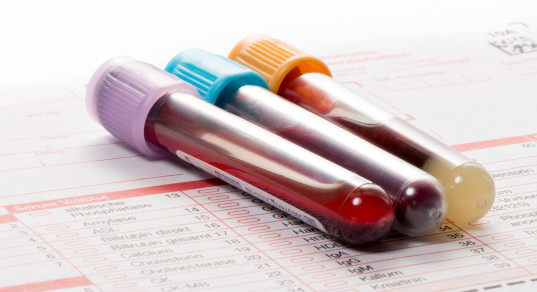
- Laboratory tests – blood and urine tests, biochemical tests of the blood, blood coagulation, determination of group and Rhesus affiliation, determination of infection status (HIV, hepatitis B and C, syphilis).
- General instruments - electrocardiography, x-rays of the lungs and heart if more than 6 months have passed since the last chest x-ray.
- Special instruments – X-rays and CT of the hand.
If there are concomitant diseases (venous diseases, stomach ulcers, etc.), the diagnostic program is expanded and consultation with a specialist is mandatory.
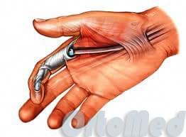
Anatomy of the flexor tendon
The flexor tendons are divided into superficial and deep tendons. The tendons of the superficial flexor tendons attach to the middle phalanges of the fingers, the tendons of the deep flexor tendons attach to the distal phalanges of the fingers (nail tendons). All tendons are located in channels in which they slide. When the muscles contract, the tendons pull on the corresponding phalanges and the fingers bend. These muscles are located on the forearm.
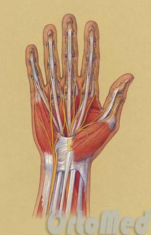
The tendons on the back of the hand and forearm are called flexor tendons.
The flexor tendons are held in their canals by the annular ligaments. This allows for smooth flexion without pulling on the skin.
Damaged tendons in the forearm, wrist, hand or finger are characterized by an inability to bend.
Tendons can be damaged very easily because they are very close to the surface of the skin. If a wound on the hand is relatively superficial, there is a high probability that the flexor tendons are damaged.
The tendons are under constant stress from the muscles to which they are attached. When a tendon is damaged, the contracted muscle pulls on its proximal end (which is closer to the forearm). The damaged ends spread far apart so that they can no longer fold up on their own.
It is very important to close the tendon ends within the first few days after injury, otherwise the damage to the tendon sheaths and the tendon itself is irreversible and a two-stage plication may be required, requiring four to six months of treatment.
Because the nerves and vessels in the hand and forearm are close to the tendons, they can be damaged by superficial trauma. Nerve damage results in numbness on one or both sides of the finger, but damage to both arteries of the finger has more serious consequences - severe ischemia of the finger (lack of blood supply), which can cause necrosis of the finger. Of course, this requires immediate surgery – revascularization of the finger (suturing the vessels).
Causes of flexor tendon injuries
The main cause of flexor tendon injuries is, of course, trauma.
This can occur through puncture wounds, saw blades, glass, etc., but closed injuries also occur.
But there are also closed injuries - tendon tears caused by excessive stress. Sudden lifting of a heavy object and sports injuries.
Rheumatoid arthritis, for example, can weaken flexor tendons and make them more susceptible to tears. This can happen without any obvious cause, without trauma - the patient may just notice that the finger no longer bends, but cannot remember how this might have happened.
Soft tissue diseases of the hand

pDamage to the finger flexor tendons or the annular ligament at the interphalangeal joints. Trigger finger syndrome arises from a knot in the flexor tendon (primary or secondary dystrophic inflammatory process), which prevents the tendon from sliding into the tendon sheath at the level of the articular surface and can completely block movement.
tThe patient complains of a clicking sound when bending and extending the finger. As the process progresses, the finger becomes stiff in flexion or extension. During examination, a lump may be felt on the palm or palmar surface of the finger at the base of the flexor tendon.
GA pea-sized or slightly larger nodule on the back of the hand or wrist in the projection of the extensor tendon sheaths. It is a degenerative bulging of the synovial sheath. Pain or discomfort occurs with finger movements. The contents of the ganglion consist of synovial fluid.
Exudative tenosynovitis of the extensor fingers
EExudative tendinitis of the upright fingers is often associated with inflammatory arthropathies of the hand. It manifests as limited swelling on the posterior surface of the wrist. The differential diagnosis is carpal and radial sulnar arthritis, which are also associated with swelling on the back of the hand. The patient is asked to flex and extend the wrist. In extensor tendonitis, the swelling shifts distally when the hand is stretched and repeats the movement of the tendon.
Stenosing tenosynovitis

- name of the fingers.
- Why are a teenager's toes crooked?.
- Why are the fingers curved?.
- Square soleus muscle.
- Short finger.
- ectrodactyly.
- The child has a shorter leg than the other.
- structure of the toes.