The ankle ligaments are the most commonly injured. Only a traumatologist can determine the extent of the injury after examining it using various diagnostic methods.
- 5 principles of sprain treatment
- First-class care: what modern diapers can do
- Swelling and other symptoms of a sprain
- Causes of Dislocations
- degree of dislocation
- How is a dislocation treated? MBST therapy as an innovative treatment method
- What is MBST?
- Symptoms of a dislocated foot
- diagnosis
- Causes of shoulder dislocations
- What are the symptoms of a shoulder dislocation?
- Rehabilitation after injury
- symptoms of a dislocation
- diagnosis
- What is a sprain?
- How do you distinguish dislocations from other joint injuries?
- Inexpensive Arnica Sprain Ointments
- treatment of sprains
- recovery period
5 principles of sprain treatment
- Swelling and other symptoms of a dislocation
- Rule 1: Diagnosis by a traumatologist
- Rule 2. First aid: rest and cold
- Rule 3. Immobilization of the injured area
- Rule 4: Medication to reduce pain and inflammation.
- Rule 5. Prevention of further sprains
A sprain is often accompanied by acute pain, swelling and stiffness of the muscle. Athletes most often suffer this type of injury when the joint is heavily loaded and the muscle is not warmed up enough. Therefore, warming up before exercising is an essential precaution against these types of injuries. However, sprains can also occur in everyday life – lifting weights, falling, and even sudden movements often damage muscles and ligaments.
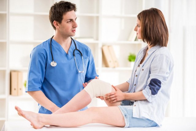
Like other injuries, dislocations require immediate attention. And the result of treatment depends on how correct it is.

First-class care: what modern diapers can do
Swelling and other symptoms of a sprain
A sprain can be recognized by a number of characteristic symptoms that appear immediately or shortly after the injury.
- Severe pain at the site of injury.
- Swelling or swelling of tissue (often around the joint).
- hematoma, redness.
- Restriction of mobility of the injured area.
Most dislocations occur in the muscles of the limbs, but improper lifting of heavy objects, sudden jerks, and other things can also damage the back and neck muscles.
Causes of Dislocations
The main cause of this type of injury is improper loading of the joint, forcing the ligamentous apparatus to perform movements that do not correspond to physiological amplitude. As a result, the nerve fibers and blood vessels that make up the ligaments become overstretched, twisted, and damaged.
The most common factors that lead to sprained ligaments in a child's legs, arms, and other body parts are:
- Sudden jumps, twists and turns;
- Unusual sprains of the joints;
- falls on the head, arms, or legs;
- severe blows to the side, e.g. B. in a collision;
- Sudden strong pressure on a joint;
- Sudden change of direction when walking quickly, running;
- Stumbling or getting your foot caught in a depression.
Sprains of the cervical spine in children are most commonly caused by jumping headfirst into a pool or open body of water from a height, or attempting somersaults or other acrobatic feats without proper preparation.
There are also a number of medical conditions that increase the likelihood of ligament injuries. This includes:
- hypodynamia, that is, the child spends most of his time in a sitting, lying position; lack of minimal physical training;
- Excessive body weight, which increases the stress on the joints;
- Uncomfortable, unsuitable footwear that does not adequately support the foot;
- high arch;
- Various degenerative joint diseases (arthritis, arthrosis);
- Congenital weakness of the musculoskeletal system;
- claudication due to the difference in length of the lower limbs;
- Disorders of the peripheral nervous system.
Children who are overly active and toddlers who try to exercise or dance without the supervision of a coach are at greater risk of dislocations and other injuries.
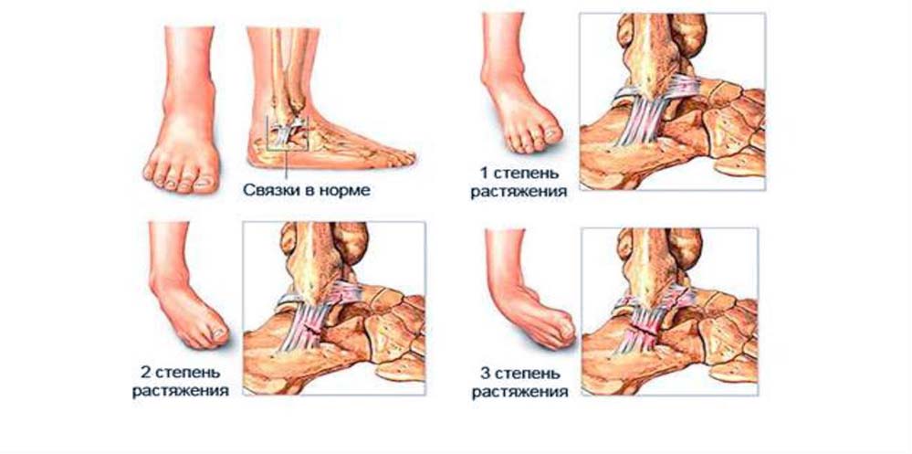
degree of dislocation
Experts distinguish between three degrees of dislocation injuries.
- Slight dislocation. Damage to a small number of fibers that keep the joint fully mobile. Pain is moderate, swelling is not pronounced, and full functionality is restored within 1-2 weeks.
- Moderate grade sprain. It affects at least half of the ligaments. It is characterized by acute pain lasting several days, moderate swelling and soft tissue edema. Motor skills are severely limited and attempting to move the injured area leads to increased pain. Recovery from this type of injury takes at least 5 weeks.
- Severe sprain. Complete tear, destruction of almost all fibers, accompanied by severe and unbearable pain, severe swelling, hematoma under the skin. The joint is completely immobile. With correct treatment, the recovery time is between 3 and 6 months.
If the treatment is wrong or untimely, it can take years before full mobility of the joint is restored.
How is a dislocation treated? MBST therapy as an innovative treatment method
MBST is a therapeutic MRI that is used in clinical practice in the form of several programs encoded on a chip card. These programs aim to restore the cartilage in the spine, hips, knees, shoulders, feet and hands. They are mainly used to treat osteoarthritis, rheumatoid arthritis and sports injuries. The dislocation treatment includes 7 or 12 sessions of 1 hour per day. During this time the patient lies on the device and 'absorbs' the energy without feeling uncomfortable.
There are special 3-day programs that are suitable for athletes after strenuous competitions and training sessions, as well as for the prevention of cartilage damage. In magnetic resonance therapy, the energy generated by our own hydrogen atoms is used for treatment. The treatment of sports injuries is carried out with the help of bioenergy, so its use does not entail any consequences for the body and the risk of developing complications. The treatment can be repeated without restriction.
What is MBST?
The principle of this therapeutic method is based on the fact that the metabolism in the tissues is regulated by electric and magnetic fields. While a healthy organ or system is in a state of regeneration, ie renewal, when it becomes dysfunctional it requires external pulsed stimulation to heal it. MBST therapy uses magnetic resonance treatment as such a stimulus, which sends signals to the diseased tissue and reproduces the signals from the healthy body system. By normalizing the metabolism, the regeneration process in the cells of cartilage, bone tissue, tendons, etc. can be resumed. In this way, the cause of the musculoskeletal disorder is eliminated instead of just relieving the patient's uncomfortable symptoms.
Based on careful research, physicists and engineers have managed to develop devices for non-surgical, highly effective treatment of sports injuries and various diseases of the musculoskeletal system. Scientific and clinical studies have fully confirmed the possibility of repairing cartilage and bone using this method, and the method itself has been patented under the name MBST. It is currently the first and so far the only therapeutic method that uses magnetic resonance imaging to treat chronic and osteoarticular diseases. It is officially approved for the treatment of osteoarthritis and osteoporosis and has been used for more than 15 years in Germany, Switzerland, Austria, Israel, Spain, the United Kingdom, Italy, Croatia, Romania, Slovenia, the Czech Republic and more recently in Turkey, Malaysia, the Philippines, the Netherlands, China and India.
Symptoms of a dislocated foot
Pain, limited mobility, swelling, and hematoma at the affected area that appear gradually are the main symptoms of a dislocated foot.
At first there is a stabbing pain that increases when trying to stand on the foot. Gradually, the intensity decreases, but after some time there is swelling, which squeezes the nerve endings and provokes a constant sensation of pain in the lower limb. When the swelling goes down, a large bruise forms that lasts for a week. The blood cells that have leaked from the damaged vessels slowly dissolve, causing the hematoma to turn yellow-greenish in color.
Symptoms depend on the degree of stretching:
- Mild. These injuries are characterized by partially torn fibers. The pain is present only at the time of injury and then gradually decreases in intensity. After 2-3 hours there is swelling in the injured area. If a hematoma develops, it is only mild. Pain is felt when trying to stand on the foot, but the affected person moves without inhibitions.
- Moderate. The grade is determined when at least 50 % of the fibers are broken. There is an acute pain syndrome, which intensifies when walking. The injured person is limping slightly and is able to walk a short distance unaided. After 5 hours, the injured foot swells, resulting in even more limited mobility. And after 1-2 days, a large hematoma forms at the site of the injury.
- serious. This condition is diagnosed when the fibers have completely detached from the bone or when most of the structure has torn. There is not only a sharp and stabbing pain, but also a characteristic crackling sound. The pain subsides and then worsens with swelling of the entire foot and lower ankle. The hematoma spreads to the sides of the foot and heel. Because of the pain and instability of the foot, the sufferer is unable to take a step.
diagnosis
There is no single guideline for diagnosing this injury. Traumatologists do not indicate when examining which ligament is damaged. X-rays are usually taken to rule out fractures or other structural damage to the bone.
The following methods are currently used to diagnose a ligament strain
- X-ray picture. Used primarily in trauma. The examination can be carried out in any medical institution. However, the soft tissues are difficult to see on the X-ray, so this method can only rule out fractures and bone injuries. Nowadays digital devices are used. Their main advantage is that they provide results in a short period of time. The images are printed out with a special printer and also stored on diskettes or memory cards.
- ultrasonography. Used for injuries of the musculoskeletal system. Ultrasound diagnosis relies on the use of a special device that emits sound or ultrasonic waves. These penetrate the tissue irregularly, whereupon a result (image) is generated from the reflected waves. However, the accuracy of ultrasound diagnosis is not very high. It depends on the diagnostician and the devices: most of the parameters have to be set. The advantages of ultrasound are its accessibility, the ability to assess the condition of tissues through movement and the low cost of the examination.
- COMPUTED TOMOGRAPHY (CT). In CT diagnostics, the X-rays penetrate different types of tissue and at different intensities. The bone structure is quite dense, so its tissue can be clearly seen in the pictures. Visualization of soft tissues is much worse, which is why traumatologists rarely use this method. Computed tomography has its advantages: it allows for quick diagnosis, which is important when diagnosing and examining children. Also, this method makes it possible to determine the effectiveness of any surgical intervention.
- MRI – used to visualize diseases of the musculoskeletal system. MRI makes it possible to more accurately identify the condition of soft tissues and determine the extent of damage to ligaments, bone marrow, tendons, cartilage and muscles. The recordings are made in all planes and adapted to the respective injury.
Causes of shoulder dislocations
- fall on shoulder ;
- mechanical injury to the shoulder from an impact;
- sudden jerky shoulder dislocation;
- a careless fall with an outstretched arm.
What are the symptoms of a shoulder dislocation?
Immediately after a shoulder injury, the following symptoms appear
- Acute pain at the site of injury;
- Increased discomfort when trying to move shoulder;
- limitation of shoulder mobility;
- slight swelling and swelling at the site of injury.
In addition, the body temperature can rise - this can indicate both a ligament injury and an incipient inflammation. If the person doesn't take action - doesn't see a doctor, continues to actively move the shoulder, the pain will increase and the swelling will get worse. All of these symptoms can indicate tendinitis. In this case, it is urgent to go to the nearest health center and get treatment from a specialist.
Even if the symptoms of a shoulder sprain are not so blatant that you rush to the doctor, complications can set in over a long period of time. Inflammation can develop at the site of the injury (tendonitis, bursitis), and shoulder osteoarthritis can sometimes develop. Any of these complications will prevent you from moving freely, exercising, and engaging in normal daily activities.
Further investigation is required to confirm the diagnosis. In most cases, magnetic resonance imaging (MRI), X-rays, and sometimes arthroscopy of the joint are done. During an arthroscopy, the doctor can simultaneously perform a microsurgical procedure in which partially or completely torn tendon fibers are brought together. X-rays of the shoulder joint should be taken to rule out fractures and broken bones, which require treatment entirely different from dislocations. It is important to have a comprehensive diagnosis carried out as early as possible - then the treatment measures taken will be as effective as possible.
Rehabilitation after injury
Ligaments in the shoulder joint can heal quite quickly after a dislocation. A lot depends on the individual's ability to regenerate, the extent of the injury and whether the patient follows all the recommendations of the specialist. Certain diseases such as diabetes, immunodeficiency, thyroid disease, etc. can delay the process of tissue regeneration and rebuilding. During rehabilitation, the body needs a lot of vitamins, proteins and micronutrients - proper nutrition, which covers all the body's needs during this period, is very helpful.
For first and second degree sprains, rehabilitation usually lasts no more than 7-10 days. In the case of a complete rupture of a ligament, the recovery time is of up to six months..
After the basic treatment, the trauma surgeon will recommend physical therapy and movement rehabilitation to the patient. The aim of rehabilitation is to restore the mobility of the joint, strengthen the ligaments and maintain their flexibility.
In the rehabilitation period after a shoulder injury, physical therapy is very important: inactivity causes connective tissue fibers to form in place of the damaged elastic tissue. This is scar tissue that cannot stretch like the ligaments should normally. Any training should be gentle, without sudden movements and with a gradual increase in load. Under no circumstances should you train when you are in pain, as this can lead to renewed damage to the ligaments of the joint. The first two or three times it is advisable to train under the supervision of a specialist: he or she will control the technique and avoid unnecessary loads.
After the rehabilitation period, certain preventive measures should be taken to avoid re-dislocations. This includes keeping your weight within limits, exercising regularly and eating sensibly.
symptoms of a dislocation
At the time of injury, there is severe pain. When a large portion of the fibers are torn, a loud popping sound can sometimes be heard. Swelling occurs, and in more severe injuries, hematoma and subcutaneous bleeding may develop. Pain occurs and increases rapidly when attempting to rotate the limb in the direction it was rotated at the time of injury. The degree of impairment in support and movement depends on the severity of the injury and ranges from mild limitation in mild sprains to inability in severe lacerations and complete tears.
Swelling is evident on examination. A mild sprain is associated with localized swelling around the ligament. With moderate injuries, the swelling spreads to the entire joint. In severe injuries, the swelling is extensive, extending beyond the joint to the distal portion of the limb. In moderate to severe injuries, hemorrhage and hematoma predominate.
Palpation of the injured area is painful, and a local increase in skin temperature is noted. There is no crepitus. In mild and moderate injuries, passive movements are limited due to pain, in severe injuries there is excessive mobility, which differs from the pathological mobility due to the fracture. In fractures, pathologic mobility occurs in the area of the fracture where it should not normally occur. With ligament injuries, movement occurs where it should (in the joint) but at a greater amplitude than normal.
diagnosis
Diagnosis is based on symptoms and, if possible, MRI or ultrasound of the joint, or arthroscopy. Because ligaments are soft tissue structures that cannot be seen on x-rays, x-rays can only be used to rule out a fracture, as fractures and strains have very similar symptoms and are sometimes linked. In addition, characteristic clinical signs are taken into account in the differential diagnosis from fractures.
Unlike fractures, dislocations do not cause pain when pressure is applied to the bone (outside the area of the damaged ligament). A cracking sound is heard at the time of injury, not a crunching sound of the bone. At rest, as a rule, there is no obvious pain syndrome that interferes with the patient's sleep and recovery. When palpating, there is no crunch, and the deformation is mainly due to swelling, and not to displacement of the fragments.
In contrast to fractures, in which damage to the ligaments of the joint can only be observed in isolated cases, a dislocation is always accompanied by a ruptured ligament or a sprain. Dislocations are also ruled out based on X-ray images and the absence of typical clinical signs. Unlike a sprain, a dislocation never involves acute and gross deformation of the joint, shortening of the limb, and springy resistance to passive attempts at movement.
What is a sprain?
Ligaments are strips of connective tissue that surround and reinforce a joint. Their ends are attached to the interacting bones. Thanks to the ligaments, the joint can only bend in a certain direction. Any external force applied to the joint in an unnatural direction can cause a strain; the first signs and first aid for them will be discussed below.
The connective tissue that blocks the joint is very strong and not elastic. It is unable to stretch. When subjected to tensile stress, the fibers of the ligament tear, which can range from a small fragment to a complete rupture of the ligament, depending on the severity of the injury. Therefore, the term 'ligament strain' does not exist in traumatology, but the term 'tear' is used.
In the event of an injury, in addition to the tearing of connective tissue fibers, blood vessels usually also tear, which leads to internal bleeding with the formation of hematomas and edema.
Below we will cover all aspects of the definition, causes, types, symptoms and first aid for this condition.
How do you distinguish dislocations from other joint injuries?
Although the principles of first aid for different types of dislocations are the same as for other injuries, it is advisable to assess the extent of the injury before consulting a trauma surgeon. This allows you to take additional measures and choose appropriate tactics to avoid complications.
The simplest type of injury is a bruise. There is a rupture of blood vessels and the formation of hematomas, but the joint structures, bones and ligaments remain intact. The symptom is post-injury pain, which quickly subsides and is felt only when pressure is applied to the injured area. A bruise is usually accompanied by swelling, but the mobility of the injured limb is not restricted.
A dislocation is caused by the displacement of the articular surfaces relative to each other. It can be recognized by an abnormal position of the bones, a change in the shape of the joint, and severe pain when trying to move it. First aid for sprains and strains should be provided to the injured person, taking care not to disturb the deformed joint. This should only be done by the trauma surgeon, who should be seen within the first 24 hours of the injury.
A fracture is usually characterized by severe pain with movement, numbness at rest, and swelling at the injured area. When pressure is applied to the injured area, a sharp pain is felt. For sprains and dislocations, the injured person should be placed in a splint and other life-saving measures immediately, and then taken to a medical facility.
The suspicion of sprains and torn ligaments can arise from the direction of the traumatic load. if e.g. For example, in a fall, the foot is severely twisted and the knee twisted in or out, a sprain is likely. Symptoms are a stabbing pain at rest and a stabbing pain with movement that only gets worse over time, and swelling may also occur.
Inexpensive Arnica Sprain Ointments
Inexpensive products based on arnica extract will help speed up the healing of bruises, sprains and other injuries. They eliminate inflammation and muscle pain. They have almost no side effects. When choosing the best ointment for an ankle sprain, you should choose this product.
The best ointments for muscle and ligament sprains with anti-inflammatory properties will relieve pain and reduce swelling. They penetrate directly into the injured area and quickly stop the inflammation. Even the best sprain ointments should be used with caution: overdosing is not advisable. The ointment should be applied to the skin 2-3 times a day for no more than a week.
Now you know what ointment is good for a sprain or bruise. Sprains in the ankle are often accompanied by swelling in the feet. We have compiled a list of effective measures you can take when your feet are swollen.
treatment of sprains
In the treatment of dislocated knee ligaments, several phases can be distinguished:
Immediately after the injury, the patient needs absolute rest. To prevent excessive movement of the leg, a tight bandage with elastic bandages is applied and the patient is placed on any firm surface - standing or walking is not allowed. The bandage should not be too tight - the patient should not experience numbness or pain in the extremity. The pulsation in the foot should be checked when the bandage is applied - it should be maintained and the foot should not be bluish. A cold compress should always be applied over the bandage - it should not be applied for more than 48 hours.
Painkillers are recommended to relieve discomfort. Non-steroidal anti-inflammatory drugs can be used: they not only relieve pain, but also prevent aseptic inflammation in the popliteal fossa.
If the pain and swelling worsen and body temperature rises, it is advisable to see a doctor immediately for further diagnosis. If synovitis is confirmed, surgical intervention is performed to remove the pathological fluid.
Even if the clinical symptoms have subsided after the dislocation, it is advisable to wear a special orthosis for 3-4 weeks. This is necessary to prevent re-injury of the knee ligaments as they take time to fully recover.
recovery period
The rehabilitation period includes a complex of measures aimed at improving tissue regeneration and restoring the full range of motion of the knee. Therapeutic exercises, physiotherapy and massage are used.
Chiropractic techniques help improve local blood circulation and speed up the regeneration of muscle structures and ligaments in the joint. After completing the main treatment, each patient is recommended to slowly train his leg with therapeutic exercises. It is recommended to do the first exercises in the presence of a medical professional who will monitor the correctness of the exercises. Therapeutic exercises should also not be carried out under pain: exercises are only indicated if there is no inflammation.
The duration of the rehabilitation period after a dislocation depends on the individual characteristics of the body and its ability to regenerate. Age, co-existing diseases (diabetes, immunodeficiency, connective tissue diseases, etc.) and the condition of the body play a role. On average, recovery takes anywhere from 2-3 weeks to six months.
Ligament strains in the posterior part of the knee joint are considered to be one of the most serious injuries in this area, as they have an unfavorable prognosis, especially if proper treatment is not provided. Inadequate treatment or failure to follow medical advice can lead to complications such as inflammation and degenerative/discopathic changes.
Read more:- Ankle ligament strain, ICD.
- Treatment of torn ligaments in the ankle.
- Injury to the ligaments of the ankle.
- Damaged ligaments of the ankle.
- How to distinguish a fracture from an ankle sprain.
- Rupture of the ligaments of the ankle.
- How do you treat a sprained ankle?.
- ligaments in the ankle.