Even for what appears to be a mild sprain, it is important to seek medical attention. Despite the obvious signs of a sprain, you shouldn't jump to conclusions, let alone treat the injury yourself. Self-treatment can lead to repeated sprains due to ankle instability. X-rays provide an accurate picture of the injury. The extent of the dislocation can be determined using x-rays and a visual examination.

- ligament strain
- Causes of sprains
- Description
- causes
- Severity of dislocation
- Symptoms of an ankle sprain
- How long should I wear an elastic bandage for a sprain?
- FEEDBACK FROM OUR PATIENTS
- Help at home for Achilles tendon sprain
- Possible risks
- What else you should do if you dislocate your ankle
- Supportive measures
- physical therapy
- Surgery for ankle sprains
- Bandage for the ankle
- Knee (or elbow) bandage
- MRI, ultrasound or CT for ankle ligament tears – which is better?
- Which is better: ultrasound, MRI or CT?
ligament strain
This type of injury is very common but does not require superficial treatment. Ligament strains of varying severity and location Injuries of varying severity and location pose a certain risk to the normal functioning of the injured part of the body and cause significant discomfort during the healing process, which you always want to heal quickly.
sprain of the ligaments – is an injury to the dense and elastic connective tissue (ligaments) that connect bones to one another. Although ligaments are the fibers that connect bones to bones, and tendons are the fibers between bones and muscles, ligament sprains are predominantly equated with tendon sprains.
contortions are usually caused by sudden movements that exceed the normal range of motion of the affected joint. Tendon strains are most commonly caused by twisting movements in a semi-flexed position. A partial tear of the ankle tendon is e.g. This is not uncommon, for example, in basketball and football players due to sudden rotational movements. In bodybuilding and powerlifting, partial tears occur with heavy lifting and rapid movements, as well as with an unprepared tendon apparatus.
Depending on the severity, each sprain is classified as a torn ligament:
- Microscopic elements, in this case it is a mild form of dislocation;
- Collagen fibers, in this case it is a moderate form of dislocation;
- The ligament has a high regenerative ability, so it will most likely heal with minimal medical intervention.
Ligaments have a high regenerative capacity, so they are most likely to heal with minimal medical intervention. Mild forms of sprains are completely unproblematic and do not require any special treatment.
The most common ligaments that become sprained are the ankle, knee, shoulder and finger ligaments. On average, treatment lasts between 3 and 5 weeks and full recovery can take up to 9 weeks. If tendons are involved in the injury, extensive evaluation and reconstructive procedures, and sometimes surgery, may be required.
Causes of sprains
causes contortions A sprain occurs when the deep connective tissue layers of the joint are put under so much strain that they break at their weakest point. Depending on your sporting preference or physical activity, the shoulder, lumbar, knee and ankle joints are at varying degrees of risk.
Some of the riskiest sports include. contortions are:
- Weight lifting - bending the barbell puts a lot of stress on the knee, which you should not do without first stretching, bandaging or relieving the area;
- Bench press - here you are at risk of tearing the lumbar ligaments, which can be confused with a sprain of the back muscle and therefore requires similar treatment;
- Bench press and shoulder press – if you overdo these exercises, you risk tearing ligaments in your shoulder;
- Push-ups – there is a high risk of injury to the elbow joint, which should be bandaged as a precaution.
- Pulling up on a bar - a state of hand inversion is created, which entails a high risk of injury to all parts of the forearm and wrist.
Repetitive movements from the above sports exercises and movements that a non-athlete performs daily are equally responsible for the risk of contortionsAn equally unpleasant, painful and restrictive risk of dislocation.
Description
'There is an effective and inexpensive remedy for joint pain. ' .
The ankle ligaments are very strong. Their task is to fix the joint of the shinbone (tibia, fibula) and ankle bone (talus) of the foot and at the same time keep them mobile.
It has a complex structure. The ankle bone is surrounded by the tibia bones like a fork. The tibia attaches to the upper articular surface and the fibula flanks the outer and inner ankle. This joint is quite stable and maintains its integrity due to the configuration and attachment of the joint tissue to the bones and allows various movements of the foot.
causes
A sprained ankle is one of those injuries that everyone has suffered at some point in their life and often tries to treat at home. The injury occurs when the elasticity of the connective tissue is exceeded and several fibers tear.
- Fall.
- Increased physical activity.
- Sudden rotation of the foot.
- Wearing uncomfortable shoes.
- Mechanical trauma to the foot.
- Frequent damage, microtears of ligaments and muscles due to falls, sharp turns, rotation of the foot;
- Unnatural movements in the joint;
- Constant stress on underdeveloped ligaments by athletes;
- overweight;
- connective tissue weakness (both congenital and acquired);
- flat feet;
- inflammatory and arthritic changes in the joint;
- Differences in length of the lower limbs;
- Diseases that block nerve and muscle conduction (myasthenia gravis, etc.).
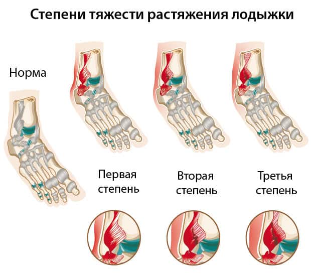
Severity of dislocation
Depending on the severity, sprains of the ankle joint are divided into:
- Mild (first degree) – there is a partial tear of the soft tissue at the ligament-muscle junction. The pain is mild and occurs when the joint is stressed or moved because its mobility is limited. The support function of the leg is not affected.
- Moderately severe (second) – a large number of fibers of the ligament are damaged. At the beginning there is acute pain, which usually subsides over time and can last for several days. Stepping on the foot is almost impossible. The mobility of the ankle joint is partially blocked by pain and severe swelling.
- Severe (third) – characterized by complete rupture of ligaments or tendons and persistent severe pain. The symptoms are similar to those of broken bones of the joint - it completely loses its mobility and support function.
Symptoms of an ankle sprain
After a minor injury, the pain may not appear until the next day. There may be slight swelling of the joint. Local bleeding may occur at the injury site. Supporting the leg is hampered by slight pain. The joint's mobility is only slightly restricted.
In more severe cases with severe pain syndrome, a specialist should be consulted immediately to determine the exact cause and avoid serious consequences from repeated trauma in the event of a rupture.
In a second or third degree sprain, the acute pain may be accompanied by a characteristic pop or click at the time of injury. This pain does not go away even when the injury rests. When you put pressure on the injured area or turn your foot, the pain increases sharply. A complete rupture of the ligament leads to rapid swelling and hematoma, as well as a local increase in temperature. The joint becomes abnormally mobile. Any movement is blocked by severe pain and a change in the relative position of parts of the joint. The leg partially or completely loses its support function.
How long should I wear an elastic bandage for a sprain?
The duration of wearing an elastic Intex bandage depends on the age of the patient and the severity of the injury.
For a first degree sprain (moderate swelling, tolerable pain upon exertion), the leg should be immobilized for 5-7 days. For a grade II or III sprain (severe swelling, severe pain upon exertion), treatment lasts 2 to 3 months.
Apply the elastic bandage only during active movement (for 1.5-2 hours).
Remove the bandage when sitting at a table or going to sleep. This helps To avoid blood congestion and swelling..
FEEDBACK FROM OUR PATIENTS
Thank you for helping me with my TMJ problems. Even the dentist noticed a difference after a few sessions! February 11, 2020.
The doctors at the clinic helped me return to my normal life after a car accident. It didn't seem like a big deal, but the injury I sustained, although it didn't require surgery, seriously ruined my life as I had headaches even in my sleep, I think. I'm glad to be able to see clairvoyance again. Thank you, Valentin Sklyarov August 28, 2019.
Wonderful place where doctors take care of patients. Excellent specialists in physiotherapy practice. Soulful trainers in the MTT room. They became a family within two months of treatment. Thank you for your help and recovery from a very difficult injury. Zoya Viktorovna July 22, 2019.
Many thanks to Sergey Viktorovna and Vera Petrovna for the fact that I can now walk without pain! What a blessing to live life to the fullest, move, and own my body! Health and happiness and good luck to the clinic! Igor Timofeevich July 17, 2019.
Vitaly Vladimirovich and Sergey Viktorovich literally got me back on my feet. I came to the clinic with a limp and a blank stare - my leg had been hurting for a month and a half. It turned out to be a large disc herniation in the lumbar spine (1 cm). After a visit to Sergiusz Wiktorowicz, I received an anti-inflammatory and pain-relieving drip and physiotherapy in the clinic on the same day. And I immediately wanted to continue living; there was an incentive to heal and move. During the treatment I got to know the doctors better, they are not only great professionals but also very interesting people. Thank you for all your help! Ivan Sergeyevich July 5, 2019.
Reputable clinic with competent doctors. They helped me get rid of the headaches I had suffered from for years. I always recommend this clinic to my friends and have never had to be ashamed. Review by yell.ru Michael V. July 4, 2019.
Hello. I am 55 years old and lead a sedentary lifestyle, until I was 50, sitting was very difficult and painful, pillows and belts did not help. When I was about 52, I started going to the doctor and going to different clinics. The pain temporarily subsided, but came back quite quickly. In the clinic of Dr. I finally got rid of Grigorenko. And unfortunately I didn't find it again until I was 54, with virtually no help for two years. The doctors are exceptional and competent, I recommend them. Bottom up! Sergius June 26, 2019.
Help at home for Achilles tendon sprain
What should a person who has suffered an Achilles tendon sprain do first? Of course, you should immediately consult an orthopedist who will confirm the diagnosis and make the appropriate recommendations. In the first few days, the most important therapeutic measure is to keep the ankle joint in an elevated position and cool it. The swelling of the ankle will go down and the pain will subside.
If a tendon sprain occurs, the injured person should lie down immediately and place something soft under their foot. A pressure bandage is placed on the injured area to relieve functional pressure. Elastic bandages can be applied to reduce swelling and limit movement of the leg. Once the pain has subsided, the leg must be lightly weight-bearing for several weeks. If the disease is severe, it is advisable to temporarily switch to crutches. During and shortly after rehabilitation, footwear should be carefully selected, avoiding high heels and uncomfortable, hard-soled shoes.
If bleeding occurs, it should be stopped and the wound washed and treated with an antiseptic. Cooling applications for tendon injuries are often found to be pleasant and pain-relieving by patients. Cold compresses or ice may be applied (for a maximum of 15 minutes with breaks of up to 20-30 minutes). After 3-4 days, you can remove the bandage before going to bed and take a warm salt foot bath. After that, the affected area should be gently dried and a light relaxing massage performed (only on the advice of a traumatologist).
Supportive pharmacotherapy in the form of painkillers can be helpful on an ad hoc basis. In the first few days, ointments can be rubbed in gently to promote blood circulation or to relieve pain. If the pain is severe, consultation with a specialist is required. Painkillers such as diclofenac or ibuprofen can provide significant relief for severe pain, but it is better not to overuse these medications as serious gastrointestinal side effects are possible.
Possible risks
If the pain in your leg worsens, if the swelling becomes more severe, if you develop hypersensitivity, and if your body temperature rises, you should see an orthopedist immediately! The appearance of these symptoms may indicate a complication.
- with tendon rupture;
- Calcification at the attachment point of the tendon on the heel bone;
- inflammation of surrounding tissue;
- reinjury due to negligence and misconduct;
- with the formation of a blood clot.
What else you should do if you dislocate your ankle
Supportive measures
It is better to take sick leave for the duration of the injury (remember the first rule of treatment - rest). You should not put any weight on your leg for at least the first two to three days after the injury. Later you can use crutches, a walker or a brace to stabilize the ankle. A severe sprain may require an orthopedic shoe to fully immobilize the joint during recovery.
physical therapy

Once the inflammation and pain have subsided and are no longer bothersome, mobility and strength of the tendon should be restored. If this step is neglected, repeated trauma can occur and the ankle joint remains less mobile overall.
Balance and stability training should form the basis. Start by standing on one leg for 30 seconds, then gradually add more complex exercises.
If you sprained your ankle while exercising, be sure to ask your doctor when you can return to normal exercise. You will probably need to avoid all physical activity for the next two to three months and replace it with physical therapy.
Surgery for ankle sprains
In rare cases, surgery is indicated for this type of injury - namely if the ligament does not heal after a complete healing process. The operation includes:
Bandage for the ankle

- Make two circles over the ankle to secure the bandage.
- Then pass the bandage diagonally across the top of the foot and make a double twist.
- Then wrap the bandage around the back of the foot on the other side and around the ankle again.
- Repeat the eight steps several times until the joint area is completely covered. Secure the bandage over the ankle.

- Place the bandage on the inside of the wrist and twist.
- Pull the bandage diagonally down through the outside of the palm between the thumb and forefinger. Return through the palm to the wrist.
- Twist your wrist, pull the bandage down through your palm and back toward your wrist.
- Repeat the eight steps several times and then proceed to place the bandage on the arm toward the elbow. Once you reach the elbow, start applying the bandage in the opposite direction. Secure the elastic bandage over the wrist with clips.
Knee (or elbow) bandage

- Relax the sore knee, bend it into a comfortable position.
- Place the bandage below the joint and make a double twist.
- Pull the bandage over the back of the leg above the knee joint and secure with a double twist.
- Repeat one rotation at a time with the previous position below and above the knee, gradually approaching the center of the joint. Secure the end with an elastic band.
- There is also a reverse version of bandaging: start with a double twist in the middle of the joint and then repeatedly apply the bandage one turn below and one turn above the knee.
- Place the bandage a third of its width above the previous layer.
- Tighten the bandage at the beginning of the bandage and loosen it slightly with each subsequent rotation.
- The maximum pressure should be applied to the narrowest point and the minimum pressure to the widest point.
- The bandage should be applied as gently as possible without creating wrinkles or gaps between the tissues.
MRI, ultrasound or CT for ankle ligament tears – which is better?
- Connective tissue fiber damage and micro tears up to a size of 5 mm
- contortion
- Post-traumatic lesions of the ligamentous system (hematomas, scarring, fibrosis)
- Tendonitis and other inflammatory processes in the ligament area
- Changes in the adjacent tissues vessels, veins, nerves, tendons.
Ultrasound of the ankle joint is most commonly used as the primary method for diagnosing torn ligaments in children. It allows for a quick x-ray of the soft tissues without having to completely immobilize the child and irradiate the body. If the ultrasound results are unclear or worrying, the doctor will recommend an MRI scan.
Computed tomography of the ankle is an expert method for diagnosing bone injuries. It allows a very accurate assessment of the condition of the bone tissue and is therefore used as an additional diagnostic method in traumatic bone injuries with ligament tears. Another advantage of CT examinations in trauma surgery is the short duration of the procedure – only 2-3 minutes. For this reason, CT is often used in emergency situations when the surgeon needs to act quickly.
Which is better: ultrasound, MRI or CT?
An experienced doctor should decide which method of examination is most suitable for you. The specialist doctor will prescribe a diagnostic method based on the patient's well-being, the symptoms of the disease, contraindications and the purpose of the diagnosis.
Radiation exposure is caused by computed tomography scans (MSCT) and X-rays. The radiation dose received during the examination is not a constant value. It depends on several factors: which area is being examined (the radiation dose varies with different protocols), the performance of the CT or X-ray machine. The average dose during a CT scan is between 2 and 15 mSv. The average dose for an X-ray examination is between 0.03 and 7 mSv.
Due to the negative effects of radiation on tissues and already existing pathological processes in the body, experts recommend undergoing a CT scan only on the recommendation of a doctor. Patients under the age of 18 can undergo a CT scan in St. Petersburg medical clinics only if they have a doctor's referral and are accompanied by a parent or legal guardian.
MRI and ultrasound are diagnostic methods that do not involve exposure to radiation. Patients can therefore take advantage of these examinations on their own initiative.
In medical centers in St. Petersburg, the most accessible diagnostic methods are ultrasound and x-rays. MRI and CT scans are specialized and expensive examinations. Therefore, doctors usually prescribe them as additional diagnostics if the first scans reveal serious pathology or if the patient needs further tests.
The most cost-effective tests are ultrasound and x-rays. CT and MRI are high-tech examinations and therefore significantly more expensive than ultrasound or X-rays.
Ultrasound is a harmless diagnostic method for which there are no contraindications. It is available to all patients regardless of their age. There are two main contraindications for MRI - the presence of metal and electronic devices in the patient's body. CT has the most serious contraindications due to radiation exposure. It cannot be performed on pregnant women and is not recommended for children under five years of age.
Read more:- How to distinguish a fracture from an ankle sprain.
- ankle sprain.
- Photo of the ankle.
- Ankle ligament sprain.
- ankle sprain.
- Ankle ligament strain, ICD.
- residual limb bandage.
- How do you treat a sprained ankle?.