Raising the arch of the foot is often accompanied by ataxia and gait disturbances. It is also characterized by mild sensory disturbances and pyramidal changes.
- Henny Rotman-Harmsen about different insoles for different foot problems
- saddle foot
- Causes of deformity
- Signs of deformity
- Symptoms of sickle cell anemia in children
- Who is at risk for sickle cell disease?
- The clinic today
- service
- Clinic services
- Acquired clubfoot: causes.
- Clubfoot: treatment
- diagnosis
- treatment methods
- Chromosomal diseases
- Gene mutations
- TBM is the market leader in its field
- We have been a successful supplier of components for windows, doors, insulating glass and furniture for more than 25 years.
- Harmful products
- Summary
Henny Rotman-Harmsen about different insoles for different foot problems
Cavus foot is a form of foot deformity in which the arch of the foot is enlarged. It is the opposite of flat foot and is more often acquired than congenital. It develops as a result of certain diseases of the muscular system or trauma, but can also occur in childhood and be hereditary. In addition to visual impairment, the condition is accompanied by pain and discomfort in daily life. In the initial stages, effective conservative treatment is possible, but in advanced cases, surgery is recommended.
The human foot consists of 26 bones connected by ligaments and muscles. The soft tissues provide strength, the ability to reconfigure under stress and redistribute pressure to specific areas.
The foot has two longitudinal and one transverse arches. In the sole projection, these begin and end at 3 main support points: the heads of the first and fifth metatarsals and the heel bone.
Danger!!!
An increase in the curvature of the longitudinal arch leads to an increase in its height and a lack of contact with the support of the midsole. Such a condition is called hollow foot. All support is in the toes and heel. The metatarsal is not stressed.
saddle foot
The most well-known arch of the foot is the longitudinal arch. It can be easily seen or felt by running your hand over it from the inside of the sole. An arch-shaped depression becomes visible. This is where the cushioning takes place - the foot springs back under load. When this arch is flattened, all shock inertia is transferred through the foot to the joints and spine.
Also read: Pain in the arch of the foot
The longitudinal arch begins at the heel bone and extends through the foot to the toes. It is higher on the inside than on the outside. Experts differentiate between five such arches depending on the number of metatarsal bones. They extend from the heel bone node to the toe joints. Their curved shape ensures flexibility when walking and cushions impacts. The highest arch is that of the second metatarsal, the lowest is that of the fifth. This area forms the outer edge of the foot and is the base of the foot when walking.
With normal development, the height of the arch of the foot at the inner edge should not be less than 35 mm. The angle of the arch of the foot can also be determined on an x-ray. It is formed by lines drawn from the tuberosity of the heel bone and the joint of the first toe to the lower edge of the wedge joint of the heel bone. Normally this angle should not exceed 130 degrees.

The transverse arch is located at the base of the toes and ensures the correct distribution of the load on the forefoot.
Causes of deformity
The deformity is usually hereditary or a symptom of a related pathology. However, the condition can be made worse by certain triggers which include the following
- Severe flat feet, leading to aggravation of the deformity;
- Valgus deformity of the 1st toe;
- Rheumatoid polyarthritis;
- Constantly wearing uncomfortable footwear or high-heeled shoes;
- Congenital anomalies of the toes that may become clinically apparent later in life;
- Down syndrome, Roberts syndrome, Feingold syndrome;
- Associated symptom of Fanconi anemia and fibrodysplasia.
Signs of deformity
When clinically examining the deformity, the wedge fingers are immediately noticeable, which are in an abnormal and irregular position. They are curved hammer-shaped or wedge-shaped. This is very uncomfortable as sores and blisters can form on the fingers. Another possibility is that the patient is unaware of the presence of the developing pathology. An experienced physician can then alert the patient to the presence of the deformity during a routine examination. It is therefore recommended not to neglect routine visits to the doctor and to take responsibility for your own health.
In most cases, the wedge deformity is hereditary. Sometimes this pathology is associated with syndromes such as Down syndrome, Roberts syndrome, Feingold syndrome and Fanconi anemia. Fibrodysplasia involves a curvature of the first fingers. The symptoms of the deformity are aggravated by wearing uncomfortable and tight footwear, which causes painful rubbing of the calluses.
Sometimes the patient does not complain and only finds out about his illness when he visits a doctor. However, it should be borne in mind that over the years the pathology can only worsen and lead to irreversible consequences. Timely treatment can help stabilize the deformity and restore normal function to the fingers.
The wedge toe deformity is associated with underdevelopment of the intermediate phalanges of the toes. Due to abnormal growth, the toe bones develop a curved C shape. Initially the disease may be subtle and peaks during puberty, during a major growth spurt. Therefore, it is usually difficult to predict the development of the deformity. In the first phase of pathology, constant monitoring of the patient is required to prevent progression of the deformity. This requires X-rays, compliance with all doctor's recommendations, active exercise therapy and massage courses.
Symptoms of sickle cell anemia in children
In infants, symptoms of sickle cell disease may not appear until 4-5 months of age. This is due to the presence of fetal hemoglobin type F in the first months of life.
Symptoms eventually appear when the fetal hemoglobin is replaced by abnormal hemoglobin and the red blood cells begin to take on a sickle shape.
Symptoms of sickle cell disease in children include:
- Jaundice – Yellowing of the skin and whites of the eyes caused by elevated bilirubin levels resulting from the breakdown or hemolysis of sickle cells.
- Anemia, which can lead to weakness and irritability in the child. The normal lifespan of red blood cells is 120 days, but sickle cells only live for 10-20 days, causing anemia.
- Dactylitis is painful swelling of the finger and toe joints caused by inflammation because the blood vessels are blocked by sickle cells.
- Painful orifices or recurring pain are characteristic symptoms of sickle cell anemia, which are due to blockage of the blood vessels.
- Frequent infections due to a non-functioning spleen as a result of sickle cell anemia.
- Vision problems associated with sickle cell disease, which clogs and damages the blood vessels in the retina.
- Growth retardation due to lack of blood cells.
The signs and symptoms of sickle cell disease vary and may change over time.
Who is at risk for sickle cell disease?
Incestuous marriages, i.e. relationships between closely related family members, are one of the most common risk factors for sickle cell disease. If a defective gene is present in the family, there is a greater risk of it being passed on to the next generation due to cousin crossing.
In addition, people of certain nationalities are at risk: Africans, Indians, Mediterranean peoples, people from the Middle East.
The clinic today
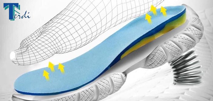
The Terdi Clinic is a center for functional diagnostics and correction of the foot. On the basis of pedographic examinations, individual therapeutic and corrective foot pads are carried out (based on the principles of Chinese acupressure massage, with the correct pressure on the foot, positive results can be achieved in various diseases).
In the Terdi Clinic, biomechanical parameters are determined and individual foot treatment is carried out using supinators, pronators and correctors.
Foot deformities cause dysfunction of certain organs.
service
Clinic services
Individual foot insoles are used for the following clinical pictures
- flat feet;
- feet with varicose veins;
- diabetic foot;
- pes cavus;
- sickle feet;
- scoliosis;
- venous insufficiency;
- heavy leg syndrome;
- lymphostasis;
- heel spur;
- Arthritis;
- overweight;
- Achilles tendon injuries;
- valgus deformity of the first toe;
- Various fasciitis;
- Professionally.
For professional and sporting activities:
Acquired clubfoot: causes.
- Neurological diseases in which the muscle supply is impaired.
- Poliomyelitis commonly caused clubfoot in the 1950s, but is not as common today due to vaccinations.
- A congenital 'open back' (neural tube defect) can lead to an undersupply of the lower leg muscles and thus to club foot.
- Injuries that sever the nerve of the lower leg muscles result in inadequate nutrition to the muscle. This leads to muscle spasms and the development of clubfoot.
- Circulation problems in the calf muscle artery (posterior tibial artery) also lead to inadequate supply to the muscle and thus to a club foot.
The inclination can usually be seen with the naked eye. To make a visual diagnosis, the doctor often makes a single one X-ray picture. This allows for a more accurate determination of the extent of the deformity.
Lately ultrasound examination increasingly used in the diagnosis of clubfoot. It is a quick and cost-effective method for detecting muscle tissue reserves.
Another examination method is Dynamic foot pressure measurement (Pedography) The measurement of the load pressure on the foot is carried out in a stable position. This method is also used in the production of orthopedic footwear.
In order to understand the causes of clubfoot, a comprehensive diagnosis must be made in every case, as the cause also dictates the treatment.
Clubfoot: treatment
For congenital clubfoot, treatment should begin immediately after birth. After birth, the body tissue is still very mobile, so the best results can be achieved. However, there are a number of less invasive (conservative) treatment options available prior to clubfoot surgery.
Newborns benefit the most Plaster therapy or taping. Babies wear a cast for long periods of time to keep their feet in the correct position. This measure may seem more intimidating to parents than it actually is. The cartilage and bone tissue is still very flexible and the child usually does not feel any pain. The soft bandage should be changed and reapplied every few days as the process is gradual.
On Bandaging First, the joints are mobilized daily through physical therapy. The affected foot is then fixed with special adhesive tapes. The resulting adjustments must be maintained. There are also special splints, orthopedic shoes or insoles that must be worn during the growth phase. Regular check-ups are also important.
Sometimes this non-invasive treatment for clubfoot does not have the desired effect or the clubfoot recurs over time. In such cases, surgical treatment with lengthening of the Achilles tendon should be considered. However, only 10 to 15 percent of children treated conservatively still need surgery.
diagnosis
The lateral displacement of brain structures is determined using echo-EEG (echo-encephalography). If the M-echo ultrasound signal reflected from the midbrain shifts by up to 4 mm, the displacement of the brain substance fragments is considered moderate. If the M-echo signal shifts up to 10 mm, the shift is severe, and above 10 mm it is critical. Other instrumental examination methods:
- MRI, CT SCAN. The most meaningful methods. Neuroimaging is able to assess individual dislocations and combinations of dislocations. Based on the symptoms - misalignment of the septum and the ventricular system (up to 4 mm - slight displacement, up to 9 mm - moderate displacement, from 10 mm - pronounced displacement), shape and degree of enlargement of the cysts - the doctor receives information about the displacement of the brain substance.
- Angiography. Deformation, compression and vascular displacement indicate a displaced brain fragment.
- Ultrasound examination.
During the physical examination, the doctor determines the neurological status of the patient (degree of impaired consciousness, presence of cerebral and focal symptoms). At the same time, pulse and blood pressure are determined and respiratory function is assessed.

treatment methods
Therapy is aimed at eliminating the cause of the disease - the underlying disease (tumor, hemorrhage, infectious lesion, mechanical injury to the skull bone). Treatment of cerebral dislocation syndrome is carried out by the doctor in a comprehensive manner, including conservative and surgical methods. If symptoms of brain dislocation are present, therapy is prescribed to remove excess fluid from the body (drainage drugs) and against the development of edema (anti-edema drugs).
The patient is immediately prepared for the operation. With glucocorticoid therapy, the patient's condition can be stabilized for a while (by reducing edema and lowering intracranial pressure values). If a life-threatening condition develops, emergency ventricular drainage and reclassification (artificial mechanical displacement correction) of the brain are performed.
With the lateral form of pathology, external decompression of the brain is performed, followed by repair of the dura. If the lesion is in the posterior cranial fossa, a decompressive cranial trephination is performed, possibly followed by an upper spinal laminectomy and dura mater dissection.
Chromosomal diseases
Pathological changes can arise both from the loss of genetic material (e.g. loss of a whole chromosome or part of it) and from the addition of new chromosomes. Clinically, they are characterized by numerous congenital defects. There are currently more than 1,000 known chromosomal abnormalities.
The exact causes are not fully understood. Scientists believe triggers include ionizing radiation, chemicals, viruses, certain medications taken during pregnancy, smoking, alcohol and the mother's age.
Chromosomal diseases can be accompanied by abnormalities:
A common feature of chromosomal diseases is the multifactorial nature of the damage. These include internal and external defects, craniofacial dysmorphia, delayed growth and development, mental and intellectual retardation compared to peers, and disorders of multiple body systems.
- cat cry syndrome (deletion in chromosome 5);
- Down syndrome (trisomy on the 21st chromosome);
- Patau syndrome (trisomy in chromosome 13);
- Edwards syndrome (trisomy on the 18th chromosome).
Gene mutations
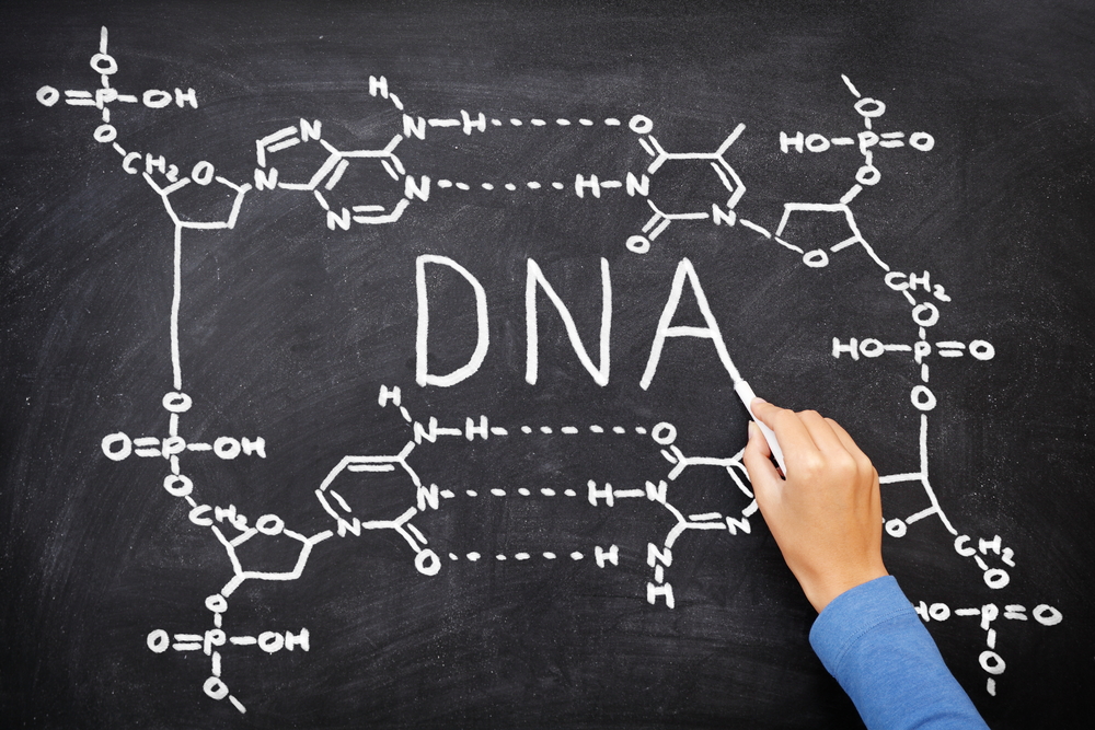
Gene mutations (point mutations) are changes in the chemical structure of a gene caused by substitution, deletion or insertion of a nucleotide. They occur more frequently than chromosome and genome mutations, but change the structure of DNA to a lesser extent. Gene mutations also include translocations (relocations), duplications (repeats) and inversions (180° reversals) of gene segments, but not of chromosomes.
Let's take the mutation GGT-CTc GGT → GTc-CTc GGT.
In the first triplet, thymine has been replaced by cytosine. The triplets GTT and GTZ code for glutamic acid, so this mutation did not result in a change in the protein structure: glu-gly-pro → glu-gly-pro.
However, in other cases, a nucleotide substitution can change the order of amino acids in the protein molecule and lead to phenotypic consequences.
In the first triplet, thymine is replaced by guanine. GTT encodes glutamic acid and GTG encodes histidine. The primary structure of the protein changes accordingly: glu-gly-pro → hys-gly-pro. There is a high probability of phenotypic changes.
TBM is the market leader in its field
For more than 25 years we are a successful supplier of components
for windows, doors, insulating glass and furniture.

More than 2,500 people work in our company who follow a lean production and procurement approach.

Our geography extends across the entire Russian territory. It is a network of branches in major cities such as Moscow, St. Petersburg, Novosibirsk, Yekaterinburg, Nizhny Novgorod, etc.
Today TBM is the largest supplier of high quality components and fittings for the production of windows, doors, double glazing and furniture. High-quality service, a wide range of over 18,000 items and favorable delivery conditions are the main advantages of working with our company.
By constantly analyzing the industry and researching the needs of our customers, we have been able to offer quality products that meet all market requirements for more than 25 years.
Working with us is a very beneficial, fruitful and open partnership.
Harmful products
It is not for nothing that endocrinologists talk about a balanced and adequate diet. According to WHO statistics, more than 670 million people worldwide suffer from various forms of hyperthyroidism and more than 1.7 billion people are at risk of endocrine diseases. Almost 70 % of them eat junk food every day. This is something to think about!
Which foods you should avoid if you have thyroid problems:
- Sugar. All popular and cheap foods contain sugar in dizzying amounts. These include sweet and carbonated drinks, cakes, pastries, candies and so on. These foods do more harm than good. Autoimmune diseases and diabetes are caused by uncontrolled consumption of sweets. Such a diet lowers hormone levels, overloads almost all systems and prevents the body from absorbing insulin.
- Fried and fatty foods contain harmful trans fats. If you like eating fried foods, try including more greens, fish and seaweed in your diet. If you have obvious endocrine problems, you should limit or even avoid eating fried foods because they reduce the production of hormones T3 and T4.
- Gluten is a substance (glue) found in grain plants and is extremely harmful to a healthy body. Foods high in gluten cause autoimmune diseases such as Addison's disease, type 1 diabetes, rheumatoid arthritis and hypothyroidism. Gluten is found in bread, bread made from wheat flour, etc.
- Semi-finished products, fast food, preservatives - all these foods cannot be called healthy for human health and the endocrine system.
Alcohol, coffee, cigarettes and strong tea should also be avoided as they interfere with the production of the hormones T3 and T4. A healthy person can afford to drink coffee or wine in moderation, but if the immune system is abnormal, such 'weakening' will lead to serious complications.
Summary
Good nutrition, exercise and a vitamin complex are the three pillars on which the health of the hormonal system rests. We have explained which foods you should include in your daily diet and which ones you should avoid.
Multivitamin complexes (e.g. Berokka plus) and even more drugs for treatment should only be purchased after consulting an endocrinologist. The specialist will also help choose a special diet and monitor the results of treatment or prevention.
THERE ARE CONTRAINDICATIONS AND A PHYSICIAN SHOULD BE CONSULTED PRIOR TO USE.
Read more:- Clubfoot in children therapeutic exercises 7 years old.
- Why does a child develop clubfoot?.
- Marfan syndrome in newborns.
- Photo of scraped feet.
- Congenital clubfoot.
- What is clubfoot?.
- clubfoot.
- baby splashing.