A lumbar S1 on MRI is not uncommon in people over 25 years of age with spinal pain. One or more transitional vertebrae form a zone of instability in the lumbosacral spine. This zone is a weak point where the sacral vertebrae undergo rotation of all lumbosacral under certain anatomical conditions and during twisting, rotation and tilting of the trunk (daily physical activities, especially asymmetrical sports such as tennis, hockey, golf, shot put, discus throw, high jump, etc.). Induce vortex. etc.) lead to rotation of all upper vertebrae, resulting in lateral curvature of the spine and corresponding muscle asymmetry and asymmetrical movement patterns. Twisted sacral vertebrae can damage the trunks of the sacral plexus. If the left leg is functional (more often due to valgus or hyperpronation), and even more so with bony shortening and pelvic lateral flexion to the left (usually in right-handed people), the sacral nerves on the left side can be affected: pain occurs in the thigh, on the outside of the hip , pain in the left hip joint, in the lower leg and even in the foot on the shortened side. Conversely, similar symptoms and complaints can also occur on the right side, with the leg on the right side being relatively shortened. Unfortunately, it is extremely rare that a radiologist, when describing an MRI of the lumbar spine, where he pays increased attention to the description of disc herniations and protrusions, pays enough attention to the diastasis of S1-S2 even in the presence of a disc (or traces of it). gives. Severe deforming spondylosis at the L5-S1-S2 level and degenerative-dystrophic changes in the sacroiliac joints can also be due to permanent recurrent rotation of the vertebral bodies and the articular surfaces of the transverse processes, as well as to permanent rotation-asymmetric damage to the ligamentous apparatus of the vertebrae at this level due to the different grades Note the lengths of the supports (legs).
- Legg-Calve-Perthes disease – diagnosis, treatment, causes and stages
- Causes of occurrence
- types of deformity
- diagnosis
- diagnosis
- Forms of lumbarization
- Conservative and surgical treatment of lower limb shortening
- This can be achieved by:
- Types of Surgical Treatment:
- Shortening of the limbs
- signs
- See also:
- ICD-10
- causes
- diagnosis
- differential diagnosis
- complications
- Treatment of achondroplasia
Legg-Calve-Perthes disease – diagnosis, treatment, causes and stages
Legg-Calve-Perthes disease (LPCP) – is a common pediatric orthopedic pathology of the hip joint, which belongs to the group of osteochondropathies. It is an aseptic osteonecrosis of the femoral head, which, if severe, leads to a functionally significant deformity of the proximal femur. Although the disease has been known in medicine for more than 100 years, the choice of surgical treatment tactics remains of scientific and clinical interest. In the vast majority of cases, the disease begins between the ages of 4 and 12, with the highest incidence occurring between ages 6 and 8. According to some Russian authors, the prevalence of the disease is 25-30 % in pediatric hip pathology and 0.17-1.9 % in the overall structure of orthopedic pathology. Foreign authors describe a wide prevalence of the disease depending on geographical location, ranging from 0.4 to 29 per 100,000 children. The disease occurs 4.5 times more often in boys than in girls, but is more severe in girls. In 8-24 % patients, the disease occurs bilaterally. Diagnosis and treatment of diseases of the hip joint is carried out in the special department No. 3 (hip pathology) of the Turner Scientific and Research Center for Children's Traumatology and Orthopedics of the Ministry of Health of the Russian Federation. The department specializes in hip pathology. Orthopedic traumatologists have developed effective therapeutic protocols for this pathology, which are regularly reviewed and improved, taking into account the latest scientific knowledge in this field.
Legg-Calve-Perthes disease – is an aseptic necrosis of the femoral head. In simple terms, it is a condition in which a partial or complete temporary loss of blood supply to the femoral head leads to infarction of bone tissue and subsequent sequestration, the size and location of which depends on the number of vessels temporarily deprived of blood.
Causes of occurrence
Although many studies have investigated the causes of impaired blood flow to the femoral head, none of the theories put forward so far are conclusive. There is no doubt that the disease is multifactorial and many predisposing factors occur simultaneously.
Predisposing factors can be congenital or acquired anomalies:
- Excessive stress (frequent microtraumas);
- Mutations in genes responsible for type II collagen synthesis, encoding factor V Leiden, increased secretion of leptin hormone, abnormal synthesis of insulin-like growth factor, excessive secretion of IL6;
- Underdevelopment of the lumbar spinal cord (myelodysplasia) resulting in impaired neuronal control of the hip;
- Hypoplasia and aplasia of arterial and venous vessels, shrinkage or narrowing of epiphyseal vessels;
- Inflammatory processes due to infections (strep throat, flu, acute respiratory infections);
- passive smoking;
- unfavorable environmental conditions;
Abnormalities of the hip joints.
Statistically, boys who weigh less than 2 kg at birth are five times more likely to develop Legg-Calve-Perthes disease than infants weighing more than 3.5 kg.
types of deformity
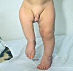
Shortening of the limbs. Depending on the location of the deformity, varying degrees of impairment of the limbs are observed. The upper limb retains its function with a shortening of up to 8 cm. The lower limb loses function with a shortening of 3 cm or more. When the lower limb is shortened, secondary deformities of the healthy limb and spine also occur.
curvature of the limbs. Can be valgus (X-shaped) or varus (O-shaped). Sometimes it is combined with shortening. The unilateral process is more often a consequence of fractures, the bilateral one is congenital.
diagnosis
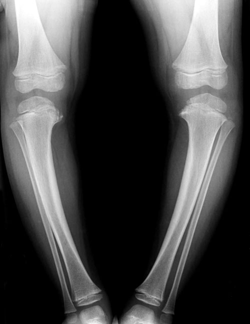
The main method for diagnosing long bone deformities is clinical examination. The type and extent of the deformity are determined and the length and angle of the deformity are measured. In addition, x-rays are used for more precise measurements. X-rays are used to determine the difference between the diseased and healthy limb, the mechanical axis (through the centers of the joints) and the anatomical axis (through the bone itself) in order to more accurately determine the tactics of treatment.
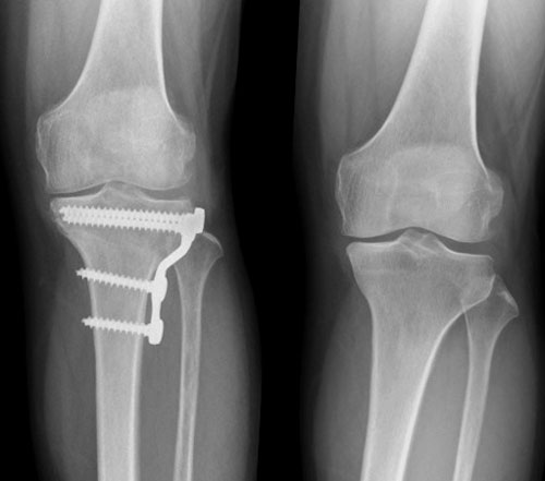
The most important principle in the treatment of long bone deformities is the restoration of the mechanical and anatomical axis of the limb. In our clinic, osteotomy and external fixation (Ilizarov) are the main methods of surgical correction of deformities.
During osteotomy, an artificial fracture of the bone at the tip of the deformity is performed intraoperatively and then fused in the correct position. This fracture can be fixed with a plate or external fixation. When fixing the plate, the skin incision is 8-10 cm. The rehabilitation period is 6-12 weeks and includes completely avoiding physical activity and walking on crutches. The metal structure is removed after 1-2 years.
The osteotomy with subsequent fixation in an external fixation device is minimally invasive. The cuts in the skin are minimal, up to 2-3 cm. The external fixator allows you to return to your normal lifestyle the day after surgery, with the exception of heavy physical work and sports. However, you must wear the splint for 1-6 months depending on the degree of deformity and carefully follow the doctor's instructions. Once the splint is removed, limb function is fully restored within 6 months.
diagnosis
To clarify the severity and nature of the pathology, the doctor measures the length of the patient's lower limbs, as well as each segment, focusing on the bony protrusions (ankles, knees, etc.). X-rays are also recommended.
Lower limb shortening is treated by specialists such as podiatrists and traumatologists. In the case of a slight shortening, conservative correction (wearing special shoes and orthoses) is indicated.
If the pathology is due to a dislocation or subluxation of the hip, treatment consists of splinting, exercises and massage. In some cases, the dislocation is corrected through surgery.
A major dislocation requires surgery. The most common method of orthopedic foot correction is the use of an Ilizarov brace.
Legs of unequal length are a serious disorder and, if not treated properly, can cause serious joint and spine problems.
- Open daily from 09:00 to 22:00
- All diagnosis rooms are open daily from 08:00 to 22:00
- Trauma and treatment rooms open 24 hours a day
- Open daily from 09:00 to 22:00
- All diagnostic practices open daily from 08:00 to 22:00
- Trauma and treatment rooms open 24 hours a day
- Open daily from 09:00 to 22:00
- All diagnosis rooms are open daily from 08:00 to 22:00.
- Trauma and treatment rooms are open 24 hours a day
- Open daily from 8:00 a.m. to 10:00 p.m
- All diagnosis rooms are open daily from 08:00 to 22:00
- Trauma and treatment rooms open daily from 08:00 to 22:00
- Open daily from 09:00 to 22:00
- All diagnosis rooms are open daily from 08:00 to 22:00
- Trauma and treatment rooms are open 24 hours a day
Forms of lumbarization
A symptom of dysplasia, lumbarization of S1-S2 and a minimal shortening of the leg by 3 mm (often the left leg) is enough to initiate the development of a lateral curvature of the spine. The severity of spinal pain often depends on the extent of the spinal deformity and the rotation of the pelvic ring in response to the shortening of one of the legs. Pain syndrome and ligamentous asymmetry of the lumbosacral region and pelvis are observed in adolescents who play a lot of sports (physiological lumbarization) or get older (transition of physiological lumbarization with shortening of one of the lower limbs to pathological asymmetry after the 23rd-25th day).
If shortening of the lower limbs is not prevented early and adequately by consistent, gradual and careful selection of the instep and dynamic height up to the age of 20-25 years, observation, application of derotation exercises, careful and persistent training of the muscles of the arch of the foot and lower limbs compensated, this pathological situation can develop into permanent changes in almost the entire musculoskeletal system.
Conservative and surgical treatment of lower limb shortening
An important step in treatment is early diagnosis, which allows possible correction of the length and deformity as well as the prevention of subsequent problems. Conservative, supportive methods are used to correct the shortening.
Treatment consists of different types of surgical procedures suitable for each age group. Additional conservative treatment is required to mechanically compensate for the shortening.
This can be achieved by:
- Compensation for shortening with an additional insert or knurling above the sole - acceptable for shortenings of up to 3 cm;
- orthopedic custom-made shoes – with length differences of 3 to 6 cm;
- Double-track walking aids – for leg length differences of more than 6 cm.
In children, topical application of paraffin and electrophoresis to stimulate blood flow to the affected limb is possible, although the effectiveness of these techniques has not been proven. With this treatment, it is important that the orthopedist carries out regular checks throughout the entire period of deterioration.
With a small difference in the length of the limbs, up to 2 cm, it is not necessary to significantly reduce the regime if the child benefits from compensation. Surgical treatment is mandatory when the lower limb is shortened by more than 4 centimeters and is recommended when it is shortened by more than 2 centimeters in children with continuous active growth.
Types of Surgical Treatment:
For congenital hip shortenings and femoral neck deformities, multi-stage treatment is performed to restore the shape and function of the hip joint and correct the length, which may be necessary one to three times during growth.
Lengthening of the limb in the external fixation device is performed by 1 mm daily to adjust the neurovascular bundle and proper callus formation at the bone junction.
Shortening of the limbs

Limb shortening is a pathological condition in which healthy body proportions are disrupted. Minor deviations from the norm do not need to be corrected. Severe deviations must be treated as they can lead to joint and spinal damage.
signs
Shortening of the upper limbs can be a nuisance in daily life, but in most cases it is a cosmetic defect. If a lower limb is shortened, it must be corrected as it can lead to pelvic tilt, joint contractures and spinal deformities (compensatory changes).
Differences in limb length can be detected through visual inspection, body measurements, and x-rays.
Different limb lengths can be compensated for in two ways:
There are two types of balancing operations. These consist of lengthening the shortened limb or shortening the healthy limb. The treatment strategy is determined on a case-by-case basis.
The Ilizarov apparatus is the most commonly used method of lengthening a shortened limb.
The orthopedic traumatologists at the Medicare Clinic in Khabarovsk treat their patients with modern, precise equipment, which minimizes postoperative complications and promotes rapid rehabilitation.
See also:
Please consult our specialists for any contraindications.
The information contained on this website does not constitute a public offer.
Make an appointment, consult all your questions
ICD-10
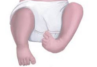
Lower limb malformations are a large group of abnormalities that occur in utero and have a wide range of severity. In the practice of orthopedics and traumatology, they are relatively common and account for 55 % of all congenital malformations of the musculoskeletal system. Gross malformations are diagnosed immediately after birth. Minor abnormalities may be asymptomatic or nearly asymptomatic and may be discovered incidentally during examination for other injuries and illnesses. Congenital anomalies of the lower limbs are treated by pediatric orthopedists and traumatologists.
causes
Congenital anomalies can be caused by both mutations and external or internal teratogenic factors. The most common external factors that negatively affect the formation of the limbs are infectious diseases, nutritional disorders, multiple medications and radiation exposure. Internal factors that can lead to abnormal limb formation include advanced maternal age, uterine diseases, severe somatic diseases, certain gynecological diseases and endocrine disorders.
Developmental disorders that can be traced back to malformations:
- Amelia – Complete absence of one of the limbs. Possibly Apus (both lower limbs missing) and Monopus (one lower limb missing).
- Phocomelia Or a limb resembling phocomelia. The middle and/or proximal limbs are missing along with their corresponding joints. It can be bilateral or unilateral. Sometimes all limbs are affected, both lower and upper. Complete phocomelia – the tibia and femur are absent, and the foot is attached to the trunk. Distal phocomelia – the tibia is missing and the foot is attached to the thigh. Proximal phocomelia – the femur is missing and the tibia and foot are attached to the trunk.
- Peromelia – A form of phocomelia in which part of the limb is missing and the distal part (foot) is poorly developed. Complete peromelia – the limb is missing and there is a skin bulge or reddish toe where it is attached. Incomplete peromelia - the femur is missing or underdeveloped, and the limb also ends in a skin protrusion or chamois toe.
In addition, there is the absence (aplasia) of the fibula or tibia, the absence of phalanxes of the toes (aphalangia), the absence of toes (adactyly), the presence of a single toe (monodactyly), as well as typical and atypical forms of cleft foot characterized by the absence or the underdevelopment of the metatarsal bones.
diagnosis
II. DIAGNOSIS AND TREATMENT METHODS, APPROACHES AND PROCEDURES
Minimum list of examinations to be carried out upon referral for elective hospitalization:
- coagulation picture (bleeding and clotting time, prothrombin, fibrinogen, platelet adhesion and aggregation reaction, antithrombin);
- blood chemistry (total protein, alanine aminotransferase, total cholesterol, bilirubin, direct bilirubin, creatinine, urea, glucose, potassium, sodium, phosphorus, calcium, chlorine);
– Coagulation values (bleeding time and clotting time, prothrombin, fibrinogen, platelet adhesion and aggregation, antithrombin);
- blood chemistry (total protein, alanine aminotransferase, total cholesterol, bilirubin, direct bilirubin, creatinine, urea, glucose, potassium, sodium, phosphorus, calcium, chlorine);
complaintsDeformity and shortening of the upper limbs, resulting in functional impairment and a cosmetic defect that affects the patient's psychological well-being.
Medical history: The disease manifests itself from birth, with increasing deformity and shortening of the limb segment with age.
– Underdevelopment of the bones of the forearm (especially the radial bone); abnormal development of the fingers and hand.
– Other changes in the hand include hypoplasia and clinodactyly of the second finger, syndactyly, flexion and extension contractures in the metacarpophalangeal and interfinger joints, especially in the second and third fingers. The carpal bones on the radial side are also affected, with aplasia or concretion with other bones.
– X-ray of the upper limbs with the adjacent joints (picture of the shortening and deformation of the forearm bones).
– CT examination, depending on the degree of limb abnormality and the type of defect, attention is paid to hypoplasia/aplasia of the bony structures of the upper limbs and their deformation.
– Consultation with an oncologist (if bone mass is present to rule out malignant disease);
differential diagnosis
Diagnosis of congenital clubfoot is not difficult due to the visually detectable deformities and obvious limb dysfunction.
• First-generation cephalosporinsSingle dose: Cefazolin, 50-100 mg/kg, intravenously, once 30-60 minutes before surgery.
Intravenous drip doses 10-20 mg/kg/day, single or multiple infusions for severe infections and in children from 1 month of age;
Vancomycin: 15 mg/kg/day, not more than 2 g/day, in 4 intravenous doses, each dose administered over at least 60 minutes.
• paracetamolParacetamol, 200 mg, tablets – at the rate of 60 mg per 1 kg of the child's body weight, 3-4 times a day. The interval between doses should be at least 4 hours. The maximum daily dose is 1.5 g to 2.0 g;
Paracetamol rectal suppositories 125, 250 mg – a single dose is 10-15 mg/kg of the child's body weight, 2-3 times a day, after 4-6 hours;
Paracetamol suspension 120 mg/5 ml, for oral administration – a single dose of the drug is 10-15 mg/kg of the child's body weight, 4 times a day, with an interval of at least 4 hours between consecutive doses (the dose for Children aged 1 to 3 months are determined individually).
Paracetamol syrup for oral administration 2,4% 50 ml - children from 3 to 12 months - ½-1 teaspoon (60-120 mg); from 1 to 6 years – 1-2 teaspoons (120-240 mg); from 6 to 14 years – 2-3 teaspoons (240-360 mg), 2-3 times a day.
The maximum duration of treatment with paracetamol as an analgesic is no more than 3 days.
• Ibuprofen Suspension 100 mg/5ml – 200 ml, for oral administration, 7-10 mg/kg body weight, maximum daily dose – 30 mg/kg. The interval between doses should be at least 6 hours. Duration of treatment no longer than 5 days, as an analgesic.
Children 1 to 14 years of age: 1 mg/kg to 2 mg/kg body weight intravenously, intramuscularly or subcutaneously. Intravenous injections should be given very slowly or diluted in an infusion solution and given as an infusion. The dose can be repeated at intervals of 4-6 hours.
complications
Patients with achondroplasia are prone to obesity. Due to the narrowing of the nasal passages, otitis media and conductive hearing loss are common. Upper airway obstruction can lead to symptoms of respiratory failure. Spinal canal stenosis is quite common in achondroplasia. It most commonly occurs in the lumbar spine, less often in the cervical or thoracic spine. Sensory disturbances, paresthesias and leg pain may occur. In severe cases, dysfunction of the pelvic organs, paresis and paralysis may occur.
The diagnosis of achondroplasia is made by a pediatric orthopedist and is not difficult due to the patient's characteristic appearance and body proportions. All children are examined in detail to determine the degree of deviation from normal skeletal development, and the data is recorded in a table. This table is periodically supplemented as the child grows and the data is compared with a standard table created specifically for patients with achondroplasia.
A comprehensive examination is carried out to assess the condition of various organs and systems and various specialists are involved. Newborns with achondroplasia are examined by a neurosurgeon to rule out hydrocephalus. If hydrocephalus is suspected, a brain MRI or a more accessible CT scan is recommended. Patients with achondroplasia are referred to an otolaryngologist to evaluate nasal and otolaryngological patency. A consultation with a pulmonologist may also be necessary.
X-ray examination of the skull reveals an imbalance between the facial and brain regions. The occipital opening is reduced, the lower jaw and the cranial bones are enlarged. The Turkish saddle has a characteristic boot shape and a flat, elongated base. The chest x-ray image in achondroplasia is usually unchanged, in some cases the sternum is protruding and slightly curved. The ribs may be thickened and deformed at the transition to the cartilaginous arches. Sometimes the normal anatomical curvature of the clavicles is not present.
Treatment of achondroplasia
Patients cannot yet be completely cured by modern orthopedics. Growth hormone treatment has been tried, but there is no reliable evidence that this technique is effective for achondroplasia. Conservative treatment is carried out at a young age to strengthen muscles and prevent limb deformities. Patients with achondroplasia are prescribed gymnastic exercises and massage, they are recommended to wear special orthopedic shoes, etc. Preventive measures against obesity are taken.
Surgical intervention for achondroplasia is indicated for severe malformations of the limbs and narrowing of the spinal canal. An osteotomy is used to correct the deformity and a laminectomy is performed to eliminate the narrowing of the spinal canal. In some cases, enlargement surgery is also performed. In achondroplasia, lengthening of the limbs is usually done laterally in two steps: first, the femur is lengthened on one side and the tibia on the other side, and then the remaining segments are operated on.
Read more:- How to determine leg shortening.
- Anatomy of the ligaments of the lower limbs.
- shortening of the hip.
- Muscles and fascia of the lower limbs.
- The muscles of the lower limbs are.
- Structure of the human lower limbs with captions.
- Manufacture of lower limb prostheses.
- Main joints of the lower limbs table.