Hip contusions are different from neck fractures. Contusions, unlike fractures, do not lead to forced alignment and shortening of the limb, and the heel strike symptom is negative (the patient can lift the heel off the floor while lying on his back). When palpating the bony structures and palpating the greater trochanter, there is no severe pain, with the greatest pain occurring in the soft tissue area. X-ray examination of the hip joint is normal. After the CT scan, the bony structures are not changed.
- fracture of the tibia
- Causes of knee injuries
- Symptoms of a knee injury
- Example 5.
- Example 6.
- symptoms
- hip bruise
- hip bruise
- complications
- Types of injuries to the shin
- fractures of the tibia
- Osteoporosis of the tibia
- Osgood-Schlatter disease
- Paget's disease of the bone
- How shin splint trauma is diagnosed
- types of injuries.
- Characteristic Symptoms.
- exercise sets
- Go
- Exercise therapy after a leg fracture
- How to do the exercises
- Treatment of bone cysts
- Periost injury (traumatic periostitis): causes, symptoms, diagnosis, treatment
- Diagnosis of periosteal injuries
fracture of the tibia
Fractures of the tibia are most commonly caused by a direct trauma mechanism, although cases involving indirect trauma are not excluded. Direct impacts and twisting of the shin when the foot is immobile are common causes of injury.
Road traffic accidents, limb crushing from moving machinery and mechanical structures, and falls from heights are considered the leading causes of PTSD.
The effects of different types of trauma, e.g. B. in the household, on the street, at school, in sports and in unorganized leisure activities are not excluded.
Extensive dislocations with muscle interposition and angulation are common. Presence of concomitant lesions, e.g. B. Fibula, usually patent, comminuted PBB requiring surgical anatomical and functional rehabilitation.
Based on the pattern of multiple human skeletal injuries, the most serious injuries are PBBB injuries. 50 % of these injuries are associated with post-traumatic shock.
The clinical manifestations of PBC injuries depend on the type of insult and the location of the TDC by traction on the quadriceps muscle of the thigh.
Palpation along the BBK ridge shows a stepped deformation of the affected bone. The pain occurs until the onset of traumatic shock and increases with axial loading.
The location of the TDC is visually visible through the tension in the overlying skin. After 1-2 hours there is swelling of the lower leg and a hematoma in this area.
Palpation usually reveals the nature and location of the fracture plane. In some cases, a cracking of the bone can be heard during the manipulation.
In cases where soft tissue is trapped between bone fragments in the PBC region, a funnel-shaped depression of the skin over the fracture, an umbilical sign, is observed.
If symptoms of PBC are found, the patient should be taken to the hospital and an appointment made with a trauma surgeon for evaluation and specialist care.
Causes of knee injuries
- direct mechanism of injury - fall on the knee, impact in a fight, sports training
- indirect – sudden twisting of the trunk while the foot is standing.
The severity of a knee bruise depends on the nature of the modifying factor:
With minor injuries, there is practically no capillary disruption. The swelling is caused by a reflex contraction and subsequent dilation of the vessels, which leads to congestion fullness. The fluid is deposited in the surrounding tissues and leaks out of the vessel wall.
With a severe contusion of the knee, massive bleeding occurs, the soft tissue structures are saturated with blood, and hemarthrosis is noticeable by its entry into the joint cavity. The pooled blood is called a hematoma. Such lesions lead to disruption of blood and lymphatic circulation and contribute to foci of ischemia and necrosis at the site of impact.
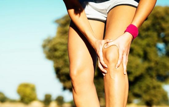
Symptoms of a knee injury
- Pain that worsens with palpation, flexing, or stretching the lower extremity
- numbness in the leg
- swelling
- Bleeding - if it is minor, it may not occur or it may not occur for several hours after the injury
- Localized fever in the joint area
- Moderate functional impairment, no deformity - patient can stand and walk, a limp is typical.
The formation of hemarthrosis and hematoma in the case of a deep contusion is indicated by swelling, which means that intra-articular bleeding is still present.
Example 5.
Patient R., 41 years old, injury resulting from a fall on ice skates. He is admitted to the emergency room of the hospital.
On examination 4 hours after admission, locking rod metal osteosynthesis was performed.
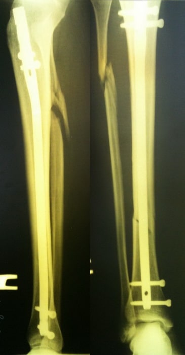
The postoperative period was uneventful. The patient was mobilized on day 5 and discharged from the hospital.
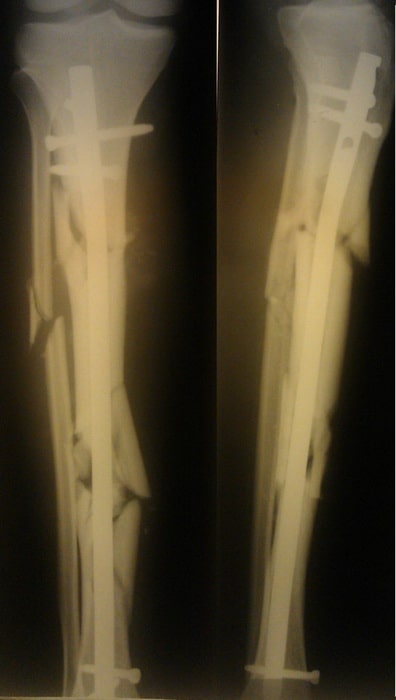
Example 6.
Patient C. Patient C. was 50 years old. He was injured in a car accident (passenger). A fragmentary fracture of the tibia was diagnosed.
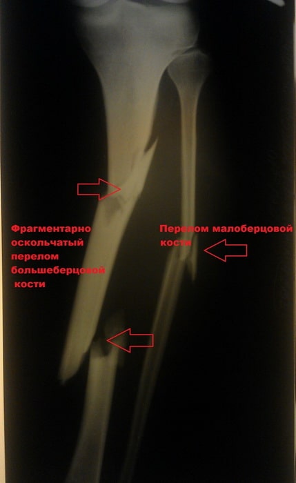
A closed reduction and internal fixation of the tibia with a locking rod was performed.
symptoms
Pain, swelling and reduced function of the limbs are common symptoms of a contusion. The pain is acute at the time of injury, subsides within a few hours, and reappears after a few days with severe swelling. The severity of the swelling varies greatly. Severe swelling usually occurs with contusions with a lot of muscle mass.
With minor bruises, the swelling is minor and subsides within 3-4 days, while with severe bruises, it is widespread and increases within 2-3 days. The swelling can be accompanied by a change in skin color and the formation of subcutaneous and intradermal hemorrhages. The injured area first turns purple and then blue. The functional impairment is usually moderate.
hip bruise
Resulting from a sideways fall or direct impact. The soft tissues around the greater trochanter are most frequently injured; other bony prominences such as the ischial tuberosity, iliac crest or pubic ramus are injured less frequently. This is accompanied by varying degrees of pain, localized swelling and restricted movement, from mild stiffness from fear of increasing pain to limping. The hold is retained. Bleeding in the injured area is common. Hematomas are rare.
hip bruise
Hip bruises are more commonly caused by direct impact with a heavy object. Sports falls, car crashes, and other high-energy impacts can also cause injury. The thighs contain large, massive, and well-perfused muscles, which tends to squeeze some of the peculiarities of this anatomical area. Such leg injuries are accompanied by significant swelling, extensive bleeding and the formation of hematomas. Bruises can be both under the skin and in the muscles.
The patient complains of discomfort and pain. With minor injuries, the pain syndrome is local, with severe injuries, the pain is diffuse and spreads along the entire thigh (front, back or side), with the pain being strongest at the point of impact. The thigh is enlarged and bruising may be visible on the skin. Movement is restricted, support is usually maintained, and there is a limp. Soft tissue palpation is painful, crepitation is absent, and the axial loading sign is negative.
complications
Minor bruises usually do not lead to complications. Serious injuries to the leg can result in hematomas and soft tissue detachments. Major joint injuries, particularly of the knee, are often complicated by hemarthrosis, which sometimes progresses to synovitis. Muscle and tendon tears are rare and require surgical treatment.
In the long term, muscle fibrosis and ossifying myositis can occur after severe injury. Muscle fibrosis is caused by muscle fall syndrome and subsequent replacement of muscle fibers with connective tissue. In ossifying myositis, small ossifications - ossicles - form in the thickness of the muscle. The quadriceps muscle is often affected.
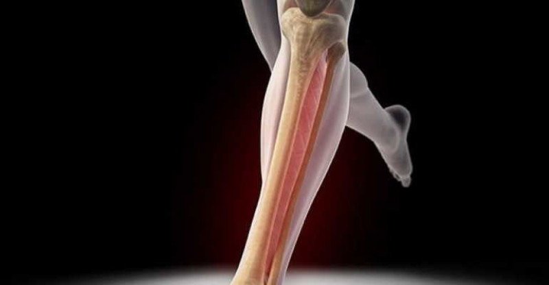
Types of injuries to the shin
The most common types of tibia injuries include fractures, osteoporosis, Osgood-Schlatter disease and Paget's disease.
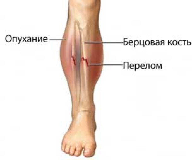
fractures of the tibia
Shin fractures are breaks in the integrity of bone tissue. Symptoms of the injury include shinbone pain, shinbone swelling, tenderness, inability to move the leg normally, bruising, deformity, or bulging.
Osteoporosis of the tibia
Osteoporosis weakens bones, making them more susceptible to sudden and unexpected fractures. Many patients are unaware that they have osteoporosis because there are often no obvious symptoms. The risk of developing tibial osteoporosis is higher in women over the age of 50.
Osgood-Schlatter disease
Osgood-Schlatter disease causes pain in the knee and upper shin when tendons pull on the upper part of the shin. It usually affects children and adolescents. The condition is also sometimes referred to as 'jumper's knee'. Symptoms include swelling, tenderness, and pain just below the kneecap.
Paget's disease of the bone
Paget's disease is a chronic bone disease in which the affected bones are constantly breaking and healing. Patients over the age of 50 are particularly at risk. Symptoms include bone or joint pain, enlargement of the head, curvature of the spine, and broken bones.
How shin splint trauma is diagnosed
Bone density measurement (densitometry) is the most common method for diagnosing shin splint osteoporosis. Bone densitometry uses low-intensity X-rays to measure bone strength. This allows the age-related loss of bone mass to be measured.
Your doctor will do the following tests to differentiate between broken bones, Osgood-Schlatter disease, and Paget's disease of the bone:
types of injuries.
The musculoskeletal system is attached to a thickening of the bone called the condyle. There are two such thickenings on the bone:
These areas are the most sensitive and are covered by cartilage. He is naturally prone to injury. The injury can be either an inward or outward displacement of the tibia. In the first case, the outer condyle is damaged, while in the second case, the inner fragment of the bone is broken.

The classification of injuries is quite extensive. Total and partial injuries, fractures of both tibial segments are diagnosed. The injuries are mainly divided into:

Condyle injuries are often associated with a meniscus and ligament tear, a fracture of the intercondylar process, and hyperextension of the knee.
Characteristic Symptoms.
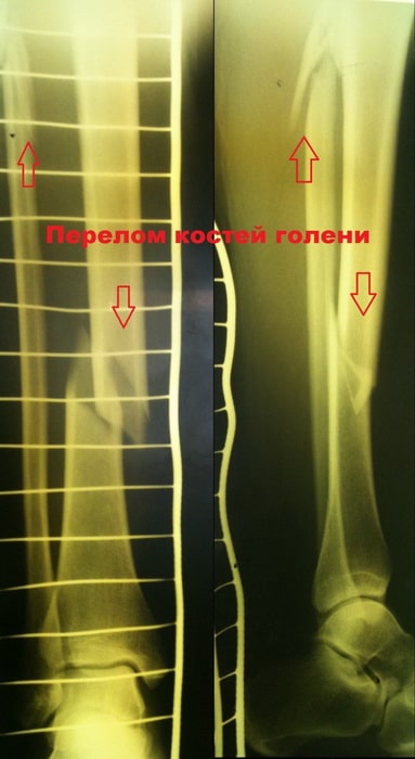
A fracture is characterized by pain and discomfort. The discomfort is so severe that sometimes there is no doubt that the fracture is serious. An X-ray can be used to confirm the diagnosis.
Even a layman can easily recognize a fracture:
- The knee joint may be laterally displaced and deformed;
- The function of the joint is impaired;
- Severe pain in leg and knee;
- The fracture is often accompanied by hemarthrosis.
There may also be enlargement and swelling of the joint. There is a large swelling. This indicates poor blood circulation in the joint apparatus. Hemarthrosis is a characteristic feature of trauma.

The necessary palpation examination is carried out for the diagnosis. In this way, the doctor determines where the severe pain is located and clarifies the nature of the problem. The doctor should be consulted whenever pain occurs when pressure is applied to the knee.
The problem can be determined by tapping on the axis of the lower leg. Often, an x-ray will show an oblique fracture, where the bone is injured at an angle. Closed fractures are often not diagnosed until the knee is injured, so it is important to seek treatment early. X-rays show how far the fracture is displaced.

exercise sets
The type of exercises is selected by the rehabilitation doctor depending on the type of injury. The exercises in rehabilitation after a broken tibia differ from the LFC for a foot injury.
The following moves are useful for any limb injury:
- Bending the leg at the knee can be done before getting up in the morning;
- Standing, shifting the weight of the body alternately on the different legs;
- swinging with maximum amplitude (forward, backward, sideways), holding the leg in the final position for 2-3 seconds, holding a support with your hand if necessary;
- squat down, starting with a flat squat and crossing your arms at your sides;
- shift body weight from heels to toes;
- Scissors exercises, alternating limb raises, cycling from the supine position.
Assisted training on a stationary bike, swimming and walking.
Go
The best and easiest way to train the foot with natural movements is to walk. Start off the car on a comfortable, flat trail and gradually increase the effort. It is advisable to change the pace, from fast to slow step. You should also climb up and down small hills.
When walking, you should also incorporate back and side steps. If there is no pain, you should start jogging gradually and end your walk with a light step.
Exercise therapy after a leg fracture
Here is a set of sample exercises for physical therapy after a leg injury or fracture. Ask your physical therapist for advice before beginning the exercises.
How to do the exercises
Each type of injury has specific sets of exercises that take into account the location and type of fracture, dislocation and surgery, as well as the condition and age of the patient.
The exercises for rehabilitation after a broken tibia are performed as follows
- morning and evening gymnastics – 10-15 minutes per day;
- Physical exercises – 45 minutes 2 times a week;
- Walking the recommended amount (distance) daily;
- Warm-up exercises – 3-4 times a day for 3-5 minutes each;
- Swimming Lessons – 45 minutes twice a week;
- LFC Complex - 45 minutes 2 times per week.
Similar exercises are also essential in ankle fracture rehabilitation, only the specific exercises may differ.
The prerequisite is that the exercises are repeated several times a day in order to continuously stimulate the muscles.
If you cannot perform any of the recommended exercises, increase the duration and difficulty of the possible exercises. It's a good idea to get an elastic band or leg stretcher at home.
If you only work out in the gym, you will not achieve the required load; You also need to exercise at home.
Rehabilitation after a fracture of the little finger or second toe is faster. In this case, more fine motor movements are performed - lifting small objects with fingers, rolling balls, walking barefoot on uneven ground.
A prerequisite for successful recovery is that all joints and muscle groups of the injured limb are fully involved and not just the injured area.
Rehabilitation and movement after a broken leg following osteosynthesis surgery begins as soon as the stitches are removed. After using an Ilizarov clamp or traction, the movement begins after 10-14 days after removing these tools.
Important! Don't remain immobile for long periods of time, especially in the right position. Don't cross your legs or strike your favorite pose with your legs.
Treatment of bone cysts
In most cases, treatment of bone cysts occurs in a surgical hospital. A series of intramural punctures are performed there. These serve to remove excess fluid and relieve the increased pressure in the cavity. Various pharmacological drugs are injected into the cyst to reduce enzyme activity. It is important for the doctor to stop the process of bone destruction and start regenerating the normal structure.
Chiropractic care is required during the rehabilitation period. It is very important to restore blood and lymph microcirculation in the affected area. Osteopathy and massage techniques are used for this purpose. It is also necessary to restore lost muscle tone. This requires a specially designed course of therapeutic exercises and kinesitherapy.
With the help of reflexology, the formation of normal bone tissue can be accelerated. Physiotherapeutic techniques are also used.
You can take advantage of a free initial consultation at our chiropractic clinic. Your doctor will examine you, review your medical records, and explain how and what chiropractic techniques are appropriate for a speedy and full recovery.
D. Medical Candidate, Chief Physician of the Clinic
Periost injury (traumatic periostitis): causes, symptoms, diagnosis, treatment
All iLive content is reviewed by medical experts to ensure it is as accurate and fact-based as possible.
We have strict rules for selecting information sources and only cite reputable websites, academic research institutes and, when possible, peer-reviewed medical studies. Please note that the numbers in brackets ([1], [2] etc.) are interactive links to such studies.
If you think any of our information is inaccurate, out of date or otherwise questionable, please highlight it and press Ctrl + Enter.
Diagnosis of periosteal injuries
In the acute period, swelling, bruising and pain at the site of injury are noted. In the following days, weeks or even months, local tissue swelling and a pronounced pain syndrome persist. Thickening of the bone in the area of \u200b\u200bthe injury can be felt on palpation.
Laboratory and instrumental studies
In the acute period, x-rays of the tibia (the most common localization of periostitis) do not reveal any pathology.
After treatment, the lesion regresses and the tissue structure returns to its original appearance, but in some cases ossifying periostitis may develop. X-rays then show a dark band parallel to and adjacent to the cortical layer of bone, which then merges with the bone's shadow, forming a wavy or jagged surface overlap.
[1], [2], [3], [4], [5]
Read more:- tibia and fibula.
- The lateral ankle is.
- Fracture of the lateral condyle.
- Мыщелка берцовой.
- Cracked metatarsal.
- Rupture of the radial-clavicular joint.
- fibula.
- Closed Foot Injury.