Cardiovascular System (CC): occasionally - high blood pressure; rarely – tachycardia, bleeding, hot flashes.
- pharmacodynamics
- indications
- Which muscles are most commonly shortened?
- Why do muscles shorten and shorten?
- Hyperextension at home without a trapezius machine
- Benefits and advantages of exercise
- Practice principles for full recovery
- Application of massage treatments
- Difference between horizontal and oblique hyperextension
- Horizontal hyperextension 1.
- 2. Oblique hyperextension
- Hyper-extension exercises: 3 correct techniques
- Anatomy and function of the lower leg muscles
- Anterior muscles of the lower limbs
- Lateral shin muscle group
- Recommendations for training the shin muscles
- The 4 best exercises for the shins
- Causes of pain syndrome
- Types of pain after a stroke
- Why do the joints in children under one year crunch?
- diagnostic methods
- Common diseases
- tendonitis
- Podiatric Keratoderma
- Possible causes of pain
pharmacodynamics
Non-steroidal anti-inflammatory drug (NSAID) from the sulfonanilide class. It is a selective, competitive inhibitor of cyclooxygenase-2 (COX-2) and inhibits prostaglandin (PG) synthesis at the site of inflammation. The inhibitory effect on COX-1 is less pronounced (undesirable effects related to the inhibition of PG synthesis in healthy tissues are less frequent). It has anti-inflammatory, analgesic and significantly antipyretic effects.
Absorption after oral ingestion is high (food reduces the rate of absorption without affecting the extent of absorption). The time to peak concentration (TCmax) – 1.5-2.5 ч. Binding to plasma proteins - 95%, to erythrocytes - 2%, to lipoproteins - 1%, to acid alpha.1-Glycoproteins – 1%. Changes in dose do not affect the level of binding. The value of the maximum concentration (CMax) is 3.5-6.5 mg/l. The volume of distribution is 0.19-0.35 l/kg. It penetrates into the tissues of the female genital tract, where its concentration after a single dose is about 40 % of the plasma concentration. It penetrates well into the acidic environment of the focus of inflammation (40 %) and synovial fluid (43 %). It easily penetrates the histohematological barriers.
It is metabolized in the liver by tissue monooxygenases. The main metabolite is 4–Hydroxynymesulide (25 %), which has similar pharmacological activity but is able to rapidly pass through the hydrophobic COX channel due to its smaller particle size–2 to reach the active methyl group binding site. 4–Hydroxynymesolide is a water-soluble compound that does not require glutathione and conjugation reactions of phase II metabolism (sulfation, glucuronidation and others) for elimination.
The elimination half-life (T1/2 ) of nimesulide is 1.56-4.95 h, 4–Hydroxynimesulide - 2.89-4.78 h.
4–Hydroxynimesulide is excreted renally (65 %) and in the bile (35 %), undergoing enterohepatic recirculation.
The pharmacokinetic profile of nimesulide is not significantly altered in patients with renal insufficiency (creatinine clearance 1.8-4.8 l/h or 30-80 ml/min) as well as in children and the elderly.
indications
articular syndrome with exacerbation of gout;
myalgias of rheumatic and non-rheumatic origin;
Inflammation of ligaments, tendons, bursitis (including post-traumatic soft tissue inflammation);
Pain syndrome of various etiologies (including postoperative, traumatic, algodysmenorrhea, toothache, headache, arthralgia, lumboischialgia).
The preparation is intended for symptomatic therapy, that is, it relieves pain and inflammation at the time of ingestion and does not affect the progression of the disease.
Which muscles are most commonly shortened?
The back and psoas muscles (iliopsoas, quadriceps, erector spinae) are most prone to shortening.
In the upper body – Trapezius muscle of the upper back, pectoralis minor muscle, sternal part of the pectoralis major muscle (which raises the scapula), transversus thoracic muscle, rectus abdominis and obliques, nuchal lattice muscles, suboccipital muscles, sternoclavicular muscles, flexor joints of the upper limbs.
In the lower part of the body – Calf muscle, hamstrings, gluteus medius muscle, thigh adductors, camel toe muscle, tibialis anterior muscle.
But basically every muscle in our body can shorten if its biomechanics are disturbed.
Why do muscles shorten and shorten?
Now we come to the heart of the matter. The cause of the problem is the same in most cases. Hypotonia (weakness), reduced functioning of the antagonist muscle.. The weakness of at least one muscle in the body inevitably leads to a tightening of another muscle, which takes over the load of the weaker muscle. The result of chronic overstretching is spasm, shortening of the muscle.
A clear example. When the gluteus maximus is weak, it leads to overstretching and shortening of the lumbar spine muscle. Why? The main antagonist of the gluteus medius in walking is the gluteus maximus. When we take a step, the gluteus medius muscle contracts and pulls the leg back, while the psoas muscle brings the leg forward. Walking is basically an alternating contraction and relaxation of the glutes and the psoas. The worse the gluteus maximus works, the more the lumbar muscles shorten.
What is the danger? There is a gradual destruction of the cartilage of the hip joint (coxarthrosis), micro-injuries of the intervertebral discs in the lumbar spine, which entails the risk of disc bulging and herniation.
Or this example. Shortening of the small pectoral muscles results from weakness of the rhomboid and middle trapezius muscle, which causes what is known as small pectoral muscle syndrome, numbness in the fingers and pain in the hand.
Hyperextension at home without a trapezius machine
The hyper-extension can be done at home on the sofa. You need a partner to do this. The starting position is that you lie on the sofa and put your legs and thighs on a support. Your partner sits on your legs. Your face and body hang down. hands behind the head. Perform the exercise in classic technique.
To perform hyper-extraction at home without a partner, you need a fitness ball. Since your body is resting on the cushioning ball, your spine is not put under undue stress. This variant of the exercise is also suitable for people with back problems and pregnant women.
- Place a ball in the center of the room and lie on your back so that your feet are on the floor, your pelvis is on the ball, and your upper body is in front of the ball. Extend your arms in front of you for support , if the ball rolls forward. Plant your heels on the floor and hold this position with your leg muscles.
- Lower your body as long as the ball allows you. In this way you bend at an angle of 45-60°.
- Maintain your posture by keeping your arms straight and your lower back arched.
- Perform an average of 2 sets of 15-20 repetitions. On the last rep, stretch.
You can replace the hyperextension at home with raising opposite arms and legs at the same time by lying face down on the floor.
Benefits and advantages of exercise
Proper technique for performing the hyperextension not only helps in isolated pumping of the posterior surface muscles, but also:
- Improves well-being in spinal pathologies – if the exercise is performed non-acutely and without weights (on average 2-3 approaches of 10-15 times, preferably on a fitball, 2-3 workouts per week).
- Effective as a warm-up exercise before strength exercises for the back muscles, e.g. B. before the deadlift (average of 2 sets of 15 repetitions).
- Indispensable for preparing the back muscles of beginner athletes for more intense exercises - in this case, the exercise is performed at each training session for a month.
- Recommended to prevent back problems in sedentary lifestyles and standing activities.
Hyper-extraction engages large muscles, saturating the body with oxygen, improving blood circulation and accelerating metabolic processes. This strengthens the musculoskeletal system.
Practice principles for full recovery
An important place in toe rehabilitation after a torn tendon is occupied by movements performed in a hand bath filled with warm water.
The patient is asked to take turns squeezing out a sponge or picking up small objects from the floor. This treatment has a relaxing effect on the ligaments and the resistance of the water greatly enhances the effects of such simple movements. The water temperature should not be higher than 34-35 degrees, as higher temperatures can cause swelling in the fingers and make movements difficult.
In the later healing phase of a distal phalanx fracture, it is advisable to carry out simple, targeted movements. The patient may be asked to tape something together and thread onto a spool. Carving, knitting, and woodcarving can speed up regeneration of the injured tendon.
Application of massage treatments
Massage is of great importance in the complex of rehabilitation measures after a quadriceps tendon rupture.
In the case of circulatory disorders during immobilization, which are accompanied by swelling and congestion in the area of the palate and hand, the specialist recommends massaging the proximal parts of the upper limbs.
If the muscle strength is reduced when moving the fingers, the forearm is massaged. This means that the muscles of the flexor or extensor group are stressed, taking into account the area of tendon damage.
If there is a tendency to form adhesions, a massage along the course of the tendon using various rubbing techniques is recommended a few weeks after the operation. Movements are chosen in which the skin and subcutaneous tissue are moved and stretched.
Water and other rehabilitation treatments allow for a quick return to a full life. Comprehensive treatment includes taking medication to speed up the healing process and activate the body's immune system.
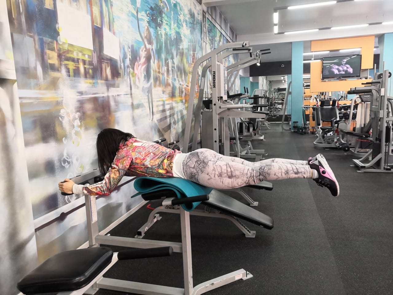
Difference between horizontal and oblique hyperextension
There are different techniques for performing hyperextension exercises, the difference being in the configuration of the exercise machine itself. The main exercise on this machine is body flexion. Different loads are used and body position is varied to target different muscle groups. Let's look at the trainers and the techniques for performing hyperextension exercises on them.
Horizontal hyperextension 1.
This variation is best for engaging the glutes, but it's important to follow the execution technique and not lose mental focus on the glutes. The design of this machine is higher on the back with rollers designed to keep the legs in place, allowing the lower back - the muscles that straighten the spine - to work harder, followed by the glutes and hamstrings. If you want to tone your glutes and hamstring biceps, this exercise is for you.
2. Oblique hyperextension
This variant of the machine is the most popular in gyms. There are no fundamental differences to the first variant. However, since the rollers are placed lower, as already mentioned, there is less strain on the body. There are no fundamental differences in exercise technique between the first and second variants. However, there is a basic principle for proper technique: the body must be positioned on the trainer so that the lower abdomen is on the platform and the upper body is hanging freely down. Otherwise, your abs will prevent you from doing a full incline, resulting in improper loading and affecting the quality of your workout.
Hyper-extension exercises: 3 correct techniques
Now let's take a closer look at the 3 basic exercises for hyperextension:
- Pump up the glutes
- To strengthen the biceps thighs.
- Separately, let's consider the exercise for testing the back flexors (spinal girdles).
In order to perform the exercise correctly, you need to adjust the trainer to your parameters, and especially the bench to your height.
- The top edge of the trainer should be right in the loin crease, ie just below the crook of the trunk.
- The bottom of the brace should end at hip level (top of thighs). In simpler terms, the pelvis should be on the platform of the exercise machine.
- The lower rollers (on a horizontal bench) should be positioned so that they are slightly above the Achilles tendon. The incline bench does not have such casters, but there is a special leg platform that you must place your feet on.
Again, the most important thing is that you adjust the upper position to your height. If the support line is higher than the indicated lumbar flex line, your abs are in the way and you won't be able to complete the incline.
After adjusting the device to your size, you need to 'lie' properly on the device. Lie on a horizontal bench and support your hips and torso on the trainer pad just below the flex line. Then place your feet on the platform, feet straight and parallel to each other.
On the horizontal bench, fix your feet slightly above the Achilles tendon, the rest of the body position is the same as on the incline bench. Bend your knees slightly and keep them in this position throughout the exercise, as you put unnecessary stress on your knee joints when your legs are completely straight. Another note: if you perform the exercise straight-legged, the hip biceps will take over 80 % of the load, which means the glute benefit from the exercise is very small.
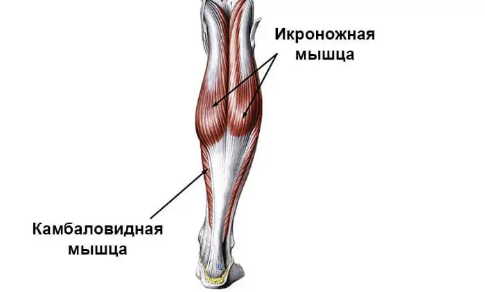
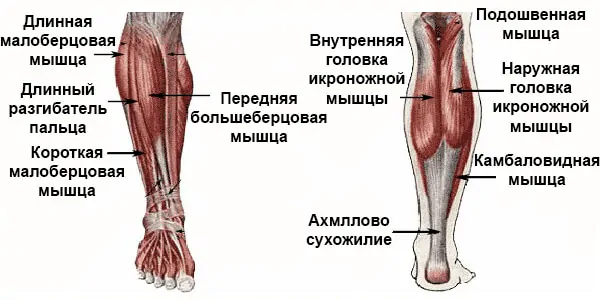
Anatomy and function of the lower leg muscles
Although the calves are known as a small muscle group due to their small number of functions, the lower leg area has a large number of muscles. They have a similar structure to the close-meshed bundles around the tibia and fibula. All of the muscles of the lower leg and foot are generally classified into three groups based on their structure, attachments, and function:
- anterior;
- Laterally;
- The backstage area.
Each area has a different function, with the focus being on the rear area in terms of importance in sport. Most exercises for the lower leg muscles are specifically designed to develop them.
Anterior muscles of the lower limbs
The entire group of anterior tibial muscles consists of three components:
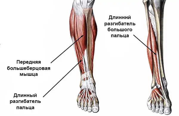
- the tibialis anterior;
- Long toe tendon;
- The long extensor of the big toe.
All three muscles of the lower limbs function as a single mechanism in anatomy, with each muscle responsible for a different edge of the foot.
The main functions are extension, elevation, supination and pronation of the foot.
Although the anterior shin muscles are not commonly exercised in strength sports, they play an important role in cycling sports (running, cycling, etc.).
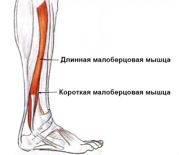
Lateral shin muscle group
Recommendations for training the shin muscles
The problem with calf training lies in the physiology of the individual athlete. For a long time it was assumed that the triceps (as the dominant muscle of the entire lower extremity) was dominated by slow muscle fibers. Therefore, the multi-rep mode was appropriate for this workout. However, the MMW setting and type of training did not favor hypertrophy, so even in some proscenium athletes, calf volume was significantly delayed.
Later studies have shown that in some people as many as 50-60 % fast-twitch muscle fibers are located in the posterior aspect of the tibia. This led to the use of strength training (i.e. working with heavy weights) as well as repetition training. So when it comes to how to pump up the lower leg, the ratio of GMV to OMV (fast and slow fibers) is key.
In addition to the physiological features, there are general recommendations that will significantly speed up progress:
- Alternate workout types, track progress. This helps in choosing the best method for a particular athlete.
- The most important sign that the calf muscles are working properly is the burning sensation.
- When exercising, all other muscles in the body must be completely out of action. Only the ankle should be moved.
- All exercises should be performed in an elevated position to increase range of motion.
- As you stand on tiptoe, push your body up as far as you can, but the movements should be smooth and without jerking.
- The load time for shin training should be 36-45 seconds.
- Like any other muscle, the calves need rest. Therefore, you should not train this muscle group more than 2-3 times a week.
The 4 best exercises for the shins
The muscles of the lower leg and foot don't have many functions anatomically, so the movements are all similar. The only difference is in the effect, the weight and the posture.
Causes of pain syndrome
Arm and leg pain occurs either immediately after a stroke or a few weeks afterward. One of the causes of pain after a stroke is joint and muscle damage due to changes in the peripheral nervous system.
Sometimes a paralyzed limb can be painful due to contractures. The brain damage causes the flexor muscles to be more active than the extensor muscles. In this case, an attempt to change the position of the limbs will lead to a strong spasm.
The pain is caused by an increase in muscle tension. An increase in the tonic neck reflexes plays the main role. Increased muscle tension occurs in response to:
- extended supine position;
- Excessive exertion when sitting;
- the need to exert force when getting out of bed;
- discomfort - cold or pain;
- a feeling of dirt on the body;
- mental stress.
Our ambulance can diagnose the cause of pain after a stroke and provide a comprehensive diagnosis of the nervous system and the entire body. It is not necessary to determine the cause of pain after a stroke yourself, as this can cause further damage to the patient.
Types of pain after a stroke
Pain in the arms or legs can be local or central. Local pain is mild to moderate in intensity and does not interfere with daily functioning.
Central pain is caused by damage to the brain due to poor circulation. This organ can turn even light touch into pain. The pain can be:
- paroxysmal;
- persistent;
- Tingle;
- burning pain;
- of varying intensity due to temperature fluctuations.
Severe pain after a stroke disrupts people's lifestyles and prevents them from performing even the simplest activities. In some cases, they lose the ability to take care of themselves.
Why do the joints in children under one year crunch?

Infants of this age have a unique skeletal structure that must be considered to determine the cause of the crunch. In young children, the joints are not yet developed and are more flexible, but this does not prevent the child from exploring the world and does not cause any discomfort. However, there are other causes of the crunch as well:
- Active growth when muscles cannot keep up with bones;
- Not enough calcium in the child's body;
- dysplasia;
- Poor calcium absorption due to vitamin D deficiency.
Doctors also speak of 'growing pains'. This diagnosis is made when muscle tissue cannot keep up with bone growth and ligaments and tendons are under constant strain. Sometimes the pain is so severe that the children refuse to walk. Medical specialists prescribe different types of massages to relieve the pain.
diagnostic methods

To find out why children's feet hurt for no apparent reason, it makes sense to consult a doctor. They will prescribe a series of tests to diagnose the problem and recommend an investigation. The main diagnostic methods include:
- general urine and blood tests;
- biochemical tests for rheumatic fever;
- tests for infections of the urogenital tract and intestines;
- immunological tests;
- Ultrasound examination of the movable joint;
- X-rays.
Common diseases
The soleus muscles of the foot are very susceptible to diseases and various pathological processes in the foot, as they are subjected to significant stress during movement.
tendonitis

Plantar tendon disease involving muscle tissue.
- trauma, severe overload of the foot;
- Degenerative damage to articular cartilage;
- Wearing uncomfortable, tight, high-heeled shoes and minor ankle injuries;
- Calcium deficiency and metabolic disorders;
- infectious processes affecting the tendons;
- flat feet;
- side effects of certain medications.
Symptoms of foot tendonitis include severe pain with active movement and at rest. Congestion, swelling and a localized increase in temperature occur in the area of the sole of the foot. The painful joint grinds or cracks.
If treatment is not started on time, the pathology will become chronic, permanent overload will occur, and the muscles and feet may ache even with normal movements.
- Complete immobilisation of the ankle (elastic bandage);
- taking NSAIDs to reduce inflammation;
- In case of infection, antimicrobial drugs are prescribed;
- Physiotherapy, therapeutic massage, gentle exercise;
- In advanced cases, surgical intervention.
Podiatric Keratoderma

Keratoderma is the colloquial term for dermatoses characterized by skin abnormalities on the feet and hands. It can occur as a symptom of an underlying disease or as an autonomic condition.
Possible causes of pain

Possible causes of pain in the foot and in the plantar muscles of the shin are.
- sprain of the ligaments;
- Joint diseases (arthritis, arthrosis, gout);
- ankle and foot injuries (fractures, dislocations);
- neuroma (growth of nerve tissue in the foot);
- calluses, warts;
- Diseases of the musculoskeletal system (herniated disc, sciatica, radiculitis).
If you have lower limb discomfort that does not go away even with rest, you should see your doctor to determine the cause and correct the problem. Regular prevention and moderate physical activity reduce the risk of foot pain and maintain joint mobility for years.
Read more:- The flexor muscles of the foot.
- The muscles that move the foot.
- How muscles and bones are related.
- muscles in the legs.
- Tibialis posterior muscle.
- Walking in medium heels.
- Anterior tilting of the pelvis.
- muscle work while running.