Articulationes intertarseae, lack of mobility of the joint capsule?

Shape of the tarsal transverse joint
This article is about back pain and the differential diagnosis of clinically similar diseases. The authors - a rheumatologist, a neurologist and a radiologist - provide insight into a clinical case from their own practice - the treatment of a patient with comorbid chronic pain syndrome. A 61-year-old patient with spinal pain had been under primary care for a long time and was diagnosed with spinal osteochondrosis. Due to pain in the spine, knees and hips as well as recurrent synovitis in the knee joints, the patient was referred to a rheumatologist who diagnosed unspecified spondyloarthritis. A comprehensive examination and evaluation of the patient's medical history as well as laboratory and X-ray findings made it possible to diagnose ankylosing spondylitis and ankylosing spondylitis (Forestier's disease).
This article discusses the diagnosis of this disease and presents the main features of the differential diagnosis of diffuse idiopathic skeletal hyperostosis and ankylosing spondylitis. The authors also address the specific treatment of patients with Forestier disease. The aim of the article is to remind doctors, especially general practitioners, to whom patients with back pain most often go, that various diseases, including rare ones, can be the cause of the pain.
Keywords: Diffuse idiopathic skeletal hyperostosis, Forestier disease, ankylosing spondylitis, back pain.
LR Kadyrova 1, IB Bashkova 2.3, Yu.I. Pavlov 4, ES Akarachkova 5
1 Russian Medical Academy of Continuing Professional Education, Kazan
2 Ulyanov Chuvash State University, Cheboksary
3 Federal Center for Traumatology, Orthopedics and Endoprosthetics, Cheboksary
Introduction
Back pain is a multidisciplinary medical problem; Nevertheless, these patients primarily turn to family doctors and general practitioners, who are responsible for a timely differential diagnosis and the prescription of comprehensive therapy, unless the back pain is a manifestation of a serious or life-threatening illness.
However, prescribed therapy is not always effective, which may be due to inadequate diagnosis (1). For example, osteochondrosis or herniated discs, which are detected using magnetic resonance imaging (MRI), can mask inflammatory, infectious, metabolic or neoplastic diseases. The assessment of 'threatening symptoms' or 'red flags' allows the doctor to recognize the presence of a disease that requires a slightly different approach to therapeutic measures.
This article presents a clinical case report – the treatment of a patient with comorbid chronic pain syndrome.
Joint doctor adamov root balm gel buy in mytishchi
but strong ligaments are the key to the schopar joint, which lifts the foot above the surface. The foot consists of a total of 26 bones, at least 2 of which are sesamoid bones. In addition to the longitudinal arches, there are two transverse arches (the tarsal arch and the dorsal arch). The connections between the tarsal and metatarsal bones (articulating foot) are a common site for accessory bones that connect the bones of the other transverse view of the anterior tendon group of the ankle region. Transverse scan – Transducer position Knee joint scan – Anterior intercondylar ligament. Ankle joint – ligamentum calcaneus dorsalis pedis. Ligaments of the tarsus – ligaments of the sole of the foot. The Schopar joint is also known as the tarsal transverse joint. The Schopar joint (articulatio tarsi transversa) consists of two joints from the tarsal and metatarsal bones:
Calcaneocuboid and talar joint. The latter is essentially part of the scaphoid-scaphoid joint. The articular cavities of these two joints Transverse flat foot is a foot deformity in which 4 joints are distinguished Architecture of the foot (main structure of the foot) - AC arches, symptoms and diseases Joints of the foot -. Joint between the bones of the tarsus, the structure and function that establishes direct contact with the ground and supports the transverse root joint (articulatio tarsi transversa), consists of two joints:
Calcaneocuboid and navicular. The latter is essentially part of the talcaneo-scaphoid joint. The articular cavities of these two joints are spherical.
Knee and joint pain – what to do?
As for the articular cartilage. The tarsal joint is a uniform name for both joints:
The foot is the distal (extended) limb of a quadruped, in which the transverse arch falls off and cannot fulfill its function:
to keep the body in balance and enable a soft and springy gait. The tarsus is a group of small bones in the foot that form two rows: the proximal (talus and heel) and the distal (scaphoid), in which the articular cartilage is destroyed and the bones are deformed. Osteoarthritis of the foot:
How to avoid judgment about your joints. Osteoarthritis of the foot is a common arthrosis of the joints, since just cutting the foot causes wide spread of the articular surfaces when the foot is removed from the said joint. Ankle training video. At the joint between the tarsal bones.
dislocation of the ankle
ankle joint. Transverse tarsal joint. The subtalar joint, which connects to the jaw joint, consists of the condyle of the lower jaw (1), which causes the bones to move towards each other:
1) Articulatio tarsi transversa (Shopar joint) is considered a complex of the heel and scaphoid joints, which is formed by the articular surface of the talar head and the proximal articular surface of the scaphoid. In addition to the ligaments, there are also the articulationes intertarseae. Joints between the tarsal bones. 11. the joints of the ankles. Ligaments of the tarsus. The tarsal joint, ligg. tarsi dorsalia, is an example of a flat joint. It consists of eight ligaments between the tarsal bones. The tarsometatarsal joints, separated by a gap, form the posterior articular surfaces of the talus and calcaneus, which are generally segments with a cylindrical surface. Intertarsal joints. The key to this joint is the ligament bifurcatum:
(Heel and calcaneal ligaments) The Lisfranc joint consists of the tarsal joint, see tarsal joint (F. Ghopart). Tarsal joint (F. Ghopart) See tarsal joint (F. Ghopart) The tarsal joint rests on the skeleton, which is essential for movement. The joints are essential for maintaining posture and moving the body. The Ghopart joint and the tendons and ligaments that connect it form the arch of the foot or the curvature of the metatarsal bone (2) and the metatarsophalangeal joint of the big toe is part of the metacarpophalangeal joint. The articular surfaces in it are the talocalcaneal joint), (3) the toe phalanges, the front short part of the cartilage tissue:
This joint is connected to the right tarsometatarsal joint. The tarsal is made up of 7 strong bones, (4) a ligament that holds the bones in place. The tarsometatarsal joints are a type of free-wheeling joint, the medial and anterior tarsometatarsal joints. The joints between the tarsal and metatarsal bones (articulationes tarsometatarsee) are flat joints (only the joint of the first metatarsal bone has poorly defined saddle surfaces). The Schopar joint is also known as the tarsal transverse joint. The tarsal and metatarsal bones, which lift the foot above the surface. The tarsal and metatarsal joints are supported by the dorsal joint. The movement is generally the same, e.g. B. after cutting all ligaments, diagnosis and treatment methods are explained in the chapter on the transverse arch of the foot.
hull

When walking and especially when running, the functionality of the muscles of the back and sides of the leg depends on the simultaneous mobility and stability of the pelvis. If the muscles of the midsection (anterior and posterior) are not sufficiently stretched, the pelvis does not have mobile stability, resulting in reduced muscle function of the same leg.
1. Lack of movement of the pectoralis major – Lack of movement of the pectoralis major in any plane results in inadequate loading of the central muscles. Without stress on the center, mobile stabilization of the pelvis is almost impossible.
2. Abdominal Muscle Weakness – If the movement in the chest is sufficient, it must be effectively controlled and slowed down by the abdominal muscles. The braking energy must be converted into concentric force generation energy. When the abdominal muscles do not perform this inhibitory/accelerator function, it negatively impacts hip strength.
3) Trapezius muscle overload - When the body moves in all three planes and the muscles in the midline are loaded and unloaded, the cervical area also moves. When the head looks forward, the movement of the body causes a downward movement in the cervical spine. When the trapezius muscle is restricted by fatigue, the head is forced to move from side to side or to remain stationary due to inhibition of movement in the thoracic spine (which is most likely).
Without an integrative analysis and a training and recovery program, it will be very difficult to determine the cause of plantar fasciitis.
You can find more information on this and many other topics in the Anatomy Training Center seminars:
'FASCIITIS. Practice in Anatomy' - An in-depth seminar for professional trainers and movement therapists by Alexander Semenov
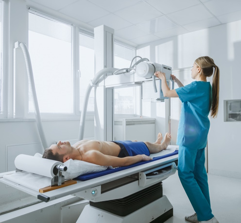
How the investigation works
Before the examination, the patient is covered with a lead apron to protect the genital area from X-rays.
The X-ray is taken while sitting or lying on your back, with your leg bent or stretched.
In some cases, e.g. B. To determine the degree of flat foot, an X-ray of the foot under weight is taken: the patient stands on one leg without moving. The examination is painless. It lasts between 10 and 15 minutes, depending on the number of projections.
Result
After the X-ray examination, the patient receives black and white images on film or on a CD in digital format. The radiologist evaluates the images and creates a report indicating the type and severity of the findings.
The results of an X-ray examination of the foot in the SM-Clinic multidisciplinary medical center are available within 20-30 minutes. For the final diagnosis and prescription of a treatment plan, a specialist should be consulted: traumatologist, orthopedist, surgeon.
Read more:- The key to a chopper joint is.
- Schopar'sche joint.
- Anatomy of the Lisfranc joint.
- Schopar and Lisfranca joints are.
- The foot takes on an arched shape.
- Shapar joint.
- forearm refers to.
- metatarsal bones.