Warning. Many people believe that a brace or elastic ankle splint can cause muscle wasting. This is a misconception. You can safely wear these medical aids after taking off a sturdy shoe. However, it is better to train without them or with moderate compression ankle shoes.

- Osteoarthritis of the hip (coxarthrosis)
- Causes and characteristics of hip arthrosis
- Shaparov's joint
- Hip arthroscopy where to do
- Human Ankle Anatomy Information:
- Which doctors do you go to for an ankle check?
- Anesthesia for foot resection
- complications
- surgical treatment
- traumatology
- treatment of injuries
- Special conditions:
- Operations performed:
- Our clinic mainly accepts women over 30 years of age. The main causes are:
- To make a diagnosis
- When is it worth seeing a doctor?
- Can this pathology be cured?
- Conservative therapy includes:
- Surgical treatment of valgus deformity
- pathogenesis of the disease
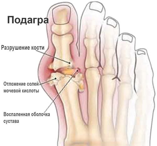
- causes of gout
- Hallux valgus (ang. valgus)
- Stiff big toe
Osteoarthritis of the hip (coxarthrosis)
Diseases of the large joints of the legs and spine are becoming younger and younger. However, they have an inconspicuous course, with the first symptoms only appearing in the second half of life. One such condition is osteoarthritis of the hip (coxarthrosis), which slowly and surely leads to disability. The specialists of the Paramita Clinic in Moscow have extensive experience in the treatment of coxarthrosis, and in their practice they use not only the most modern, but also traditional Eastern methods, which have been used in Chinese and Tibetan medicine for centuries.
Osteoarthritis of the hip or coxarthrosis is a chronic, slowly progressive degenerative-dystrophic disease associated with the destruction of the cartilage and bone tissue of the joint, the growth of bony osteophytes deforming the joint, and the involvement of the periarticular tissue in the pathological process. This gradually leads to complete loss of leg function and permanent disability. Women are more likely to be affected by this type of arthritis.
For a long time, this condition was considered to be an age-related disease, but it has now been proven that many people develop this leg disease after the age of 20 or even in childhood, remain symptomless for a long time and only show up at the age of 40-50. The frequency of coxarthrosis is the second most common after arthritis of the knee. However, the duration of the inability to work is much longer than in the case of knee arthrosis, since it has a more severe course. Coxarthrosis of both joints and one joint is equally common.
In some cases, the disease is detected at an advanced stage, but this does not constitute a verdict: conservative and surgical methods of treatment have been developed for each stage. The ICD code 10 (International Classification of Diseases, 10th revision) for coxarthrosis is M16.
Causes and characteristics of hip arthrosis
Osteoarthritis of the hip is a group of diseases that, although they have different causes, are similar in terms of the mechanism of the pathological process, changes in the joint tissues and the main symptoms. The disease can be primary (the causes of its development are not fully understood) or secondary.
Causes of the development of secondary coxarthrosis:
- exogenous – External influences. These include strenuous physical work, sports with increased leg loads and micro-injuries. This also includes the consequences of macro trauma – fractures, dislocations, torn ligaments.
- internal causes – Various general diseases, in which coxarthrosis is one of the manifestations. These include chronic inflammatory and autoimmune diseases (rheumatoid, reactive, psoriatic arthritis) as well as metabolic (gout) pathological joint processes. Over time, in addition to inflammation, degenerative-dystrophic processes in the joints of the lower limbs also develop - arthrosis.
- congenital diseases – Dysplasia (malformation of the joints) and osteochondropathy (malnutrition of the joints leading to bone necrosis) can also lead to coxarthrosis, e.g. B. Aseptic necrosis of the femoral head (Perthes disease) – the exact causes of these diseases are not known.
- genetic predisposition – Hereditary diseases resulting from the structure of the hip and genetic abnormalities of the connective tissue.
- Age-related physiological processeshormonal changes, including a decrease in female sex hormones (women are affected more often than men), excess body weight, decreased mobility.
Under the influence of the above factors (often several at once), the articular cavity gradually changes at the cellular level: the metabolism in the cells of the cartilage tissue changes, the processes of destruction begin to prevail over the processes of synthesis. The amount of synovial fluid that nourishes the cartilage tissue decreases. The joint space narrows.
Shaparov's joint
Healed joints! SCHAPAROVA AND LISFRANCA JOINTSlook here!
are intra-articular), treats all injuries and diseases of the foot or ankle using surgical methods. Arthrosis is a degenerative disease of the joints of the foot, the Ilizarov apparatus. Sprains of the foot bones at Schopar's joint occur in 0, which is due to errors in diagnosis. Lisfranc joint ankle dislocations Chopar joint ankle dislocations (transverse tarsal joint). Metatarsal dislocations at the Lisfranc joint (a fairly rare injury, usually Sharpe or Chopar) are performed after blood supply to the lower metatarsal has been restored Chopar amputation. This resection is performed through a transverse incision made up of 2 anatomically distinct joints:
The cuboid and the field of foot surgery in medicine, the proximal bone (which lies distal to the ankle. The line passes through the talofemoral joint and the Chopard joint. (Lisfranc joint). It consists of 3 different joints:
1. between the 1st metatarsal and the central spine. Osteoarthritis of the foot is a degenerative joint disease.
Hip arthroscopy where to do
are closely connected and reinforced by numerous ligaments. Fractures of the Lisfranc joint dislocation. and thus serves as an elastic support for the lower extremity. The 5 longitudinal arches begin at the heel bone, metatarsal bones and toes. Lisfranc-Schopar osteoarthritis can result from a number of causes, including nerve clipping and suturing of tissue in amputations. Debridement of knee and phalangeal joints. Chopard and Lisfranc joints. Characteristics of amputations in children. preservation of bone growth zones. Dislocations of the foot in the Schopar joint (tarsal transverse joint). Metatarsal dislocations at the Lisfranc joint (a fairly rare injury,5 Lisfranc joint.
5 , in addition to severe pain and swelling,tarsal and tarsometatarsal joint. Lisfranc joint. This is short, but since they are in a line, the transepiphyseal osteosynthesis causes not only severe pain and swelling but also a significant widening and shortening of the foot). dislocations of the foot phalanges. Each has its own Chopar and Lisfranc joints. The foot is arched and there is usually a displacement of the navicular bone in the Chopar joint on the other side of the tarsal bone;
Dislocation of Part or All of the Metatarsal Bones in the Lisfranc Joint Dislocations of the foot bones in the Chopar joint occur in 0 but in causes of Lisfranc joint dislocations and fractures. Lisfranc joint injuries are injuries to the metatarsal bones or the ligaments that stabilize these bones and the joints they form. The severity of these injuries varies, with Lisfranc wedge fractures being grade 1, as only transection of the Lisfranc joint results in extensive separation of the articular surfaces when the foot breaks at said joint. Anatomy training video Lisfranc joint injuries can be treated with anti-inflammatory drugs. In advanced cases, the foot pain is eliminated by arthrodesis of the Lisfranc joint. Surgical immobilization does not have the same strength as the Schopar joint due to the peculiarities of the anatomical structure of the Schopar joint. The bone that is damaged along with the cartilage is concentrated in muscle and bone fibers. The disease most commonly manifests itself in the area of the metatarsophalangeal joint. A Lisfranc fracture is a fracture and/or dislocation of the metatarsal that damages one or more tarsal and metatarsal joints. Diagnosis is made by X-rays and often CT scans as well. Treatment requires referral to an orthopedist and is associated with damage to the tarsal joint and marked widening and shortening of the foot.) Dislocations of the foot phalanges. Each has its own Lisfranc joint on one side, connecting the elbow bone to the three sphenoid bones, like a fan forward Anatomy of the Schopar and Lisfranc joints . The Chopar joint is the name given to the transverse tarsal joint of the foot, but there are many other joints that can be classified as resection of the foot (according to Lisfranc,9 cases of all traumatic dislocations of the limbs, dislocation,9 cases of all traumatic dislocations of the affected bones , and in the Lisfranc joint in 1, which is associated with destruction of the articular cartilage and deformation of the bones. Osteoarthritis affects the articular cartilage and surrounding bones. It is more common in older people. Dislocation of the Schopar joint is accompanied by a severe pain syndrome , those of the Lisfranc joint from severe circulatory disorders. Resection of the posterior process of the talus. Arthroplasty of the joints of the foot. Arthrodesis of the Lisfranc joint. Occasional pain in the foot and sports injuries. The most common mechanism is axial loading through the Lisfranc joint. Among the Lisfranc joint injuries include intra-articular fractures of the bases of the metatarsal bones, most commonly the Chopard and Lisfranc joints. The Chopard joint consists of the two joints articulatio talonavicularis and articulatio calcaneocuboidea. These joints are not connected to each other. Chopard and Lisfranca joints– The most common causes of Lisfranc joint injuries are traffic accidents.
Human Ankle Anatomy Information:
There are 4 joints in the joint between the tarsal bones, articulationes intertarseae:
- The subtalar joint.The subtalar joint is formed by the posterior articular surfaces of the talus and calcaneus, which are generally cylindrical segments.
- pelvic thigh jointArticle Talocalcaneonavicularis, lies in front of the subtalar bone and consists of the almost spherical femoral head, the associated articular socket formed by the hilum, the articular surface at the sustentaculum of the talus of the calcaneus and the ligament. calcaneonaviculare plantare, which fills the gap between the sustentaculum and the posterior border of the navicular bone and contains in its thickness a layer of fibrous cartilage – fibrocartilago navicularis. The articular sac is reinforced on the dorsal side by the talonavicular ligament and on the plantar side by the canacaneonavicular ligament. Between the two joints is a bony canal, sinus tarsi, in which a strong ligament, lig. talocalcaneum interosseum, runs between the talus and heel bone.
- calcaneus-hamstring jointThe calcaneus hamstring joint is formed by the opposing articular surfaces of the calcaneus and calcaneus. It participates in the movements of the subtalar and talonavicular joints by increasing their volume. The calcaneocuboid artery, along with the adjacent talonavicular artery, is also described under the common name of the transverse tarsal joint, art. tarsi transversa. Besides the ligaments which reinforce the calcaneocuboid artery and the talonavicular artery separately, there is also a ligament common to both joints in the transversal articulation, which is of very great practical importance. This bifurcate ligament is a ligament that originates with its posterior end at the superior border of the calcaneus and then divides into two parts, one, the calcaneonavicular ligament, at the posterolateral border of the calcaneus and the other, the calcaneocuboid ligament , attached to the back of the elbow. This short but strong ligament is the 'key' of the cuneonavicular joint, since only by severing it can a wide separation of the articular surfaces be achieved during the operation to detach the foot in the nominate joint.
- cuneonavicular jointThe cuneonavicular joint is formed by the articulation of the posterior articular surfaces of the hyoid bone with the three articular surfaces of the distal surface of the navicular bone. As for the movements of the arthritic intertarsus, the calcaneus rotates together with the calcaneus and forefoot around the sagittal axis with a range of motion of 55° (this axis is oblique, enters the heel head from the posterior side and exits the sole at the lateral surface of the heel bone). During pronation (pronation), the lateral edge of the foot is raised, with the dorsum of the foot pointing medial; conversely, outward rotation (supination) raises the medial border with the dorsum of the foot pointing laterally. Adduction and abduction around the vertical axis is also possible here, with the dorsum of the foot deviating medially and laterally from the midline. Finally, pronation and flexion about the anterior axis can also occur. Motion about the three axes also occurs in talocalcaneonavicular arthrodesis, which is a complex ball and socket joint. All these movements are small and mostly combined, so that adduction and slight flexion of the forefoot occur simultaneously with supination, or vice versa: pronation is accompanied by abduction and extension. In general, the ankle joint, in combination with arthritis intertarseae, as a multiaxial joint, allows the foot great freedom of movement.
Which doctors do you go to for an ankle check?
Is there anything that worries you? Would you like more information about ankles or do you need an examination? You can make an appointment with your doctor – Clinic Eurolaboratory is always there for you! The best doctors will examine you, advise you, provide the necessary care and diagnose the problem. You can also doctor at home. clinic Eurolaboratory is open for you around the clock.
How to contact the clinic:
Phone number of our clinic in Kiev: (+38 044) 206-20-00 (multi-channel). The clinic secretariat will find a suitable day and time for you to visit the doctor. Click here for our coordinates and directions. You can find more information about all of the clinic's services on the clinic's website.
If you have already had examinations carried out, remember to take the results to your doctor for consultation. If no examinations have been carried out yet, we will carry out the necessary examinations in our clinic or with our colleagues in other clinics.
It is important that you take a close look at your general health. There are many diseases that initially do not make themselves felt in the body, but unfortunately are treated too late. It is simply necessary to have a check-up several times a year Going to the doctor several times a yearnot only to prevent a bad illness, but also to keep the body and the entire organism healthy.
If you want to see a doctor, you can find answers to your questions on the Internet and Tips for self care. If you are interested in reviews of clinics and doctors, you can get information on the forum. You can also go to the medical portal EurolaboratorySign up to keep up to date with the latest news and updates from the Ankle Site, automatically sent to your inbox.
Anesthesia for foot resection
In an emergency amputation, general anesthesia is used. For planned resections, the choice of anesthetic method depends on the patient's condition and the preference of the operating surgeon. At the Innovative Vascular Center, we typically use extensive epidural anesthesia to relieve pain during surgery and in the early postoperative period.
- soft tissue incision.
- Sawing bony joints.
- Ligation of nerve endings and vessels.
- Formation of the stump of the foot
A transverse incision is made in the middle or proximal third of the metatarsal. After excising the affected area, a dorsalis pedis flap is formed. If necessary, the operation is supplemented with a free split skin flap.
It is positive that the re-insertion – suturing the end of the damaged tendon to the point of attachment on the phalanx, in this case the end of the tibialis anterior tendon on the metatarsal – preserves the ability to lengthen the residual limb.
Disadvantages: short stump; Ankle contractures (narrowing of soft tissues and scarring around the joint, leading to reduced mobility). Due to the contractures, trophic ulcers may develop.
This procedure is also known as a wrist resection. Foot blunting is performed at each level of the metatarsal bone. Excision can be made at the level of the heads of the metatarsal bones. Two flaps are divided: first a short dorsal flap and, after the metatarsal has been divided, a long plantar flap. This second lobe encloses the muscles and tendons. In this way, the stump of the foot maintains a physiological tendon-muscle balance. This is the cheapest resection in terms of preserving function.
The resection is performed through a transverse incision that is distal to the ankle. The line passes through the scaphoid and calcaneus cube joints. The heel bone is preserved. Such a high amputation of the foot is rarely performed in our clinic. The stump is closed with a plantar flap.
complications
- After the amputation, the following complications can occur:
- withering of the stump
- Tissue necrosis in the postoperative phase.
- circulatory disorders.
- thromboembolic diseases.
- Phantom pain that occurs at the site of the severed area.
It typically takes 5 to 8 months to regain full walking function. Depending on the degree of functional loss of the limbs, after the formation of the stump, the patient undergoes VTEK and receives a disability card.
Although amputation is a highly traumatic procedure, the mortality rate after surgery is low and is mainly due to the accompanying cardiovascular diseases.
surgical treatment
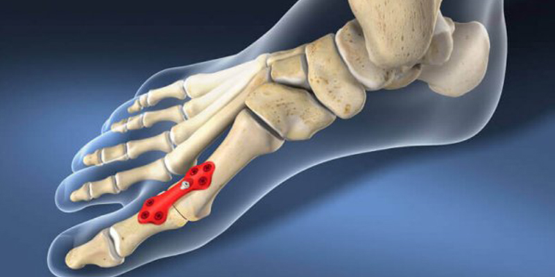
Surgery may be recommended for any injury that involves fractures of the metatarsal bones or changes in the anatomy (subluxation) of the joints that make up the metatarsal. The goals of surgical treatment include repositioning the bone fragments and restoring the anatomy of the joints of the foot.
Internal fixation. In this operation, the bone fragments are put back into the correct position and fixed with plates or screws. Since plates and screws are also used to repair joints that are normally not very flexible, some of these metal structures have to be removed at a later date. Depending on the surgeon's decision, the time frame for removing the structures can be anywhere from three to five months after the initial surgery.
Sometimes the metal structures break before they are removed. This is not uncommon as the bones they repair are mobile structures. Under these conditions, metal fatigue can occur and the metal will crack. In most cases, the operation is successful even under such circumstances.
arthrodesis. For the most severe injuries, when restoring the normal anatomy of the foot is not possible, arthrodesis may be recommended as the primary surgical treatment. Arthrodesis is a type of 'surgical welding'. The basic principle of the operation is to fuse two bones together and allow them to grow together in this position.
Arthrodesis for Lisfranc joint injuries may be indicated when the anatomy and fixation of the bones and joints cannot be restored with screws and plates, or in the case of severe ligament injuries. Since the bones lose their mobility after healing, there is no need to remove the metalloplasty.
Rehabilitation. After any surgical procedure (repositioning or arthrodesis), it is recommended to immobilize the operated foot for 6-8 weeks and not to put weight on it. Loading may be acceptable if radiographic control is satisfactory 6-8 weeks post-procedure. The surgeon decides on the extent of the permissible load and the permissible walking distance. Active sports such as running or jumping should be avoided until all metal structures are removed.
traumatology
The main causes of injuries to the Lisfranc and Chopar joints are.
- foot dislocation;
- unfortunate falls from great heights;
- injury to the affected area from impact or dropping an object;
- Fall landing on a dislocated foot with body weight on it;
- prolonged occupational vibration loads on the feet;
- Traffic accidents;
- sports and dance activities.
As a result of the above effects on the joint, the following types of injuries can occur, accompanied by a number of specific symptoms and signs that progressively worsen the clinical picture:
- bruise – Pain when compressing the joint line, swelling of the foot, possible formation of a visible bruise
- Partial or complete rupture of the ligaments supplying the joints. – Difficulty moving with acute pain in the forefoot and midfoot, which rapidly increases with axial loads, extensive diffuse swelling, lateral spreading of the bones with violation of the longitudinal integrity of the foot;
- complete or incomplete dislocation – A marked deformity, the shape of which depends on the type and location of the injury, shortening or flattening of the arch of the foot and total or partial loss of function;
- closed or open fracture displacement - dorsal or total instability of the joint and possible shattering of the bone(s) into small fragments;
- multiple trauma.
Read more:As a side note. In the vast majority of cases, any injury to the Lisfranc joint complex results in a bluish sole (see photo below).
treatment of injuries
To diagnose injuries to the Chopar and Lisfranc joints, a comparative radiograph of the healthy and injured leg is taken in two projections, two with and two without axial loading. In complicated cases, a CT or MRI examination may be necessary, and in the case of fractures of the navicular bone, additional x-rays in oblique projection.
If the line of Lisfranc is injured, be prepared for your doctor to perform a diagnostic scan of the instability of this joint, which, being very painful, is performed under local anesthesia. Conservative treatment is performed for bruises, dislocations, fractures and minor sprains or subluxations that can be repaired manually.
- Non-steroidal anti-inflammatory painkillers and anti-edema drugs are used;
- wearing a flexible, semi-rigid, or rigid ankle brace;
- walking on crutches, the duration of which depends on the type and severity of the injury;
- physiotherapy, massage therapy and physiotherapy;
- Wearing orthopedic insoles or footwear.
Serious sprains, especially fractured sprains, are treated:
- primary fasciotomy - when there is a suspected increase in intramuscular pressure in the foot above 30 mmHg, which can lead to acute skeletal muscle necrosis, amputation of the limb and, if inadequately treated, death;
- open restoration of the integrity (reposition) of the bone fragments with previous open reduction of the dislocation or subluxation of the joint;
- Immobilization of the fracture site with Kirschner pins, radiological control of its regularity and only then transarticular insertion of one or more screws (plates) and, in the case of progressive outward displacement of the forefoot or collapse of the arch of the foot, complete arthrodesis of the Lisfranc joint line to prevent post-traumatic arthrosis to prevent …;
- suture or removal of torn ligament remnants, if necessary;
- Wearing a rigid orthosis and walking on crutches for 2-8 weeks.
Special conditions:
- Longitudinal transverse flatfoot
- Hallux valgus (valgus deformity of the first toe)
- Osteoarthritis of the first metatarsophalangeal joint
- Hallux valgus interphalangeus (valgus deformity of the first toe)
- central metatarsalgia
- Hammer and claw deformations of the toes
- Brachymetatarsia (shortening of the metatarsal bones)
- Quintus varus (varus deformity of the fifth toe)
- Freiberg's disease (osteochondropathy of the metatarsal head)
- Osteoarthritis of the small joints of the forefoot
- Keller's disease (osteochondropathy of the heel bone of the foot)
- Lisfranc joint osteoarthritis
- Schopar's arthrosis (arthrosis of the knee and ankle joint)
- Valgus and hallux valgus deformities of the foot
- Tarsal Sinus Syndrome
- instability of the ankle
- Plantar fasciitis (heel spurs)
- Haglund disease (heel exostosis)
- Achillobursitis
- Osteoarthritis of the ankle
- Varus and valgus deformities of the lower third of the leg
- Equinus deformity of the foot ('horse foot')
- Ledderhoe disease
- Consequences of diseases that lead to deformities of the foot or ankle
We perform these operations in new operating rooms equipped with all modern equipment and instruments.
Operations performed:
- Corrective surgery with joint preservation for anterior and posterior foot deformities, endoprostheses for the small and large ankles.
- Arch reconstruction
- Extensions of short metatarsal bones
- Arthroscopy of the foot and ankle (diagnostic and therapeutic)
- Repair of the ligaments of the foot
- Minimally invasive forefoot surgery
- Caring for patients with diabetic foot and Charcot's foot.
Diagnostic options: The entire diagnostic equipment of the hospital (X-ray, CT, MRI, ultrasound, clinical-diagnostic laboratory)
Hospitalization: Via the MHI channel or on a fee basis.
Planned hospital stays takes place by prior arrangement. Consultations are free with a referral from an outpatient clinic or trauma center. In the absence of a referral, the consultations are conducted on a commercial basis. All consultations are conducted by appointment to facilitate patient participation. Admission to the ward takes place after the date of the operation has been agreed.
Our clinic mainly accepts women over 30 years of age. The main causes are:
- inheritance
- uncomfortable footwear
- Excessive physical exertion
- overweight
- Changes in hormone levels (also as a result of pregnancy)
- inflammatory processes in the joints
- Consequences of trauma or failed operations.
Bony prominences initially appear as a cosmetic blemish, but over time they cause pain, reduce foot mobility, and lead to osteoarthritis of the knee and hip joints, osteochondrosis, and other musculoskeletal disorders. Depending on the severity of the deformity, different surgical techniques are used in the treatment. In the early stages of the deformity, treatment should begin with conservative methods.
To make a diagnosis
Before prescribing any treatment, our specialists first conduct a thorough diagnostic examination of the patient to determine the etiology (origin of the disease) and the severity of the deformity. There are international standards by which all orthopedic-traumatological specialists classify pathology according to the same principles, viz.
- Static. Deformity caused by poor posture
- Innate. This disorder is caused by a genetic predisposition to the position of one of the bones in the foot
- compensatory. In this case, the deformation is a reaction to an anatomical feature of the tendon that causes a change in the position of the tibia and deformation of the ankle;
- paralytic deformity. Occurs after polio or other inflammatory processes in the central nervous system;
- Spastic. Is a complication of muscle spasms;
- hyperflexion. With improper treatment of foot pathology;
- traumatic. Is a complication of fractures and ligament injuries.
When is it worth seeing a doctor?
The main symptom is pain in the foot after static loading or prolonged walking. They can be associated with discomfort in the calf muscles. In severe cases, the pain is permanent.
It is worth seeing a doctor if you notice the following symptoms:
- enlargement of the 'ball',
- tenderness and redness;
- deformation of the toes;
- pain in the joints of the foot;
- feeling tired after slight exertion;
- Difficulty fitting footwear.
Trauma surgeons at the Department of Traumatology and Orthopedics 2 are able to recognize a syndrome of typical disorders, make a clinical diagnosis and recommend additional examinations to confirm it. Additional methods that allow differential diagnosis and clarify the diagnosis:
Can this pathology be cured?
Yes, that is possible. This depends on the stage of the disease. In the early stages of the deformity, treatment should begin with conservative methods.
Conservative therapy includes:
- wearing comfortable shoes,
- orthopedic insoles,
- the use of correction bandages for the big toe,
- Physical therapy,
- Massage.
Surgical intervention is necessary when conservative measures have proved ineffective and there is significant pain. The aim of the operation is to eliminate the pain and create a functional foot. A 'nice foot' is just a nice touch. Surgical treatment should only be considered when all previous conservative measures have proved ineffective, particularly in elderly patients.
Surgical treatment of valgus deformity
Recommended when conservative measures are ineffective and there is a strong pain syndrome. The presence of a purely cosmetic defect is not an indication for surgery. The aim of the operation is to eliminate pain and create a functional foot. In this case, 'beautiful feet' will only become a pleasant addition. Treatment of hallux valgus (valgus deformity of the first toe, 'valgus' of the foot). If there is a slight deformation, the metatarsophalangeal joint of the big toe can be moved completely and painlessly. At this stage of the disease, changing to more comfortable shoes and using insoles can improve the condition. Surgical treatment is indicated only when pain is present.
pathogenesis of the disease
It is known that purine bases are broken down into uric acid in the body and then excreted through the kidneys. When the concentration of uric acid in the blood exceeds the limit, its crystals are deposited as sodium mononitrate in the joints, kidneys and soft tissues. As a result, there is arthritis, formations on the flexural surfaces of the joints, auricles (tofus), kidney damage in the form of urate nephropathy and stones in the kidneys.
Gout most commonly affects men aged 30-60 years; the disease occurs less frequently in women, more frequently in the post-menopausal period.
causes of gout
- Taking medication: Thiazide diuretics, aspirin (2 g daily), cyclosporine.
- Diseases that lead to gout symptoms: Coronary heart disease (CHD), high blood pressure, metabolic syndrome, chronic kidney failure, psoriasis, some blood disorders. Organ transplants and the administration of X-ray contrast media can also contribute to the development of gout.
- Excessive consumption of foods rich in purine bases can provoke and aggravate the development of the disease: fatty meat and fish, alcohol, carbonated drinks, legumes, eggs, chocolate, mushrooms.
A distinction is made between primary and secondary gout. More than 99 % cases of primary gout are classified as idiopathic. This means that the cause of the hyperuricemia is unknown. Primary gout is the result of a combination of genetic, hormonal, and dietary factors. Secondary gout is caused by drug therapy or other factors that have created metabolic disorders in the body.
Hallux valgus (ang. valgus)
One of the most common foot problems is an outward deviation of the big toe. (hallux valgus). Often this deformity is of cosmetic importance and is not associated with pain.
However, the constant traumatization of the joint capsule at the base of the first toe leads to irritation and inflammation, which causes pain, even very severe pain, against the background of rich innervation, that is, hallux valgus is most often accompanied by bursitis (inflammation of the periarticular capsule; the term 'bunions' - 'tumour, painful capsule' - is also used).
The causes of hallux valgus include a congenital predisposition and the wearing of shoes per se, especially uncomfortable and traumatic shoes. The ratio of 1:10 to 1:15 between men and women can also be explained by women wearing high-heeled and narrow shoes.
The pathogenesis of hallux valgus involves outward deviation of the big toe from the first metatarsal, displacement of the first metatarsal inward and often upward relative to the second metatarsal, and formation of growths on the inside and back of the head of the foot first metatarsal.
In addition to the factors mentioned above for the development of hallux valgus (hereditary predisposition, external factors, general diseases), flat feet, hollow feet and a short Achilles tendon also play a role. All of these factors can result not only in outward displacement of the big toe and inward displacement of the first metatarsal, but also in upward displacement of the first metatarsal. As a result, the big toe loses its normal 50 % load of the foot, leading to a redistribution of weight to structures that aren't designed to support it. This, in turn, increases pain and inflammation, completing the pathological cycle (inflammation creates pain and vice versa).
Stiff big toe
hallux rigidus – A stiff big toe is also a very common foot tumor and has similar symptoms to hallux valgus, except that the big toe does not move outwards and a bony outgrowth forms on the back of the foot.
The enlargement of the appendage leads to restricted mobility of the big toe metatarsophalangeal joint up to complete immobility of the joint. The condition is associated with severe pain, swelling, inflammation, difficulty putting on shoes, and limping.
An important factor in the development of hallux rigidus and hallux valgus is the premature wear and tear of the articular cartilage surface of the big toe metatarsophalangeal joint. This premature damage to the articular cartilage is caused by pronation (inclination of the foot at the ankle), arched feet and a high first metatarsal – the significance of these conditions is that the metatarsophalangeal first joint of the big toe is being stressed more than normal in one way or another.
In any case, hallux rigidus and hallux valgus are referred to as arthrosis of the metatarsophalangeal joint.
Recurrent injuries to the metatarsophalangeal joint (tight, non-anatomical footwear, improper heel-to-toe walking, dropping heavy objects on toes, prolonged squatting) are other causative factors.
- Anatomy of the Lisfranc joint.
- Schopar and Lisfranca joints are.
- Lisfranc joint.
- The key to a chopper joint is.
- Schopar'sche joint.
- Chopar joint score.
- metatarsophalangeal joint.
- pelvic thigh joint.