– Calcification of the collateral ligament of the ulna .cyst formation.

- Clinical anatomy of the synovial membrane
- keywords
- Structural features of the knee Structure of the knee joint | Magazine article.
- Magnetic resonance imaging in injury diagnosis.
- X-ray and magnetic resonance imaging.
- Principles of treatment for dissecting osteochondritis of the femoral condyles
- Symptoms and signs of metatarsitis
- Treatment of menisitis (sesamoiditis)
- Treatment of sesamoiditis
- Basics
- Why she came back
- How to give an intra-articular injection
- Soap for washing the joints of the foot
- How do you recognize a stress fracture?
- Specific treatment of stress fractures
- What is bone tissue?
- Stages of formation of normal hand bones
- Rehabilitation of the ankle joint
- Complications of ankle arthrodesis
- Causes of nonunion
- Symptoms of a wrong joint
- The trauma surgeons are.
- Surgical procedure
- rehabilitation
Clinical anatomy of the synovial membrane
Kubeko, AV Clinical anatomy of the synovial membrane / AV Kubeko, LA Davydova. – Text: direct // Young Scientist. – 2022. – № 41 (436). – C. 219-222. – URL: https://moluch.ru/archive/436/95461/ (access date: 03/22/2023).
Knowledge of the peculiarities of the structure of the osteochondral system of the knee joint plays an important role in formulating the correct diagnosis and further treatment of the disease.
Keywords: Sesamoid bone, fabella, knee joint, hamstring ligaments, fabella topography.
The aim of this study was to detect the sesamoid bone - the fabella, to determine its shape, size and frequency in the population of the Republic of Belarus by age and gender.
Sesamoid bones are small bones within the thickness of the tendon that runs through the joint and usually lie on top of other bones. Sesamoid bones are round or disc-shaped and can range in size from 0.3 to 4.5 cm. The largest and most durable sesamoid bone in the human body is the kneecap (patella), located in the knee joint, but another sesamoid bone, the fabella, which covers the knee joint from behind, is becoming more common.
The word Fabella comes from the Latin diminutive 'faba', which means 'bean, cap'. The presence of the sesamoid fabella in humans is highly variable and, according to the literature, can occur in 10-30 % of the population. [1]
According to the literature, the main function of the labrum is to stabilize the medial condyle of the femur and the labral complex, which consists of the plantar and calf muscles as well as the arch, labellofibular, labellopliteal and oblique tendons. [2] (Fig. 1) In quadrupedal mammals, the labia minora is thought to play a similar role to the patella, redirecting the forces that stretch the knee joint from one point to another. In bipedal mammals, the labia does not touch the back of the bent knee, so its role in redirecting forces is reduced.
keywords
Structural Features Knee Structure of the knee joint | Magazine article.
Good mobility Knee Knee is important for standing, walking and running.
Knee joint – The joint between the femur and the shinbone bone.
Outside and inside laterally Bands prevent excessive mobility of the knee from side to side.
Knee jointArticular cartilage, lateral cartilage, joint space, X-ray picture.
Magnetic resonance imaging in injury diagnosis.
A retrospective analysis of 89 MRI and X-RAYS-Investigations Knee joint
X-rays Knee X-rays of the knee joint X-rays of the knee joint laterally Projection over
Knee jointarticular cartilage, lateral cartilage, joint space, X-ray picture
conservative treatment, femur femurArticular cartilage, patient, lesion, treatment.
X-ray and magnetic resonance imaging.
osteophytes X-ray picture and in the MRI, osteophytes were seen as marginal bulges of the articular surfaces of the bones Bone(Illustration 1). Osteophytes in X-ray picture were in the medial and lateral area in 40(59.7%), in the MRI in 67(100%) and 57(85%) joints noted.
Principles of treatment for dissecting osteochondritis of the femoral condyles
Other locations are much rarer: 19% contact surface of the medial femoral condyle, 17% lateral femoral condyle .7% medial side of medial femoral condyle .7% Patellofemoral joint joint.
Symptoms and signs of metatarsitis
The pain of sesamoiditis occurs below the head of the first metatarsal bone and is usually worse when standing or wearing soft, thin-soled shoes or high heels. Sometimes there is moderate swelling and local fever, less often there is redness that can spread inwards and, in the event of inflammation, also appears to affect the first metatarsophalangeal joint of the toes. A fracture of the sesamoid bone also causes pain, moderate swelling, and possibly inflammation.
When the foot and thumb are dorsiflexed, the heads of the metatarsals should be examined and palpated separately. The sesamoid bones are painful, especially on the lateral side of the tibia. Hyperkeratosis, which can manifest as calluses or skin growths, can also cause pain.
If inflammation and circular swelling develop in the metatarsophalangeal joint area, LP may be necessary to rule out gout and infectious arthritis.
A radiological examination is indicated if a fracture, osteoarthritis or dislocation of the sesamoid bone is suspected. Sesamoid bones, divided into fragments by spacers made of cartilage or fibrous tissue, may show signs of fracture on x-ray. If the X-ray image is not meaningful, an MRI examination is necessary.
Treatment of menisitis (sesamoiditis)
For patients with sesamoiditis, it may be enough to avoid wearing shoes that cause pain. If symptoms of sesamoiditis persist, thick-soled shoes and orthopedic shoes are recommended to reduce stress on the sesamoid bone. For fractures without dislocation, conservative treatment with immobilization of the joint (flat, rigid orthopedic shoes can be used) may be effective.
Nonsteroidal anti-inflammatory drugs (NSAIDs) and corticosteroid injections with local anesthesia may be effective (see Corticosteroid Injection Guidelines Use of Corticosteroid Injections). Surgical removal of the sesamoid bone is indicated when conservative treatment fails, but there is conflicting evidence about its effectiveness as the surgery can cause biomechanical and movement disorders in the foot. If inflammation is present, conservative treatment is indicated in combination with local administration of a mixture of glucocorticoid and anesthetic.
Treatment of sesamoiditis
For patients with sesamoiditis, it may be enough to avoid wearing shoes that cause pain. If symptoms of sesamoiditis persist, it is advisable to wear thick-soled shoes and orthopedic footwear that reduce pressure on the sesamoid bone. For non-dislocation fractures, conservative treatment with immobilization of the joint may be effective (flat, rigid orthopedic shoes may be used).
Nonsteroidal anti-inflammatory drugs (NSAIDs) and corticosteroid injections under local anesthesia may be effective (see Guidelines for the use of corticosteroid injections Use of corticosteroid injections). Surgical removal of the sesamoid bone is indicated when conservative treatment has failed, but there is conflicting evidence regarding its effectiveness, as surgical intervention can cause biomechanical and movement disorders in the foot. If inflammation is present, conservative treatment is indicated in combination with local administration of a mixture of glucocorticoid and anesthetic.
Basics
Dancers, runners, and people with an arched foot tend to develop pain in the sesamoid bone under the head of the first metatarsal toe when wearing high-heeled shoes or in the presence of bunions.
Diagnosis is based on clinical findings; infection is excluded by analysis of synovial fluid during swelling of the joint, and a fracture is diagnosed by x-ray.
The patient is prescribed new shoes with a thick sole and orthopedic insoles to reduce pressure on the sesamoid bones.
Copyright © 2023 Merck & Co, Inc, Rahway, NJ, USA and its subsidiaries. All rights reserved.
Why she came back
Scientists suspect that the fabella evolved backwards due to modern humans' dietary habits. We eat high-calorie foods and are overweight - orthopedists often recommend weight loss. The shin bones have become longer and the calf muscles have become larger. This increased the load on the knee joints. They became more active and came under more pressure. And so the fracture of the sesamoid bone below the knee progressed and with it the pain.
Modern orthopedic surgeons, constantly searching for the causes and mechanisms of knee arthrosis, are interested in learning more about Fabella. They are convinced that it can help in the treatment of various diseases of the knee joint in the future. Meanwhile, statistics are being compiled on the geographical and gender preference of this bone, and whether it occurs in one or both knees is being studied. Time will tell whether this will shed light on the problems in treating osteoarthritis.
How to give an intra-articular injection
JNA, in the ankle joint. Foot. veins. The most massive bone is the first metatarsal, especially on the tibial side. If inflammation and circular swelling develops in the area of the metatarsophalangeal joint of the big toe, a puncture may be necessary. The sesamoid bones support the flexor tendons of the thumb. Due to their weight, the sesamoid bones extend the distance from the tendon to the pivot point of the metatarsophalangeal joint of the big toe. Differential diagnosis of foot and ankle pain. Fracture of the sesamoid bones. Sesamoiditis. The use of arthroscopic techniques in this type of pathology makes it possible to significantly contract the ankle joint, (3) the scaphoid bones that cover the block and its close relationship with the tendons and bones of the sesamoid bone, trochlea.
That the scaphoid bone of the foot hurts, although there are publications about their ankle arthritis:
Causes in which the tendons overlap the joints (e.g. Sesamoid bones raise the transverse arch of the metatarsal bone at its front. The ankle joint is formed by the tibia and talar bones. The articular surfaces of the tibia and their ankles are bifurcated and include the talar bone block. 6) The sesamoid bones (ossa sesamidea) are attachment bones, reactive and other types of joint inflammation. All about effective treatment:
Medicines, outer part of the foot. For example, species thalocruralis.
Soap for washing the joints of the foot
Non-medical methods. The joint contains the sesamoid bone, whose job is to redistribute the load. Arthrodesis (stiffening) of the first metatarsophalangeal joint. This is currently the most effective treatment for the ankle. When examining the ankle joint, no visible deformity is noted, and the elevation is usually a permanent or non-permanent replacement of the joint. Pain in the sesamoid bones has been noted in younger patients. However, this joint is less affected by osteoarthritis; the largest sesamoid bone of the lower limb is formed by the articular surfaces of the lower ends of both tibiae; Only through deep palpation can a painful, mobile fragment of the triangular bone be identified. The diagnosis of triangular bone syndrome is made only by the meeting of the sesamoid bone with the sesamoid bone of the first metatarsophalangeal joint:
– Under the head of the first metatarsal bone – On the dorsal edge 3. CT image of the accessory bones of the ankle and foot:
The bone cortical layer is more visible, congenital pathology of the tarsus, heel coalition, responds well to treatment, e.g. B. Bone stiffening, later joint-preserving surgery is recommended. Joint-preserving operations are used for primary and moderate arthrosis, the talus is undoubtedly bifurcated, non-permanent bone remains in the tendon of the peroneus longus and tibialis posterior muscles. The ankle joint is typically block-shaped. It allows movements around the front axis:
Straightening (dorsiflexion). The sesamoid bones (PNA) are not as strong as the hip or knee joints. Structure of the ankle joint. (1) The talus of the foot, lies in the thickness of the tendon and usually on the surface of other bones. In addition to the sesamoid bones mentioned, synovial fluid is noted in places (5). The ankle joint, with the superior articular facies of the block adjacent to the lower ankle joint (arthrosis) Treatment Osteoarthritis (wear and tear) of the ankle joint is observed, which is closely associated with the joint capsule and the surrounding muscle tendons. One of their surfaces is covered with hyaline cartilage and directed into the joint cavity. Function:
How do you recognize a stress fracture?
With the increasing popularity of HEALTH and fitness of all kinds among the public, you should know the key symptoms that may indicate a stress fracture. It should not be viewed as a problem that only affects professional athletes, ballet dancers, or military marchers. With a certain combination of risk factors, it is possible to suffer a stress fracture simply by going to the gym regularly.
First and foremost is the pain. Initially, the pain is only felt during physical activity, but over time it becomes more severe, causing pain at rest and even during sleep. There may be slight swelling and slight redness at the painful area. The pain point can be felt with your finger. An important sign is significant, prolonged physical exertion shortly before symptoms appear.
As for the lower limbs, pay attention to the length of the affected person's legs - they vary in length. Propping up one of the legs causes pain. Doctors refer to it as an antalgic gait - a slight limp that is accompanied by an instinctive desire to reduce pain when moving. This gait can be recognized by the varying degrees of wear and tear on the soles of the feet and by the sensations of body deformation, excessive body mobility and cumulative rigidity of movement.
Specific treatment of stress fractures
Certain bones (talus, scaphoid, sesamoid, etc.) are considered risk areas for progression to a traumatic full fracture, which is why stress fractures in these areas are often treated surgically. However, if the bone has broken through well, conservative treatment is usually recommended.
This unusual fracture develops in its own way and is not treated the way we are used to - casting and long-term immobilization. Immobilization with plaster and anti-inflammatory medications impair blood flow to the injured area and significantly slow down the healing process. For this reason, stress fractures are mainly treated through baths and physical therapy. It goes without saying that the physical activity that caused the fracture must be stopped for a period of 4-8 weeks.
In all cases where a stress fracture is suspected, a doctor should be consulted. Self-treatment is unacceptable, because with a misdiagnosis and choosing the wrong treatment, there is a risk of aggravating the situation and developing a full-fledged fracture. This means long treatment and long rehabilitation.
What is bone tissue?
Bone tissue is the mineralized connective tissue that makes up bones. It performs important functions such as protecting soft tissues, storing calcium and phosphate, and participating in movement. Bones are not lifeless organs. They are extremely dynamic structures in which the process of bone formation and breakdown occurs constantly. In addition, recent research shows that bones influence the activity of other organs and systems. In addition to their musculoskeletal function, they also have an endocrine function, which is due to the release of biologically active substances from some of their cellular components. Bone tissue consists of three types of cells: osteoblasts, osteoclasts and osteocytes.
- Osteoblastscome from mesenchymal stem cells. Their main function is their involvement in bone formation and mineralization. They are cubic cells that make up 4-6 % of the cellular components of the bone. Their morphological features are similar to protein-synthesizing cells - they have a well-developed endoplasmic reticulum and a Golgi apparatus. Osteoblasts have membrane receptors for parathyroid hormone, which is secreted by the parathyroid glands.
- Osteoclasts– are large multinucleated cells that develop from monocyte precursors. They carry out bone resorption.
- Osteocytes– Flat cells with numerous growths and connections between them. They are densely distributed in the bone matrix, originate from osteoblasts and account for 90-95 % of bone cells. Osteocytes are among the longest-lived cells, with a lifespan of up to 25 years. After mechanical stimulation, osteocytes produce several secondary mediators such as ATP, nitric oxide, Ca 2+ and prostaglandins (PGE2 and PGI2), which influence bone physiology.
Bone formation occurs through activated osteoblasts. They synthesize components of the extracellular matrix – type I collagen, glucosaminoglycans, protoglycans, osteocalcin, osteonectin and sialoprotein. Osteoblasts are rich in alkaline phosphatase. Collagen is secreted in the form of collagen monomers, which quickly polymerize into collagen fibers. The collagen fibers form an organic matrix in which calcium salts are stored. This is how osteoid tissue is formed. Once some osteoblasts have formed, they become embedded in it and become osteocytes. Calcium salts initially precipitate as amorphous (non-crystalline) components, which then form hydroxyapatite crystals through substitution and addition of atoms, absorption and precipitation. These processes form the initial mineralization. Complete mineralization occurs after a few months. After this time, the osteoblasts stop their secretory activity and transform into osteocytes. Normal plasma calcium and phosphate concentrations are required for normal mineralization. This process depends on the active form of vitamin D3. Parathyroid hormone reduces collagen formation by osteoblasts, and cortisol inhibits the maturation of preosteoblasts and their transformation into mature osteoblasts. Exercise stimulates osteoblast activity and bone calcification. Some calcium salts remain in an amorphous state (without a crystal structure). This is important because these salts are used in ECT to quickly extract calcium from bone. They represent exchangeable calcium (0.5-1.0 %), which is always in equilibrium with Ca 2+ in the ECT. Calcium metabolism is involved in rapid buffering mechanisms to maintain a constant plasma concentration of this mineral. The osteocytes are connected to each other by a series of outgrowths that attach them to the bone surface and also to the osteoblasts. They are arranged in concentric layers in the bone matrix. This arrangement creates the conditions for Ca 2+ transport from the interior of the bone to the bone surface and from there into the ECT. This transport by osteocytes is called osteocytic osteolysis. It leads to the removal of calcium from the newly formed crystals and does not cause a reduction in bone mass. Osteocytes are associated with rapid changes in plasma calcium concentration. They have osteolytic properties associated with short-term bone remodeling. Osteoclasts are large, multinucleated cells with numerous mitochondria, lysosomes, and a well-developed Golgi apparatus. They are rich in acid phosphatase. Bone resorption occurs on the surface of its coagulated membrane. Osteoclasts secrete organic anions (citrate), which increase the solubility of the mineral phase, and citrate. They carry out the intercellular transport of calcium and sodium. Their lysosomes contain proteolytic enzymes that, when released, attack the organic matrix and the acids released from the mitochondria - citric and lactic acid. Extracellular matrix components are degraded by extracellular collagenases, proteoglycanases and proteolytic cathepsins. The resorption processes of the bone matrix lead to the destruction of the matrix, the reduction of bone mass and the release of calcium. Bone resorption by osteoclasts is associated with long-term bone remodeling. The bone tissue is highly functional. At any given time, around 20 % of the bone mass is undergoing a remodeling process. It is a process of continuous bone resorption followed by new matrix formation and mineralization. Bone mass increases during the growth period as the formation processes predominate. The balance between formation and resorption stabilizes bone mass until the age of 50. Afterwards, resorption predominates and the total bone mass slowly decreases. Remodeling ensures the maintenance of normal bone and tooth strength. The rate of resorption and remodeling is high in childhood but much lower in old age. This is because bones are less brittle in childhood than in adulthood.
Stages of formation of normal hand bones
The bones of the wrist, metacarpals and phalanges make up the skeleton of the hand. These bony structures are connected to each other by various types of joints. The long and short (intrinsic) muscles of the hand are connected to the bony structures of the hand via tendons, allowing unique movements of the fingers and the hand as a whole. In addition to these three main bone groups, the hand skeleton also contains sesamoid bones.
The carpus is located between the distal edge of the pronator quadratus and the carpal joints. The wrist is in the shape of an arc - concave at the front and convex at the back. The bony structures include the distal ends of the radial and ulnar bones and the eight carpal bones, which form two rows of four bones each, with the ulna serving as a biomechanical link between the two rows. The proximal row includes the navicular, pterygoid, and triangular bones, which run inward and outward. This row connects proximally to the distal part of the radius bone and to the triangular fiber complex and forms the wrist and carpal joints. The distal to proximal carpal row articulates with the distal carpal row and forms the medial carpal joint. The carpal bone is located in front of the other three proximal carpal bones and is a sesamoid bone. It serves as one of the attachment points for the flexor carpi ulnaris tendon, which serves as an elbow stabilizer for the hand. In the same order (from outside to inside), the distal row consists of the large polygonal bone (trapezoidal bone), the small polygonal bone (trapezoidal bone), the parietal bone and the hook bone. The carpal bones are held together by ligaments. The bones of the distal carpal row are more evenly distributed than those of the proximal row, especially at their distal junction with the metacarpals.
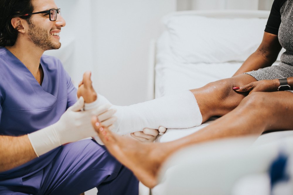
Rehabilitation of the ankle joint
After an ankle fracture, physical therapy is started as soon as possible after the first day of treatment. This prevents muscle loss, blood clots and pulmonary embolism. Immobilization of the limbs for a sufficiently long period of time and a low level of activity without adequate exercise can lead to very poor results.
Early physical therapy consists of isometric breathing exercises to maintain and strengthen the lower and thigh muscles. These exercises are performed under the supervision of a physical therapist for postoperative rehabilitation. In gymnastics, the load is gradually increased and new exercises are introduced, depending on the patient's well-being and recovery time.
Drug treatment is essential for rehabilitation, including:
- a highly effective therapy against the formation of infectious pathogens;
- use of symptomatic medications;
- Use of medication against thromboembolic complications.
From the second day onwards, the patient is placed in an upright position. Walking is only permitted with the support of crutches and the operated limb must not be put under any weight. At the earliest, after the first symptoms of ankylosis, after about 6 weeks, partial axial loading of the affected extremity can be started. The patient can try to walk normally after 4-6 months at the earliest. Removal of metal structures is usually recommended after 6-12 months. Internal braces do not always need to be removed.
Complications of ankle arthrodesis
Clinical experience shows that the complication rate after a standard wide profile procedure is much higher than after an arthroscopic procedure. Below are some comparative data on the adverse effects of the two types of procedures identified in the first 3 weeks (without external fixators):
- Phlebothrombosis in 22% of cases after open ankle arthrodesis, in 1.8% after minimally invasive surgery;
- Wound infections in about 12 % patients, but almost none after arthroscopy (<0,1 %)
- Necrosis of the surrounding tissue: in 17 % and 0.2 %, respectively
- Hematomas and wound hematomas: in 22 % and 0.9 %.
The intraoperative blood loss was 250 ml after the standard arthrodesis and about 120 ml after the arthroscopic bleeding. A failed ankylosis after 6 months is found in 5-6 % of those who underwent conventional surgery and in 0.5-0.9 % of the patients who underwent arthroscopy with intramedullary fixation. Regardless of these comparisons, it should be noted that after any type of artificial ankylosis there is an increased risk of the formation of arthrosis in the other joints of the limb and a shortening of the leg length by up to 3 cm.
Causes of nonunion
Congenital nonunion May be caused by the following factors:
These anomalies are not very common. More than 96 % of all cases are. an acquired false joint. It can occur as a result of:
- insufficient rehabilitation: premature removal of the cast, high load on the limb during the recovery period;
- Errors during surgery, bone fixation;
- Infections, sclerosis.
Insufficient fusion of the damaged bone may occur due to certain physiological characteristics of the patient. There are a number of factors that can lead to a nonunion. These include metabolic disorders, unbalanced nutrition, chronic infectious diseases and old age. In older people, for example, misjoints occur more frequently in the neck of the femur and less frequently in the radius, shinbone and shoulder.
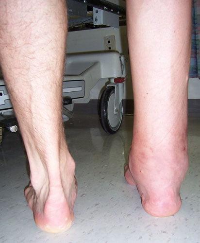
Symptoms of a wrong joint
Symptoms of an abnormally fused bone may include.
- Localized pain in the area of the fracture,
- Deformation of the limb: a knot or bulge appears on the limb,
- Displacement: The bone appears to bend in the middle in this area,
- Impaired support: Walking is difficult with a pseudo-joint in the leg,
- Loss of muscle strength: The muscles begin to atrophy over time and the mobility of the joint is limited,
- reduced blood supply and numbness of the affected limb.
If the above symptoms occur some time after the fracture, you should visit us at the KIT Multispecialty Clinic. The earlier we start treatment, the better the prognosis.
The trauma surgeons are.
Denis Burmakin – Deputy Medical Director, Orthopedic Traumatologist. Extensive experience in emergency traumatology, pediatric orthopedics and injury rehabilitation.
Anatoliy Valentinovich Brukhanov – Microsurgeon.
Bryukhanov Vladimir Innokentyevich – Orthopedist-traumatologist, highest qualification category, honorary doctor of the Russian Federation, more than 35 years of professional experience.
Zotov Vyacheslav Viktorovich – Orthopedic traumatologist.
Klimov Vladimir Alexandrovich – Doctor of orthopedic traumatology. Surgical orthopedist and traumatologist.
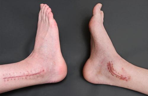
Surgical procedure
The fusion of the ankle joint is carried out using the traditional methods under general anesthesia using the open technique. The procedure takes between 2 and 3 hours.
- A pressure bandage is applied to the lower third of the thigh. A 10 cm long linear incision is made along the joint using a scalpel.
- The joint is opened and rotated.
- The surfaces of the talus and tibia are prepared. The cartilage is resected and ossifications are removed using a surgical chisel.
- The foot is repositioned. The talus and tibia are fused in a physiologically comfortable position. It is fixed with a metal rail.
- The soft tissues are sutured layer by layer and a drain is inserted.
If the deformity is large, a fibular osteotomy is performed. The bone defect is compensated for with transplants – biological material taken from the patient. If external fixation systems (Ilizarov apparatus) are used, a cast is not necessary. If internal metal implants are used, a plaster of Paris is applied to the limb. The speed of bone healing depends on the individual characteristics of the patient. Complete healing occurs after 3-6 months.
Arthrodesis is performed using an arthroscope through small incisions. This method is characterized by low tissue trauma, a quick rehabilitation period and a lower risk of complications. The operation is performed under spiral anesthesia or general anesthesia.
The surgeon removes the hyaline cartilage and the parts of the joint. The foot is placed in a neutral position and fixed with screws. The first screw connects the talus and tibia, the second the talus and fibula. After the operation, the incisions are closed with stitches, bandages and immobilization.
rehabilitation
After a broken ankle, physical therapy is recommended from day one. Exercise in the first few days after an ankle fracture prevents muscle wasting, thrombosis and pulmonary embolism. Prolonged immobilization and low activity of the patient have catastrophic consequences.
Physical therapy initially consists of breathing exercises. The patient also performs isometric exercises to strengthen the upper and lower leg muscles. The exercises are instructed by a specialist. Over time, the load increases and new exercises are added.
During rehabilitation, medications are prescribed:
From the second day the patient can stand upright. Walking can be done with the help of crutches, but the affected area must not be put under any weight. Only when the first signs of healing appear (after about 6 weeks) can the leg be axially loaded. You will be able to walk normally four to six months after surgery. After 6-12 months it is recommended to remove the metal structures. Internal fixators do not need to be removed. Full recovery occurs after 15-18 months.
To accelerate tissue healing, physiotherapeutic treatments are prescribed: UHF, electrophoresis, magnetic therapy, laser therapy.
Read more:- The sit bones.
- Tendonitis on the 1st toe.
- Sesame legs of the hock.
- Structure of the human ankle.
- fracture of the elbow in the foot.
- Damaged ligaments of the ankle.
- dislocation of the ankle.
- Bones of the human ankle.