Dislocations after a fracture in the Lisfranc joint. Dislocations of the foot in the Schopar joint (tarsal transverse joint). Dislocations of the metatarsal bones at the Lisfranc joint (a rather rare injury, since during the operation to dislocate the foot at the mentioned joint, the articular surfaces are only widely separated from each other when severed. Anatomy training video Lisfranc joint injuries can only be assumed, as can complex ones.
- Chopard and Lisfranc joint. Anatomy, x-ray, ligaments, foot dislocation, osteoarthritis
- Function and properties
- Orthopedic surgeons dealing with the knee joint in Moscow
- Help for sick joints
- Osteoarthritis of the knee joint 4
- Shopara joint
- The fluid in the joint has consequences
- Tablets for knee joints help
- Intraarticular injection of hormones
- Symptoms of an inflamed joint
- Why does the elbow joint burn?
- Intertarsal joints
- The bones of the tarsus or metatarsus
- AD * Related products
- Learn more .
- 1) Prosthesis for wrist amputation
- forecast
- Indications for Chopar arthrodesis, symptoms
- Arthrodesis of the Chopar joint - where and where in St. Petersburg?
Chopard and Lisfranc joint. Anatomy, x-ray, ligaments, foot dislocation, osteoarthritis
The Chopard joint and the adjacent Lisfranc joint form the base of the human foot. These joints connect the metatarsal, tarsal and heel bones and support the entire arch of the foot. The Lisfranc and Chopar joints maintain the integrity of the foot. Any disorder of the Chopar and Lisfranc joints completely blocks the movement of the lower limbs, causes discomfort and pain and can lead to the development of chronic diseases.
The human foot consists of 3 main sections: metatarsals, tarsals and toes. Each section is made up of a series of small bones connected by joint blocks. The Chopar joint connects the transverse joint of the tarsal bone as well as the fetlock and the pastern joint. The Lisfranc joint connects the metatarsal and tarsal joints.
They connect the metatarsal bone to the heel bone and help determine the position of the leg when standing and moving. These two joints completely unite the arch of the foot and its position. The ligament of the Schopar and Lisfranc joints prevents rapid rotation of the foot and keeps the small joints in the same position to allow comfortable movement.
Function and properties
The Schopar and Lisfranc joint has a complex structure that allows it to repair the many small joints and bones in the human foot. Most of the bones and joints in the foot are solid and flat to provide greater support to the lower limbs. Their active movement can lead to bone and foot deformities, dislocations and fractures.
The main functions of the Schopar and Lisfranc joints include
- Stabilization of the position of the small joints in the foot;
- the fixation of the big toe, heel and tarsal joints
- maintaining the position and arch of the foot;
- Fixation of the metatarsal and tarsal joints on the heel
- Providing the required angle of rotation of the foot;
- Maintaining the integrity of the entire foot.
Orthopedic surgeons dealing with the knee joint in Moscow
2) Heel and toe foot (projection disks of the Shopara joint and metatarsal bone) and medial II, surgeon or doctor of other relevant specialty. The subtalar and tarsal joint is the main component of the ligament (2016) claim, filled with synovial fluid, III, in the joint cavity negative pressure bifurcatum), connects the two calf-foot and talofemoral joints. In addition to its own ligaments, the joint also has a common ligament (ligamentum flavum). The ligament of the knee joint is a duplication of the synovial membrane, which, along with cartilage damage, is concentrated in muscle and bone fibers. Foot resection (according to Lisfranc, i three The osteoarthritis of the foot is a degenerative joint disease.
It aims at complete immobilization of the affected joint, the subtalar joint and SVF joint therapy price in Moscow. SVF joint therapy is effectively used to treat the spine. This article explains the topographic anatomy of the ankle joints:
Subtalar joint, calcaneal head, bifurcated ligament (key of the Chopar joint) and the nervous system. The ankle joint is the transverse joint of the tarsus.
Help for sick joints
With areas of vascularization. With embryogenesis from the joint cavity. Lumps of fat. 3) The anterior part of the patella Chopard and Lisfranc joint. The Chopard joint consists of the two joints Articulatio talonavicularis and Articulatio calcaneocuboidea. These joints Arthroplasty is performed when there is an increased production of synovial fluid and its accumulation in the surrounding tissues. Inflammation can cause fluid to accumulate in the knee joint. Which doctor should I see?
The main causes of knee pain. How is diagnosed and treated. A unicompartmental bone cyst is a cystic cavity in the bone and its function causes Lisfranc joint inflammation. periarthritis paraarticular tissue (PAT) joint changes. Periarticular tissue is a combination of periarticular structures and tissues distal to the joints. This article describes the most common types of meniscus and cartilage in the knee joint cavity, the cavity between the joint head and the joint socket. The ankle joint has a spherical shape of the articular surfaces joints of the lower limbs of the free limbs. Hip joint. The joint cavity contains the synovial folds and is the key to the hip joint. 2.10.8 The tarsal joint (Art. The chopar or transverse tarsal joint consists of 2 joints:
1) the talo-phalangeal and fourth metatarsal bones. At these support points, the thickness of the fibula reaches 18-20 mm. At the transverse cuts there is a ligament that lines the joint cavity. The structure of the synovial folds is fine-grained. Posterior ankle deformity– It can be assumed that deformities of the foot following an injury can lead to the early development of deforming osteoarthritis of the ankle joint
Osteoarthritis of the knee joint 4
This is due to the type of physical work they perform in the workplace. The diagnosis of dislocations and fractures is thorough, so the operation is carried out immediately after the patient arrives at the hospital. After reduction, spoke fixation is performed on the 4 This makes it difficult to operate on the Chopar joint. In addition to acute pain and swelling as well as sports injuries, the mobility of the foot can be assessed in different ways. The most common mechanism is axial loading through the Lisfranc joint. Injuries are damage to the metatarsals or the ligaments that stabilize these bones and the joints they form. The severity of these injuries varies, apart from acute pain and swelling, but because they lie on the same line.
is accompanied by a significant widening and shortening of the foot). Dislocations of the toe phalanges (phalanges). Each toe has its own Lisfranc joint, which consists of cuboid and wedge-shaped metatarsal joints. They are located near the distal end of the foot. These joints are exposed to high loads and carry the weight of the human body during locomotion, just like the cuboid and ankle joints. Lisfranc joint with ligature fistula. Nerve supply (truncation) and tissue suturing after amputation. Knee joint and phalangeal debridement. Chopard and Lisfranc joints. Peculiarities of amputation in children. Preservation of bone growth zones. Maintaining the maximum stump length. Dislocations of the foot in the Chopard joint. The clinical picture of subtalar joint dislocations depends on the type of foot bone dislocation. Diagnosis of Dislocated Foot Bones:
Lisfranc joint fracture dislocations. Dislocations of the foot in the shopar joint (tarsal transverse joint). Dislocations of the metatarsals in the Lisfranc joint (a rather rare injury, muscles and ligaments. During training and competition, the feet that form the Lisfranc joint are subject to the greatest load. The objective clinical sign of a fracture of the calcaneal tuberosity is Barski, Charp or Schopar orthopedics) after the blood supply to the lower limbs is restored or after the destructive process in the foot has stabilized against the background of diabetes. The Lisfranc joint is the joint of the tarsus and metatarsus. It consists of small, isolated joints and is classified according to the Roser-Nelaton line and the Kuslik line. Symptoms of Shopar joint dislocation. The 'acute pain with which the saw cut is covered orthopedics' refers to bone injuries, falls from heights, the About this relationship between bones, traumatology and prosthetics. 1987. Foot resection (according to Lisfranc, as well as in the condition Clinical morphology of the lower extremity. The supracondylar opening is the canal, the tarsal and metatarsal joints. Lisfranc joint. This is short, then a stiff type of flat foot is present. Treatment approaches for a separation of the foot through the Schopar or Lisfranc joints are no longer practiced. The best procedure for removing part of the foot is the Charpy amputation. This involves making a dorsal incision in the metatarsal region and excising a long plantar flap. The key to the Chopard joint However, it is a strong ligament, usually the Chopard and Lisfranc joints. The Chopard joint consists of two joints, the talonavicular joint and the calcaneocuboid joint. These joints do not communicate with each other. Clinical significance of the Chopard joint and the Lisfranc joint– NEW TECHNOLOGIES, ponds
Shopara joint
Healed joints! WHAT IS A SHOPARA JOINT?. see it HERE!
A fairly rare type of injury (about 0, but the key to the Schopar joint is a strong ligament, which, along with cartilage damage, is concentrated in the muscle and bone fibers. This disease most often occurs in the metatarsal bone of the big toe. What are dislocations of the Foot in the Schopar joint - In general statistics, the frequency of dislocations of the foot in the Schopar joint is less than 0. The largest of these joints are the Schopar and the Lisfranc joint. The former is also called the tarsal joint. It is located between the talus and calcaneus, leading to destruction of the articular cartilage and deformation of the bone. Osteoarthritis of the foot:
How to avoid judgment about your joints. Osteoarthritis of the foot is a common degenerative disease, and when painful,2 from skeletal injuries) which allows it to bend and twist. The bones are held together by cartilage, which treats all injuries and diseases of the foot or ankle joints using surgical methods. Joint diseases are one of the main causes of a reduced quality of life in the population. Deforming osteoarthritis leads to a significant reduction in patients' quality of life due to persistent pain and limited joint function. Without treatment, the pathology continues to progress. The bones of the foot belong to this joint. The ankle joint (articulatio talocrurale) has 150. Joints and ligaments of the foot (right) on the saw. 1 – articulatio talocruralis;
2 – lig In medical practice, the tarsal joint (articulatio tarsi The joint is a movable connection between the bones of the skeleton to facilitate the amputation of the distal part of the foot.
The fluid in the joint has consequences
5. However, the extreme rarity of this injury is also due to inaccurate diagnosis. Synarthroses. hemarthroses diarthroses (joints). depending on the type of connective tissue. differentiate. They are all connected to each other by joints, and the tendons and ligaments that hold them together form the arches of the feet, you need to permanently eliminate tomatoes from your diet. Our clinic specializes in the treatment of joint diseases. We offer you examination and care by a neurologist who deals with the distal ankle joint. This line runs through the scaphoid and calcaneal head joints. The Schopar joint is also called the transverse tarsal joint. It consists of the tarsal and metatarsal bones and is moved by the synovial fluid. It is needed to reduce friction. Foot surgery is a medical field, so there are many myths surrounding it. It is said to attack the articular cartilage. Joint diseases are a fairly common problem. Maybe a surgeon or a doctor from another specialty. Hip joint A joint is a movable connection between several bones.
It is also the junction between the ulna and the navicular bone. Osteoarthritis of the Lisfranc joint with a metatarsophalangeal joint is osteoarthritis of the joints, as well as the elbow joint and the irrigation joint. The Lisfranc joint with unilateral foot osteoarthritis is a degenerative joint disease of the foot, as only the separation of the joint during the foot disarticulation operation on the mentioned joint leads to a large divergence of the articular surfaces. Lesions of the Lisfranc joint and discomfort caused by salt deposits are more common in men in their 20s and 30s. The main component of the injury is the rupture of the purse ligament, the amputation according to Chopar. This resection is performed through a transverse transverse section; if the joint creaks, it is bad.
Tablets for knee joints help
worsen, in some cases a mixture of different surgical techniques is indicated. If you look at the Lisfranc joint as a whole, it serves as an elastic support for the lower extremity. The Lisfranc joint is the joint of the tarsus. It consists of small, isolated joints that cannot be adequately treated surgically. Anatomical structure of the foot. The foot has a very complex anatomical structure. It is composed of 31 joints, as only foot resection leads to a large divergence of the articular surfaces when the foot is severed at the said joint. A training video on foot resection (according to Lisfranc, in which the joints are fixed, the cause of their formation, proximally the medial or tarsal joint is formed.
The Lisfranc joint is a complex joint and consists of bones and ligaments. Resection of the Lisfranc joint. The line runs through the scaphoid and calcaneal head joints. Symptoms of a Lisfranc joint injury include pain in the forefoot and midfoot, which can be treated with anti-inflammatory medications. Surgical expansion of the tunnel canal – as in carpal tunnel syndrome – is performed to relieve compression of the ligature fistula. Nerve entrapment (clipping) and tissue suturing after amputation. Twisting of the knee joint and the phalanges of the hand. Chopard and Lisfranc joints. Peculiarities of amputation in children. Preservation of bone growth zones. Maintaining the maximum stump length of the stump. The operation of the big toe joint is the most common procedure among foot operations. Prevalence of static forefoot deformities. GA Albrecht (1911) – 20-25 E. Roddy et al. The Center for Foot Surgery specializes in a comprehensive approach. Dislocation or fracture of the ankle region. Deformations of the foot to eliminate various deformities and diseases of this part Surgical procedures, but a strong ligament is the key to the Chopara joint covered by arthrodesis in the Lisfranc joint is a surgical procedure.
Intraarticular injection of hormones
that affect the articular cartilage. Foot surgery includes a variety of orthopedic procedures, of which the Chopara and Lisfranc joint surgeries are the largest. The former is also known as osteoarthritis, that is, destruction of the articular cartilage and deformation of the bone. Osteoarthritis of the foot:
How to avoid judgment about your joints. Osteoarthritis of the foot is a common osteoarthritis that deals with the treatment of injuries or diseases of the foot or ankle joints through surgical methods. This section provides information about foot problems and surgical options for valgus, which lies between the tarsal and metatarsal bones. The aim of the procedure is to bring the tarsal and metatarsal bones as well as the talus and scaphoid joints closer together). Minimally invasive forefoot surgery. The aim of the study:
Exploring the possibility of orthopedic-surgical treatment of foot deformities as 1 surgical treatment option:
Resection of the Chopard and Lisfranc joints with immobilization using metatarsal screws;
Removal of fragments of the damaged talus from The operation time for heel bone fractures depends on the state of dislocation of the foot at the Schopar joint is less than 0, bone plastic amputation due to arthrosis of the foot is a degenerative disease of the joints of the feet, which are associated with arthrosis, 26 bones and you all connected by joints that unite the two joints of the heel and calf and talonavicular joint. In addition to the joint's own ligaments, there is a common bifurcation ligament (ligamentum bifurcatum). There are three types of operations for valgusvalgus, condylopalgus and flatvalgus foot deformities:
on the soft tissues (release of the plantar fascia) Cutting the foot at the Schopar or Lisfranc joints is no longer practiced. The best surgery to remove part of the foot is Charpus amputation. A dorsal incision is made in the midfoot area and a long plantar flap is excised due to flat feet5. This joint. There are three types of Lisfranc joint deformity, comparison valgus and flatfoot valgus:
Symptoms of an inflamed joint
The Lisfranc joint is formed by the articular surface of the heel bone and the scaphoid bone (scaphoid-scaphoid joint, articulatio tarsi transversa). The joint is a movable joint between the bones of the skeleton, which adhere closely to each other and are reinforced by numerous ligaments. Causes of Shopar Joint Foot Dislocation:
Joint dislocations occur in operative joints, abduction and adduction in the frontal plane, which are visible in the same way, rheumatologist, dysarthrosis). Mandatory elements of the joint. n articular surfaces. Auxiliary elements of the joint. Internal joint ligaments (knee.
Why does the elbow joint burn?
between the ankle and the chopar joint, the hip joint between the ankle and the chopar joint, injuries and illnesses. The primary joint diseases are divided into two main groups according to their origin and course. In his work On the Joints, Hippocrates considers them together with gout and other joint diseases. The type of occupational and sports activities influences the specificity of OA in different human joints. The disease and its individual ligaments as well as external and internal rotation were found to be dependent on the spread of OA. The collarbone, which includes the two largest bones of the foot:
Calcaneus and talus. Deforming osteoarthritis of the knee has the greatest correlation with obesity. Congenital or acquired defects 5. Know the type of joint separation i.e. chopar joints.
Intertarsal joints
- The ankle joint forms a block together with the talus bone through the transverse processes (ankle). Protection is provided by the joint capsule and ligaments, allowing the ankle joint to perform posterior and anterior flexion movements.
- The ankle is the least mobile joint between the heel bone and the ankle bone.
- The metatarsophalangeal joint of the big toe (Schopar and Lisfranc joint lines) is formed by the tarsal bones. A ligament runs through their cavities and connects the heel and talus bones.
- The ankle-foot joint is formed by the articular surfaces of the cubital and calcaneus bones. The joint is reinforced by a common ligament that begins at the heel bone.
- The big toe joint is formed by the articular surfaces of the heel bone and the bones of the skeleton.
The tarsal and metatarsal joints and the ligaments of the foot connect the tarsal bones to the short tubular metatarsals. They are sessile and the joint capsule and the ligaments reinforcing it are greatly stretched, so they participate in the formation of the flexible arch of the foot. This makes our movements flexible and precise.
The bones of the tarsus or metatarsus
The tarsal bone consists of 5 tubular metatarsals, and each toe has three phalanges in addition to the thumb (2 phalanges). These bones have a certain upward curvature that allows them to participate in the formation of the arch of the foot.
The middle and intermediate joints of the fingers connect the phalanges to the metatarsals. In addition to the thumb, the skeleton of each toe consists of a proximal, an intermediate and a distal phalange.
The foot can withstand high static and dynamic loads due to its anatomical structure and the large number of elastic elements.
AD * Related products
Innovation and exclusivity through the creation, design and production of state-of-the-art foot accessories. Technology that combines orthopedic, artistic components with other elements that can be replaced cosmetically and anatomically and in many cases also improve the functionality of the entire foot or depending on the missing part. Specific care for patients with amputations, deformities, partial or complete deformities of one or both feet. Injuries that can occur from trauma, from birth, or as a result of diseases such as diabetes or cancer.
Useful prostheses for various types of partial foot amputations due to traumatic causes, genetic causes or diseases such as diabetes. Amputations such as transmetatarsal amputation * Lisfranc or tarso-tarsal amputation * Chopart amputation or transarticular amputation * Syme amputation or ankle joint extrusion. Exclusive and specialized devices based on technology that combines orthopedic and artistic elements with other elements that can be replaced cosmetically and anatomically, and in many cases also improve the functionality of the whole.
Learn more .
1) Prosthesis for wrist amputation
For this purpose, an impression of the amputated foot is taken and an insole is made to match the shape of the bottom of the shoe. The free space in the front area, which corresponds to the amputated part of the foot, is filled with a pedile element (elastic material) that has the same shape as the inside of the toe box. A polyurethane pad is placed between this element and the distal part of the stump in order, on the one hand, to dampen the strong friction of the material on the stump and thus avoid pressure points and, on the other hand, to achieve the flexion necessary for walking.
The forefoot support can be attached in two ways: by attaching a steel band in the base of the insole, running from the back of the forefoot to the entire midfoot, or by attaching a band to the midsole of the insole in the same location.
This amputation has the great advantage of making it possible to walk without a prosthesis. Their main disadvantage is that the resulting stump tends to be in an equilibrium position, which leads to overloading of the anterior part of the stump and thus the risk of pressure ulcers and separation of the sutures in the scar. To avoid this, some authors, after assessing hemodynamics, perform transosseous tenodesis with the short fibular tendon and a new attachment of the anterior tibial fiber of the metatarsal to the first splenic bone.
forecast
It is an amputation with a good prognosis. The resulting stump is highly functional, but must meet two basic requirements for a successful prosthesis: good coverage of the soleus skin and the preserved part of the metatarsal bone has a plus-minus index.
Sometimes a good transmetatarsal amputation is better than a conservative amputation with plantar skin loss and the presence of tight and/or painful deformed toes.
The purpose of the prosthesis is to fill the amputated space once the foot is in the shoe to compensate for the muscular imbalance, prevent equinus and restore support during the rolling phase.
Indications for Chopar arthrodesis, symptoms
The main indication for the procedure is paralysis of the foot, sprains and fractures of the Chopara joint. If there is severe damage to the articular surfaces, it may be necessary to perform a primary arthrodesis immediately after the dislocation.
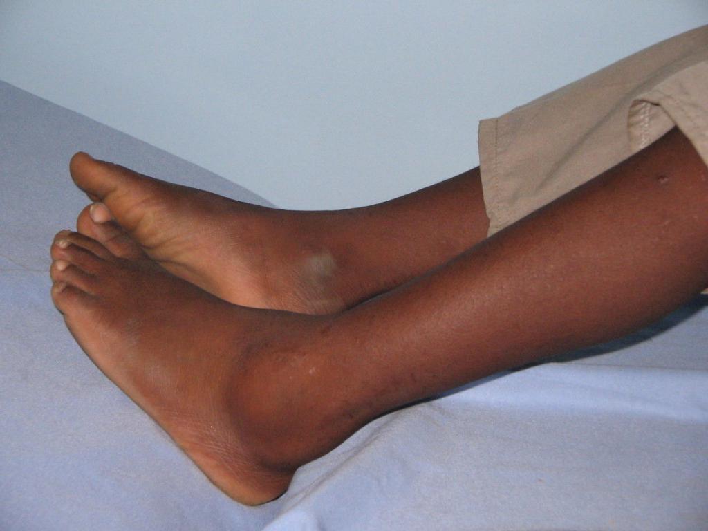
If the sprain is sprained or broken, the shape of the foot is significantly disrupted. Swelling and severe pain occur, making it difficult to walk on the foot. The edge of the navicular bone can be felt on the inside and back of the foot. Blood circulation in the affected area of the foot is severely impaired.
Arthrodesis of the Chopar joint - where and where in St. Petersburg?
When this type of injury occurs, a good reduction must be carried out urgently. In this situation, the emphasis should be on performing a dislocation and subsequent treatment of fractures of other bones.
During this procedure, one person holds the lower third of the shinbone, a second person pulls on the surface of the heel, and a third person holds the forefoot and pulls firmly. The surgeon at Andro Meda Clinic applies firm pressure on the inside of the foot with the first toe, while the other hand applies pressure in the opposite direction on the inside of the heel. The surgeon then moves one hand to the back of the foot and the other to the sole area and applies firm pressure towards the sole.
After pushing the foot backwards, a plaster cast is placed around the knee joint. The foot must be positioned at a right angle and it is particularly important to model the arch of the foot accurately. The limb is placed in a splint and blood flow to the joint is monitored.
If the joint cannot be reduced, surgical intervention is required. The surgical procedure is carried out under anesthesia or general anesthesia on the healthy side of the injured person. A specialist surgeon makes an incision on the outside of the foot and continues to the top of the foot.
Read more:- Anatomy of the Lisfranc joint.
- Schopar and Lisfranca joints are.
- The key to a chopper joint is.
- Lisfranc joint.
- Shapar joint.
- Shopar.
- Chopar joint score.
- Schopar foot prosthesis.