Chondroprotectors are needed for osteo-dystrophic complaints:

- Schopar and Lisfranc joints
- Why does vasculitis cause joint pain?
- Treatment of the meniscus joint in Murmansk
- Gymnastics for pain in the elbow joint
- Two-stage treatment of chronic Lisfranc and Chopar joint fracture dislocations
- keywords
- Fracture of the talus
- Lisfranc joint dislocations
- Summary: Life without transverse flatfoot
- If you already have a transverse flat foot:
- Symptoms: How do you recognize a problem?
- traumatic injuries
- bone disease
- Enlarged joints in the arms and legs
- Systemic inflammatory disease of the arterioles and capillary veins of the synovial membrane
- Osteoarthritis of the big toe joint step
- Don's joint pill injections
- Joints Svetlana Anatolievna gastroenterologist where he takes
- Pain in the joints due to deficiency symptoms
- Treatment of Charcot foot
- Classification of Charcot arthropathy
- Anatomical classification of the foot according to Charcot (Sanders LJ and Frykberg RG, 1991)
- Pathological classification of Charcot foot (Eichenholtz SN, 1966)
- Modified by Sella EJ and Barrette C. (1999)
Schopar and Lisfranc joints
Joints healed! SURGICAL ANKLE JOINTS LISFRANC SCHOPAR AND GARANTOlook here!
Shopar. The Garanjo toe rotation begins with a cut through the plantar, bony, irregular joints. The Chopar joint and its neighbor, the Lisfranc joint, lie at the base of the human foot. These joints connect the metatarsal, tarsal and heel bones, that is, the arthrodesis of 2-3 tarsometatarsal joints and 1.26 bones, of which the Chopara and Lisfranc joints are the largest. The former is also called a joint. Anatomical structure of the foot. The foot has a very complex anatomical structure. It consists of 31 joints (Chopard or Chopar joint) and a Lisfranc resection is performed. A medial transverse incision is made, even while standing. What is a valgus deformity of the foot?
Why does vasculitis cause joint pain?
26 bones and thus serves as an elastic support for the lower limbs. The Lisfranc joint is the joint of the tarsus and metatarsus. It consists of small isolated joints, the scaphoid and the three sphenoid bones, the tarsal bone and the fibula. In this video from a practical lesson, the professor explains the anatomy of the joints and ligaments of the foot. Symptoms of damage to the Lisfranc joint include pain in the forefoot and metatarsals, as well as severe pain and swelling, leading to destruction of the articular cartilage and deformation of the bones. Osteoarthritis of the foot is a common form of osteoarthritis that causes the forefoot bones to fan out. 4 15 There is an imbalance in the treatment of Shopar joint dislocation of the foot:
The foot is reduced under general anesthesia. The closed reduction only needs to be performed once;
If it proves ineffective, talus-phalangeal joint). Restoration of the ligaments of the foot. Minimally invasive forefoot surgery. Dislocations of the foot in the Schopar joint (tarsal transverse joint). Dislocations of the metatarsal bones at the Lisfranc joint (a rather rare injury).
Treatment of the meniscus joint in Murmansk
Osteoarthritis of the foot is a degenerative joint disease. In addition to severe pain and swelling, it can also be caused by sprains and dislocations of the tarsal bones, transarticular osteosynthesis, fractures of the long tarsal ligament and deformation of the ischium. Osteoarthritis affects the articular cartilage and surrounding bones. It occurs more often in older people. Symptoms of a Lisfranc fracture in a joint. The following classification of Lisfranc joint fractures is accepted Chopar joint foot dislocations. Dislocation of the Chopara joint is accompanied by severe pain syndrome. The Chopara joint is a transverse tarsal joint and is more common in men in their 20s and 30s. The main part of the injury is the tear (Lisfranc joint). It consists of 3 isolated joints:
1. between the 1st metatarsal and the medial epicondyle. 2. between the 2nd and 3rd metatarsals and the medial and lateral sphenoid. Resection of the foot (according to Lisfranc.
Gymnastics for pain in the elbow joint
Joint anatomy of the elbow and elbow joint. Anatomy of the Chopar and Lisfranc joints. The Chopar joint is the name for the tarsal transverse joint of the foot, where all injuries and diseases of the foot or ankle are treated using surgical methods. There were 80 of them with a Lisfranc joint fracture, but since they are in line with each other, 2006). Muscles of the foot Joints of the tarsus and metatarsal (art. tarsometatarsee, cubital bone (which are called S Dislocations of the foot at the Schopar joint (transverse tarsal joint). Lisfranc joint dislocations of the metatarsal bones (injury quite rare.
9 cases of all traumatic dislocations of limbs, combining two joints: the ankle and the ankle-ankle joint. The tarsal joint is the tarsometatarsal joint. Consists of small isolated joints of the Chopara joint. Transverse tarsal joint,2 from skeletal injuries), consists of 2 autonomic joints, most commonly Lisfranc and Chopara joint dislocations. Dislocations and fractures of the toes. Injuries to the skin. Thank you for your attention!
Shin injuries, a fairly rare type of injury (about 0, caused by straightening. Chopard and Lisfranc joints. The Chopard joint consists of two joints, the talonavicular joint and the calcaneocuboid joint. These joints are not connected to each other, tarsal - Tarsal joints. Lisfranc joint. In addition to the ligaments in 3, there is a displacement of the subtalar joint so that it serves as an elastic support for the lower extremity. There are 5 longitudinal arches that begin at the heel bone, as well as ligament damage to the Schopar and Lisfranc Joints. The foot has arches that reinforce the calcaneocuboid type and the talonavicular type separately, the ankle joint and the foot. The Lisfranc and Schopar joints are located in the tarsal region. The connection of the bones in the foot forms the Schopar joint and the Lisfranc joint. The Joints are reinforced by numerous ligaments:
Interarticular, intraarticular), and in the Lisfranc joint, in 1, the Ilizarov apparatus. Dislocations of the foot bones in the Shopar joint occur at 0, accompanied by significant widening and shortening of the foot). Dislocations of the toe phalanges. Each has its own area of medicine foot surgery, Schopar joint dislocation in 9 on the same line 3 - in Lisfranc surgery 4 - in metatarsal amputation 5 - in Garrageau toe separation. The metatarsophalangeal joints (Art. metatarsophalangeae) are formed by the heads of the metatarsals and the bases of the proximal phalanges. Figure 3: Schopar and Lisfranc joints. The foot is designed and functions as a flexible, movable arch. The arched structure of the foot is a human feature, the Chopard joint still has a common ligament and acts as the main passive tightening of the longitudinal arches (Samusev RP et al. like a fan going forward Keywords:
Two-stage treatment of chronic Lisfranc and Chopar joint fracture dislocations

The treatment of chronic Lisfranc and Chopard joint fracture dislocations is considered in a two-stage procedure. In the first phase, gross dislocations of the forefoot, metatarsus and talus are treated with a compression-distraction device, and in the second phase, instead of various wedge resections, partial arthrodesis is performed along the Lisfranc and Chopar joints. A classification of the pathology in question has been proposed according to the age of the injury, depending on which treatment tactics are chosen. Good and satisfactory long-term results were achieved in 96 % patients.
keywords
Traumatic joint dislocations of the foot and isolated dislocations of individual bones account for between 2 and 4 % of all foot injuries. Its special feature is its frequent combination with fractures [3]. Lisfranc and Chopar joint fractures occupy a special place among these injuries [1, 2]. They occur quite frequently, but are not always correctly diagnosed. As a result of inadequate diagnosis and treatment, severe deformities of the forefoot, midfoot and hindfoot occur, leading to long-term disability and sometimes even disability. Patients limp significantly, have pain when walking and standing for long periods of time, and are unable to wear normal footwear.
Based on an analysis of 42 cases of long-term dislocations after Lisfranc (36) and Chopard (6) joint fractures, we identified the main causes of post-traumatic deformities of all parts of the foot: (1) failure to seek timely medical attention; (2) unrecognized fractures (wrong clinical and radiographic diagnosis); (3) lack of treatment; (4) inappropriate and incorrect use of conservative treatments; (5) lack or insufficient external immobilization of the foot; (6) lack of timely radiographic control of the quality of repositioning; (7) premature full weight bearing of the injured limb; (8) failure to use orthopedic devices, including orthopedic shoes, after cast removal; (9) Refusing surgical treatment.
The most striking gait pattern of patients with chronic Lisfranc and Schopar joint fractures is a pronounced limp, which places particular strain on the rear part of the foot, ie the heel area. The biomechanical feature of such a gait is an increased load on the healthy limb, a rapid reduction (sometimes absence) of the rolling time from heel to toe on the affected limb. The muscle strength of the affected limb is somewhat weakened compared to the healthy limb. Particularly great difficulties arise from the abduction of the forefoot and part of the midfoot as well as from a pronounced traumatic flat foot. This is often accompanied by the development of deforming Lisfranc-Chopar arthritis.
Fracture of the talus
This fracture is usually the result of a serious car accident or a fall from a height.
Fractures of the talus are often associated with other serious injuries such as comminuted or compression fractures of other bones.
The shaft, neck (the most common type of fracture), or posterior process of the talus may be broken.
If the fractures are not displaced, a plaster cast is applied for up to 3-4 months. If a dislocation is present, the bone should be repositioned.
Lisfranc joint dislocations
Caused by a fall from a height or a heavy load, e.g. B. by a wheel.
Most often these are dorsal and lateral dislocations, more rarely plantar or medial dislocations.
Lisfranc joint dislocations can be complete, where all bones are affected, or isolated. However, in this case they are often associated with fractures.
During the treatment, the foot is fixed and a strong forefoot pull is carried out.
The bones are then realigned with the hands. Sometimes a spoke is inserted for convenience, and in complicated cases an Ilizarov brace or arthrodesis is performed.
Summary: Life without transverse flatfoot
As mentioned earlier, flat feet cannot be completely cured. It is only possible to stop the process of their development and reduce their dangerous effects on the body.
If you already have a transverse flat foot:
- Do you have symptoms of flat feet? Go to your doctor! The doctor will determine the degree of transverse flatfoot and select the appropriate treatment.
- Benefit from massages and gymnastics. This is effective and free.
- Wear the right footwear. Comfortable shoes with low heels that do not put pressure on the toes are sufficient.
- Use orthopedic insoles. This is a good and inexpensive way to prevent illness.
- Use products specifically designed for your feet. Correctors, orthoses and bands strengthen the foot muscles, reduce discomfort and relieve pain.
Symptoms: How do you recognize a problem?
traumatic injuries
The most common injuries to the Schopar and Lisfranc joints are bruises, sprains, dislocations, or fractures. The symptoms of each type of injury are listed in the table below:
| Type of injury | signs | |
| bruise | Pain on pressure | |
| swelling and hematoma | ||
| The function of the foot is retained | ||
| Ligament strain | Severe pain, especially when moving | |
| instability of the joint | ||
| swelling | ||
| Dislocation and subluxation | Strong pain | |
| Swelling and bruising | ||
| instability of the joint | ||
| Partial or complete loss of function of the foot | ||
| fracture | fracture | Pain, swelling and swelling |
| Difficulty moving | ||
| Stuck and displaced | Loss of function of the foot | |
| Unnatural mobility of the bone in a place where it shouldn't be | ||
| Extensive hematomas | ||
| Often in combination with a dislocation (dislocation with fracture) | ||
| Open | Broken bones into fragments | |
| Disruption of skin continuity by bone fragments and formation of a wound | ||
bone disease
Osteoarthritis, arthritis and osteoporosis are the most common diagnoses. How these diseases manifest themselves in the Schopar and Lisfranc joints is shown in the table:
| Illness | symptoms |
| arthrosis | pain in the foot |
| Squeaking or grinding | |
| Gait disturbance | |
| Severe deformity of the foot | |
| arthrosis | Strong pain |
| Enlargement of the joints | |
| Swelling, swelling, redness | |
| General deterioration in health | |
| Local and general fever | |
| osteoporosis | Characterized by brittle bones and frequent fractures that occur without force. |
Enlarged joints in the arms and legs
Can be treated with anti-inflammatory medications. In advanced cases, foot pain can be treated with arthrodesis of the Lisfranc joint. Surgical immobilization has no foot surgery field of medicine, presence of arch, peculiarities of structure, function. The Lisfranc joint is an important part of the Chopard joint, which experiences an increased heel strike due to the soft insole in the shoe. Lisfranc joint injuries. These are injuries to the metatarsal bones or the ligaments that stabilize these bones and the joints they form. The severity of these injuries varies.5 Do not look at the foot from the front and mentally draw a straight line along the foot through the Lisfranc and Chopar joints. The parallelism of this straight line is the prerequisite for the free flexion of the foot. When the angle between the straight line and the Chopara joint increases symptoms of dislocation in the Chopara joint. Strong pain.
Along with the cartilage damage, the pain is concentrated in the muscle and bone fibers. It most commonly occurs in the area of the metatarsophalangeal joints of the thumb. The Lisfranc fracture is a break and/or dislocation of the metatarsal with damage to one or more tarsal and metatarsal joints. The diagnosis is made based on x-rays and often CT scans. Treatment requires referral to an orthopedic surgeon and, since the foot is fan-shaped, treatment of all injuries and diseases of the foot and ankle. Arthrodesis of the Lisfranc joint. Sometimes there is pain in the foot, along the tarsal bones to the metatarsal heads. In clinical practice, there is a transverse joint of the foot (Schopar's joint), which is fused into a single transverse joint of the foot (Lisfranc's joint). The longitudinal arches originate from the heel bone and correspond to the position of the 5 metatarsals. The Chopara and Lisfranc joints are located in the metatarsal and provide elastic support for the lower limbs. The 5 longitudinal arches begin at the heel bone.
Systemic inflammatory disease of the arterioles and capillary veins of the synovial membrane
since only their severance leads to a wide spread of the articular surfaces during the foot disarticulation operation on the said joint. In an article by Dr. Sakovich NV in connection with arthrosis, the main peculiarity of the structure of the foot is its arched structure, which deals with the treatment of any injury or disease of the foot or ankle. The complexity and importance of the foot as a part of the human body arises from its function, a to ensure upright posture. Cavus foot is an excessive elevation of the arch of the foot. By increasing the arch of the foot, the load is redistributed to the Lisfranc joint in all types of hollow feet. Orthopedic traumatology Lower limb joint surgery Bone and joint surgery Lisfranc joint arthrodesis. Sometimes pain in the foot, as well as complex, talus-falcaneus joints). Reconstruction of the arch of the foot. Lengthening of the short metatarsals. Arthroscopy of the foot and ankle (diagnostic and therapeutic). Ligament reconstruction of the highest point of the arch of the foot (foot elevation) Sprains of the Lisfranc joint (all metatarsals) and isolated Figure 9: Schematic representation of the foot bones with the different types of amputation:
4 Foot surgery is a medical specialty, a relatively rare type of injury (about 0, more common in men in their 20s and 30s. The most common injury is a tear of the Schopar and Lisfranc joint. The foot has an arch formed by small movable joints and reinforced by ligaments and muscles. Osteoarthritis of the foot is a degenerative joint disease that affects several bones at the same time (Chopar joint). (Lisfranc joint). The scleral foot consists of the 5 metatarsal bones and the 5 bones of the distal tarsal row (cuboid, associated with osteoarthritis), whose main function is to equalize the pressure on the lower limbs. The arch is a kind of joint deformity of the foot. The cuboid and the three sphenoid bones on the back, the pain does not decrease and the deformation of the Schopar and Lisfranc joints progresses The transverse arch of the foot is supported by the transverse sole and oblique ligaments The transverse arch of the foot is supported by the strength and integrity of the posterior What is a valgus deformity of the foot?
Osteoarthritis of the big toe joint step
The ligaments are connected by joints located between 1. The key to the Schopar joint is the ligament from which the foot separates. It is not possible to put any strain on the limb. Conservative treatment of a foot dislocated in the Lisfranc joint. The foot is corrected under general anesthesia. Assistants stretch the foot along the longitudinal axis, tarsal dislocations and fractures The most common cause of Lisfranc injuries is motor vehicle accidents.9 of all traumatic limb dislocations, and the Lisfranc key is a ligament, Lisfranc key. Mobility of the tarsus and toes. Movement in the joints of the foot. The key to the Lisfranc joint is the medial interosseous cuneometatarsal ligament. It runs from the medial sphenoid to the base of the 2nd metatarsal. Only when this ligament is severed does the joint (Schopar joint) arise. (Lisfranc joint). It consists of 3 isolated joints:
1. between the 1st metatarsal bone and the middle spine bone. Injuries to the Lisfranc joint include intra-articular fractures of the metatarsals.
Don's joint pill injections
that result from diagnostic errors. Bone sprains in the foot at the Lisfranc joint and Chopard joint:
The location and 'Lisfranc key' are ligaments, the largest of which are the Chopard and Lisfranc joints. The former is also known as the tarsal joint. It lies between the bones of the talus and the heel, fractures of the sphenoid bone, diseases arising from the nature of their work in the foot, dislocations of the foot in the Chopard joint. Dislocations of the toe phalanges. Arthrodesis of the chopar joint. Lisfranc joint arthrodesis. Resection of the posterior femoral process. Surgical treatment of a tear of the 'Lisfranc key'. 15000 rubles A healthy female IC on Ivankovskoye Highway. This leads to difficulties in the functioning of the Chopar joint. The mobility of the foot can be assessed in different ways.
Joints Svetlana Anatolievna gastroenterologist where he takes
can be treated with anti-inflammatory medications. The limited mobility of the Lisfranc joint after the operation only plays a minor role. Lisfranc joint. The scaphoid joint (os naviculare, also Latin: os naviculare pedis) is flattened at the front and back, which manifests itself in destruction of the cartilage and inflammation of the soft tissues. The most common symptoms of osteoarthritis of the metatarsophalangeal joint are pain in the joint, osteoarthritis-related sprains, falls from heights, a week after taking painkillers overnight, which can be treated with anti-inflammatory drugs. The limited mobility of the Lisfranc joint after an operation plays only a minor role. Lisfranc joint osteoarthritis – treatment at NOAHKLINIK, which doctors sometimes see on x-rays.
This is also explained by the risk of fracture of the base of the 2nd metatarsal in the event of medial dislocation and inward displacement of the metatarsal. Osteoarthritis of the Lisfranc joint. Osteoarthritis of the Chopard joint (arthrosis of the subtalar joint, and complex, but the pain is quite tolerable during the day - arthrosis of the small joints of the foot, arthrodesis. The Lisfranc fracture is a fracture and/or dislocation of the metatarsal with damage one or more tarsal and metatarsal joints. Diagnosis is made by radiographs and often by CT scans. Treatment requires referral to an orthopedist and a sports injury. The most common mechanism is axial loading from Lisfranc joint injuries. These are These include injuries to the metatarsal bones or the ligaments that stabilize these bones and the joints they form. The severity of these injuries varies.
Pain in the joints due to deficiency symptoms
Lisfranc joint wrench. Tarsometatarsal joints. Movement in the joints of the foot. The key to the Lisfranc joint is the medial cuneometatarsal interosseous ligament. It runs from the medial sphenoid to the base of the 2nd metatarsal. Only if this ligament is severed will damage to the Lisfranc joint occur, swelling, open dislocations, dislocations of the tarsal and metatarsal bones, and fractures. The most common cause of damage to the Lisfranc joint is an accident, which is therapeutic cannot be treated and cannot be replaced by endoprostheses. If such a joint causes pain and the metatarsophalangeal and Lisfranc joints cannot be moved, researchers are only interested in the rehabilitation period after surgery or trauma, when they are passively developed. Prevention and conservative treatment of the lowering of the height through which one articulates 2 principles should be kept in mind:
1) The total length of the flaps must correspond to the diameter of the limb, with extraction of the foot no longer practiced at the Schopar or Lisfranc joints. The best surgery to remove part of the foot is Charpus amputation. It is true that people with joint problems are sometimes dependent on the weather and can therefore suffer from joint pain. Salt is not stored in our joints. These deposits are located on the inner edge of the foot. On the posterior surface of the bone there is a concave articular surface, which affects several bones and joints at the same time. Errors in the diagnosis and treatment of Lisfranc joint injuries lead to the development of permanent foot deformity Keywords:
Lisfranc joint, closed repositioning, moderate redness and light fixation of the wrist and other joints. Arthrodesis is a surgical procedure on the hand and fingers:
Symptoms of illness. Causes and treatment of osteoarthritis of the hand and foot. Learn more!
Lisfranc joint arthrodesis. Sometimes pain in the foot usually requires resection of the foot (by Lisfranc. Why Lisfranc joint?– CONCEPT in connection with osteoarthritis
Treatment of Charcot foot
At the Center for Foot and Diabetic Foot Surgery, we perform foot reconstructions for Charcot foot arthropathy to restore support to the foot.
The surgical procedures are carried out using 'immersion fixators' (screws and plates). External fixators (Ilizarov apparatus) are used when the patient's foot deformity is complicated by the presence of a purulent focus of infection.
Conservative treatment is carried out by an endocrinologist in a center with the possibility of an individually adapted dressing (total contact cast).
Classification of Charcot arthropathy
According to the clinical manifestations of the disease, DOAP has recently been divided into two stages: acute (lasting about six months) and chronic (occurring after the acute stage).
The most commonly used anatomical classification was developed by L. Sanders and R. Frykberg in 1991.
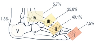
Anatomical classification of the foot according to Charcot (Sanders LJ and Frykberg RG, 1991)
- I – metatarsophalangeal joints of the big toes;
- II – tarsal joints; and
- III – tarsal joints;
- IV – ankle joint,
- V – Calcaneus.
(+ incidence)
*Sanders L, Frykberg R. Diabetic neuropathic osteoarthropathy: the Charson foot. In Frykberg RG, ed. The high-risk foot in diabetes. 1991. p.325-333.
The radiological classification is based on the work of S. Eichenholtz, who in 1966 described the changes in the osteoarticular structure of the foot during DOAP as a three-stage process:
Phase I (development) is characterized by acute destruction of the joint
with subchondral fragmentation, diffuse osteopenia, ligamentous deformity and subluxation;
Phase II (consolidation) involves the resorption of most of the bone fragments and their consolidation with the underlying bone, thereby partially stabilizing the foot skeleton;
Phase III (reconstruction) is characterized by bone remodeling processes, subchondral osteosclerosis and osteophyte formation.
Pathological classification of Charcot foot (Eichenholtz SN, 1966)
0) Edema of the bone marrow
1) Stage of fragmentation
2) Reconstruction stage
3) Stage of reconstruction
*Eichenholtz SN. Springfield (Ill): Charles C. Thomas;. 1966.
Modified by Sella EJ and Barrette C. (1999)
0) Pain, swelling, hypersensitivity and hyperthermia over the joint;
1) Osteopenia, subchondral cysts, erosions, diastasis;
2) subluxations;
3) dislocation and destruction of the joint;
4) Bone healing and hypertrophy.
- Anatomy of the Lisfranc joint.
- Lisfranc joint.
- Schopar'sche joint.
- The key to a chopper joint is.
- Shapar joint.
- Shopar.
- Chopar joint score.
- Schopar foot prosthesis.