a – upon admission; b – after open reduction of the Chopard joint dislocation with Kirschner immobilization. c – 3 years after treatment: Reversal of Chopard joint dislocation.

- Lisfranc joint and Chopard joint
- Why do pathologies of the Schopar and Lisfranc joints occur?
- symptoms
- Intertarsal joints
- 1) Metatarsal amputation prosthesis
- forecast
- Hallux Rigidus (stiff first metatarsophalangeal joint)
- Chopard amputation
- Proprioceptive information about the foot
- anatomy
- Shopar joint foot dislocation
- symptoms and diagnosis
- Stamps on a Chopard watch
- Chopard: the history of the brand
- Features of Shopar Sunglasses
- Treatment: which methods are effective?
- preparations
- prevention
Lisfranc joint and Chopard joint
The bones in the foot are connected by the Chopard joint and the Lisfranc joint. This joint is held in place by strong ligaments on the soles of the feet. Joint injuries and diseases – arthritis, osteoarthritis and osteoporosis – are common. If you have discomfort in your foot, it is worth visiting a doctor who will diagnose the problem, prescribe treatment and give preventive advice.
The Schopar joint is located in the tarsal region, near the ankle joint, and connects the joints of the scaphoid and heel bone. It is supported by a strong bicarticular ligament that begins at the upper end of the heel bone and wraps around the bony structures of the scaphoid and heel bone. This connective tissue is called 'Chopard's key'.
The anatomy of the Lisfranc joint includes the cuneiform joint and the proximal metatarsal joint, which are located closer to the distal end of the foot. The joint formations are reinforced by the intercondylar cuneiform ligaments, which connect the second cuneiform and metatarsal bones. This tape device is called the 'Lisfranc key'.
Why do pathologies of the Schopar and Lisfranc joints occur?
These movable joints of the foot are subjected to a lot of wear and tear as they support the weight of the body when walking and standing, and are the most vulnerable to injury and illness.
The main causes of diseases and injuries of the musculoskeletal system are.
- tripping over foot;
- Falling from a height
- Pinching the foot/leg directly against a hard object;
- participation in sports in which the lower limbs bear the brunt;
- Occupational activities that involve long-term exposure to vibrations and shocks;
- lack of calcium in the body;
- unhealthy habits and poor diet.
symptoms
The clinical symptoms vary from patient to patient. The degree of inflammation and destruction varies depending on the stage of the disease and ranges from mild pain with prolonged standing to gait changes and toe deformities. Many people wonder how to diagnose foot osteoarthritis and how to tell that it is not arthritis with similar symptoms.
The main symptoms of foot osteoarthritis:
1.
When the joint moves, a characteristic, rough crunch occurs, which is caused by the appearance of uneven joint surfaces.
2.
Pain occurs during physical exertion, which may subside with rest. Typically the pain is dull and inconsistent and increases in cold, wet weather and when the affected joints come into contact with water. Complete immobility is rare, but stiffness is also seen in the development of rheumatoid arthritis of the hip.
3.
The patient feels stiffness upon waking in the morning, and throughout the day joint mobility is limited and muscles are tense.
Gradual deformation of the joints, which may be a sign of the development of osteoarthritis. However, these are not caused by soft tissue, but rather by bony overgrowth.
5.
Impairment of gait and posture as the patient attempts to relieve pressure on the painful joint.
6.
Swelling, occasionally swelling and redness of the skin over the affected joints.
7.
An increase in body temperature is characteristic of any joint disease, regardless of whether it is osteoarthritis of the hip, knee or foot.
8.
Increased fatigue, reduced ability to work and the appearance of specific blisters.
9.
In the later stages of the disease, the joints become dysfunctional, thicken, and bony hypertrophy or Heberden nodules develop, which can later grow outward.
Intertarsal joints
- The ankle joint forms a block with the talus. Protection is provided by the joint capsule and ligaments that allow the ankle joint to move forward and backward.
- The ankle is the least mobile joint between the heel bone and the talus.
- The metatarsophalangeal joint of the big toe (Schopar and Lisfranc joint lines) is formed by the tarsal bones. A ligament runs through their cavities and connects the heel and ankle bones.
- The ankle-foot joint is formed by the articular surfaces of the elbow and heel bone. The joint is reinforced by a common ligament that begins at the heel bone.
- The metatarsophalangeal joint of the big toe is formed by the articular surfaces of the ischium and scaphoid bone.
The tarsal joints and ligaments of the foot connect the tarsal bones to the short tubular metatarsals. They are freely movable, the joint capsule and the ligaments that strengthen them are greatly stretched, so they participate in the formation of the flexible arch of the foot. This makes us agile and precise in our movements.


1) Metatarsal amputation prosthesis
An impression of the amputated foot is taken and an insole is made that is adapted to the shape of the bottom of the shoe. The free space in the front area, which corresponds to the amputated part of the foot, is filled with a pedile element (elastic material) that has the same shape as the inside of the toe box. A polyurethane pad is placed between this element and the distal part of the stump in order, on the one hand, to dampen the strong friction of the material on the stump and thus avoid pressure points and, on the other hand, to achieve the flexion necessary for walking.
The forefoot support can be attached in two ways: by attaching a steel band within the base of the insole, running from the backfoot of the forefoot to the entire midfoot, or by attaching a band to the midsole of the insole in the same location.
This amputation has the great advantage of making it possible to walk without a prosthesis. Their main disadvantage is that the resulting stump tends to assume an equilibrium position, which leads to overloading of the front part of the stump and therefore the risk of wounds and suture separation in the scar. To avoid this, some authors, after assessing the hemodynamics, perform transosseous tenodesis with the short fibular tendon and a new anterior tibial fiber attachment of the metatarsal to the first splenic bone.
forecast
It is an amputation with a good prognosis. The resulting stump is highly functional, but must meet two basic requirements for a successful prosthesis: good coverage of the soleus skin and the preserved part of the metatarsal bone has a plus-minus index.
Sometimes a good transmetatarsal amputation is better than a conservative amputation with plantar skin loss and the presence of tight and/or painfully deformed toes.
The purpose of the prosthesis is to fill the amputated space once the foot is in the shoe to correct the muscular imbalance, prevent equinus and restore support during the rolling phase.
Hallux Rigidus (stiff first metatarsophalangeal joint)
Of all the joints in the foot, the metatarsophalangeal joint of the big toe is most commonly affected by osteoarthritis. It is the most important joint in walking because its sole surface is one of the main supporting surfaces of the foot and the big toe is exposed to the main force of the sole impact. For this reason, stiffening of the big toe joint causes severe pain when walking and disturbances in gait. Hallux Rigidus most commonly affects people aged 30-60. The causes of this pathology are not entirely clear, and in most cases no specific cause can be identified. In cases where the cause is clear, the development of stiffness is usually due to previous trauma or abnormal anatomy of the foot that results in excessive overloading of the forefoot.
As with most other movable joints, the ends of the bones that form it are covered by smooth hyaline cartilage. Severe wear, trauma or metabolic damage causes defects of varying degrees of severity in the cartilage. Impaired joint glide and reduced congruence transfer excessive stress to the surrounding soft tissues, the inflammation and subsequent calcium deposition of which leads to the formation of bone spikes that prevent joint movement and normal walking. There are no nerve endings in the hyaline cartilage itself; from this point of view, the cartilage does not 'hurt', so the pain syndrome indicates an advanced degenerative process, since it means that the underlying bone is involved in the pathological process.
Pain in the area of the first metatarsophalangeal joint of the big toe, especially when pressing the toe while walking.
Swelling in the area of the big toe joint.
'Nodules on the dorsal surface of the metatarsophalangeal joint of the big toe.
Limitation of the range of motion of the big toe.
Chopard amputation
Chopard prostheses or transarticular amputations of the foot are based on a technology that combines orthopedic and artistic elements with other elements that can replace them cosmetically and anatomically, in many cases also improving the function of the entire foot or foot, depending on the missing part becomes.
We must first understand its main function, which is to be the necessary support for bipedal (two-foot) locomotion.
The legs perform several basic functions:
They absorb the entire weight of the body and ensure its balance.
They absorb constant shocks and soften the energy generated by the forces involved in walking.
They adapt to different types of surfaces, even the most uneven.
Functions like a rigid lever that generates the propulsive force required for walking.
Transmits the rotational forces generated by the hips.
Of course it is linked to walking.
Proprioceptive information about the foot
Your muscles, tendons and joints have special sensors that react to movement or pressure. Thanks to these sensors, your brain knows how your feet are positioned in relation to the ground.
All of the structures that make up the foot, the muscles and tendons, work together with the bones, ligaments and joints and are synchronized with the brain to enable the feet to perform these functions. If one of these elements does not function properly, all others will be affected.
Solutions recommended by leading surgeons, orthopedists, traumatologists, physiotherapists, podiatrists, psychologists, psychiatrists and relevant medical staff.
Exclusive and innovative for patients who have suffered amputations, deformities and other types of traumatic or congenital injuries.
anatomy
The ligament of the Chopard joint is bifurcated and attaches to the distal edge of the heel bone. It branches almost at the origin and forms the lateral and medial bands. The lateral calcoclavicular ligament attaches to the dorsum of the elbow, while the medial calcoclavicular ligament attaches to the heel bone.
The two-part ligament is short and very strong and ensures a stable position of the bones. It is the 'key to the Schopar joint': even if all neighboring ligaments are torn, the relative position of the bone structures remains unchanged. Only if the foramen ligamentum is severely damaged will the joint open.
The Schopar joint or transverse tarsal joint is cut in the shape of a horizontally lying Latin letter S. The common line runs 2.5 to 3 cm below the inner malleolus and up to 4 cm below the outer malleolus. This means that the medial joint protrudes forward and the lateral joint protrudes backwards.

People with weak ligaments are particularly susceptible to sprains
Shopar joint foot dislocation
According to medical statistics, injuries to the Schopar joint are quite rare. However, the statistics do not always take into account an inaccurate diagnosis, so the frequency of dislocation of the Chopar joint can be much higher than 0.5 %.
A dislocation can be caused by a sudden fall with the support of the forefoot, a sharp and forceful impact on the protruding metatarsal bone. Most of the time the injury occurs indirectly with considerable force.
As a result of the injury, the ligaments between the scaphoid and talus bone and between the elbow and heel bones tear.
The forefoot is displaced in different ways:
- In the rear direction;
- towards the sole of the foot;
- laterally;
- medial (most common);
- Combined relocation, e.g. B. dorsal-medial.
Since the Schopar joint can only be damaged by very high force, foot dislocations are often accompanied by bone fractures. Fractures of the scaphoid, cuboid or talar bone are possible. Since the forefoot is usually displaced inward, the elbow bone also breaks. A fracture of the scaphoid bone is somewhat rarer because the distal part of the foot is less likely to be displaced outwards.

A compression fracture of the ischium is caused by misalignment of the foot resulting from displacement of the talus.
symptoms and diagnosis
An injury to the Schopar joint can be recognized by the characteristic deformation of the foot - the head of the talus protrudes sharply above the surface and threatens to break through the skin. The patient suffers from severe pain syndrome and cannot support the foot. There are clear signs of inflammation - swelling and redness.
It is noteworthy that as the swelling increases, the deformity becomes less visible. To clarify the type of dislocation, an x-ray is taken. Diagnosis of the injury can be complicated if it involves a fracture of the navicular bone. In this case, x-rays are taken in unusual oblique projections.
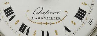
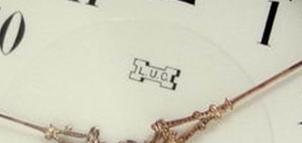
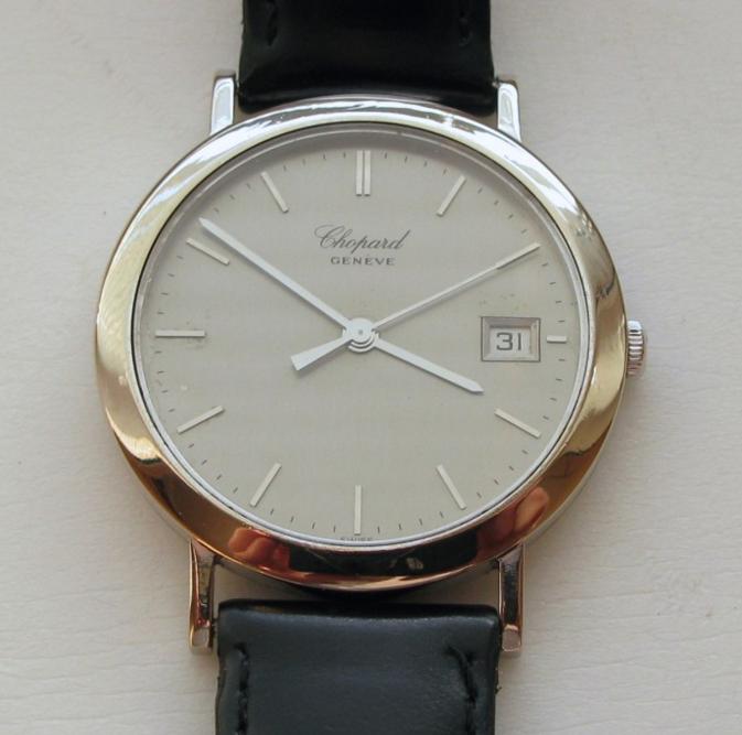
Stamps on a Chopard watch
There are several types of stamps on Chopard watches. The name of the founder and the place of manufacture are indicated on the dials of the first models: 'Chopard A Sonvillier'.
Another variant is the letter LUC, an abbreviation for the name of the company's founder, Louis-Ulysse Chopard.
There are also variations on this theme. For example, when moving to Geneva, the watch is signed: 'Chopard Geneve'.
In another case, only the first names are abbreviated and the last name is written out in full: 'LU Chopard'.
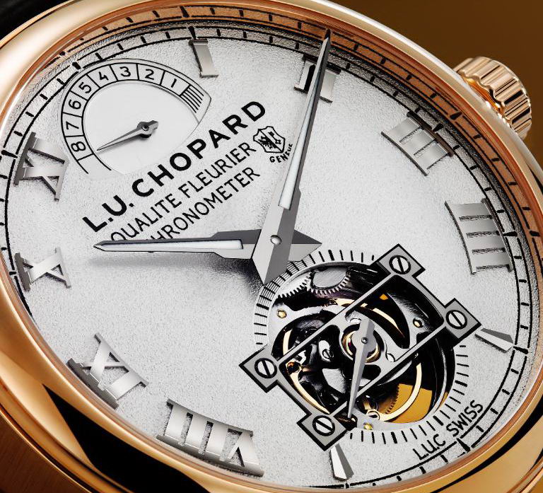
On the mechanisms and covers themselves there are various of the above options, from a short LUC with or without a numeral to a longer 'LU Chopard & Cie Geneve'.
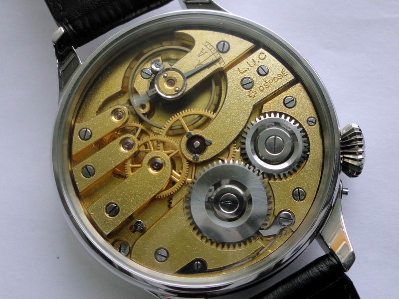
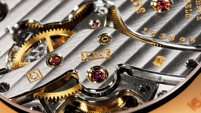
Interestingly, the long name and the brand name LUC can appear on the same part of the watch.
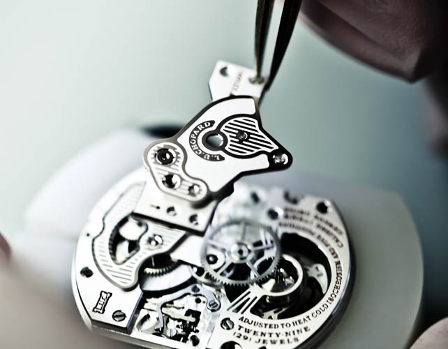
Chopard: the history of the brand
The history of Chopard begins in the 19th century. Century. In the 1860s, the watchmaker Louis-Ulysse Chopard founded his own factory to produce watches. The company quickly made a name for itself in Switzerland as it became an important supplier to the Swiss railways. A few years later, the company founder moved to Geneva, which was then a center of watchmaking. In 1963, Karl Scheffel, a member of the Chopard family, took over the company. He was not only a master watchmaker, but also a jeweler. The company's product range is expanding.
For many decades, Scheffel and his successors maintained the tradition for which Chopard products were famous. Today the brand also produces accessories, including sunglasses and eyeglass frames. They are all associated with luxury. The first models appeared in the early 2000s and were an immediate bestseller. The following organizing principles contributed:

- A tradition of fine craftsmanship preserved since the first wristwatches were made;
- Technical innovation;
- An original and unexpected approach to design;
- the use of the latest exclusive materials;
- the use of precious metals and gemstones in our work;
- Reliable protection for every product against counterfeiting.
The history of Chopard glasses has just begun. Each collection is eagerly awaited by the public. Critics have poetically described the brand's products as 'luxury that magnetically draws you into the abyss of passion'.
Features of Shopar Sunglasses
The company pays great attention to detail in the manufacture of its sunglasses and medical frames. Every single detail is there. There is nothing superfluous, just like with a watch or a piece of jewellery. Expensive materials are used for the manufacture of optical products: mahogany, leather, rubber, precious or semiprecious stones. The lenses are made of very durable materials that are hard to scratch and difficult to break. They do not lose their optical properties over the years.
Of course the branded items are expensive. However, the company pursues a relatively democratic pricing policy. Many models are not only available for people with high incomes. In this case, the quality of the cheaper goods does not suffer. It is not possible to buy Chopard sunglasses on the street, but a person with an average income can gift themselves such an exquisite accessory.

The selection of Chopard glasses is very diverse. Celebrities prefer cat-eye style models. These are glasses frames with extended upper corners. They are suitable for women. With such glasses, any image will become even more sophisticated and sophisticated. Wear them to a party, to the beach or just for a walk. The lenses protect you from the sun and the frame suits every style.
Like many other brands, Chopard makes aviator-shaped sunglasses. They are made of metal. The thin metal frames with glass lenses look very serious and stylish. There is a wide range of colors. Among the most coveted models are those with gold frames and gray lenses. This factor also depends on the time of year. However, Chopard sunglasses are timeless. They will always be relevant because they symbolize luxury and refined taste. Let's take a look at the brand's most famous models.
Treatment: which methods are effective?
preparations
Traumatic injuries are treated by immobilization and the application of a cast, brace, or tunic. To relieve pain and inflammation, it is recommended to take medications:
Chondroprotectors are needed for degenerative-dystrophic complaints:

- 'Dona';
- 'Artra';
- 'Chondroxide'.
If Chopar and Lisfranc joints are affected by osteoporosis, calcium preparations must be used:
All medications must be taken as prescribed by a doctor; self-medication is not permitted. To restore mobility to the foot, physical therapy, massage, chiropractic care, water treatment, and daily strengthening and recovery exercises should be performed. The degree of stress on the foot should be determined by the doctor to avoid recurrence of the disease.
prevention
To keep the Lisfranc and Chopar joints intact, it is advisable to avoid falls. Choose comfortable shoes with a wide, stable heel no higher than 4 cm. When playing sports, special insoles should be used to support the foot. Strengthen your immune system, eat right, give up unhealthy habits, lead an active lifestyle and control your weight.
Read more:- Anatomy of the Lisfranc joint.
- Schopar and Lisfranca joints are.
- Schopar'sche joint.
- Lisfranc joint.
- The key to a chopper joint is.
- Shapar joint.
- Chopar joint score.
- Schopar foot prosthesis.