Arthrodesis is the main treatment option for unresectable cases after conservative or surgical treatment has failed.

- Fractures of the navicular bone
- Conservative treatment
- scaphoid
- Clinical Significance
- Horse anatomy
- hammer toes
- heel spur
- diagnostic methods
- How to treat?
- Conservative treatment
- Early postoperative period
- Two weeks after surgery
- Conservative treatment of calcaneal fractures
- Surgical treatment of calcaneal fractures
- Periarticular necrosis
- arthrosis
- Rehabilitation after a foot fracture
- massage
- physical therapy
- PHYSICAL THERAPY
- Stages of rehabilitation.
- prophylaxis
- rehabilitation
Fractures of the navicular bone
The navicular bone is an important bone in the longitudinal arch of the foot. It is located at the top of the longitudinal arch of the foot between the head of the talus and the three sphenoid bones. Its special anatomy makes it a kind of unique link between the hindfoot and midfoot.
The navel is a key bone in the longitudinal arch of the foot. It is located at the tip of the longitudinal arch of the foot between the head of the talus and the three sphenoids. The special anatomy makes this bone a kind of unique link between the hindfoot and midfoot.
The lateral surface of the navicular bone is the attachment point for the tendon of the posterior tibial muscle. This muscle supports the longitudinal arch of the foot. Dysfunction of the tibialis posterior muscle leads to flat feet.
The ankle is also the attachment point for the various ligaments of the foot.
The ankle joint is covered with cartilage on almost all sides, so that the blood vessels only penetrate the bone in a few places.
There are four types of fractures of the heel bone:
Treatment for these fractures depends on the type and mechanism of injury and is discussed in more detail below.
A burst fracture is caused by forced plantar flexion of the foot.
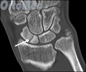
This involves tearing a bone fragment from the scaphoid to which the taloclavicular ligament is attached. This fracture is the most common type of navicular fracture.
Statistically speaking, such fractures account for 47 % of all scaphoid fractures in the foot.
Patients with this type of fracture usually report acute pain and swelling in the foot, difficulty walking, and increased pain when flexing the sole of the foot.
Examination may reveal dorsal swelling, bleeding (bruising), and local tenderness of the navicular bone and/or the navicular oval joint.
Conservative treatment
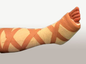
Fractures of the heel bone can usually be treated conservatively.
In these cases, immobilization is recommended, although the type of immobilization may vary.
If the fragment is small, an elastic bandage may be sufficient.
If the fragment is larger or the patient cannot tolerate an elastic bandage for various reasons, immobilization with an orthopedic shoe or even a short plaster splint may be possible.
The duration of immobilization depends on the load capacity and the presence or absence of accompanying injuries to the metatarsal and collateral ligaments of the ankle joint.
Immobilization usually lasts 4 weeks, after which the load on the foot can be gradually increased as the load tolerance increases.
scaphoid
The navicular bone is a small bone in the feet of most mammals.
In humans, the navicular bone is one of the tarsal bones of the foot. Its name derives from the human bone's resemblance to a small boat, caused by the highly concave proximal articular surface. The term navicular bone or carpal bone was previously used for the navicular bone, one of the bones of the wrist.
The human calcaneus is located on the medial side of the foot and connects proximally to the talus, distally to the three sphenoid bones, and laterally to the elbow bone.
It is the last of the foot bones to begin ossification, usually not until the end of the third year of life in girls and the beginning of the fourth year of life in boys, although a wide range of variation has been reported.
 Calcaneus. View from above.
  The pastern bone. View from below.
  Fracture of the heel bone
The posterior tibialis muscle is the only muscle that attaches to the heel bone. The main part of the muscle enters the navicular tuberosity. An additional navicular bone may be present in 2-14 % of the general population.
Clinical Significance
The navicular bone is usually not a broken bone in humans, but it does sometimes break for two reasons. The first mechanism is the stress fracture, which most commonly occurs in athletes, the second mechanism is high energy trauma. The scaphoid is the cornerstone of the foot: it is part of the foot pelvis and is connected to the talus, the first, second and third sphenoids, the ankle and the heel bones. It plays an important role in the biomechanics of the foot by supporting inversion, eversion and movement; it is the structural link between the midfoot and forefoot and part of the longitudinal and transverse arch of the foot.
Horse anatomy
In the horse, the navicular bone is located inside the hoof, on the inside of the hoof joint between the second and third toe phalanx (coffin bone). The horse's navicular bone is supported by the distal sesamoid ligament and the two lateral sesamoid ligaments. The pocket of the navicular joint is located between the flexor side of the navicular bone and the deep flexor tendon of the finger, which passes between the pocket and the distal phalanx. The middle tarsal bone in the horse's hip joint is homologous and similar to the navicular bone of the human foot, so the horse's navicular bone has a different structure than the human hyoid bone.
The navicular area is an important structure in relation to lameness, particularly in the forefoot, and is involved in a significant pain process called navicular disease or navicular syndrome. Much of the original literature on scapular disease has recently been questioned, particularly the importance of radiographic changes as the sole diagnostic criterion. Scapular syndrome may account for up to a third of all cases of lameness in horses, but radiographic changes in the scapula do not always allow a definitive diagnosis. New imaging techniques have shown that soft tissue damage in this area can be a significant contributing factor to lameness and that multiple causes can lead to visible lameness.
hammer toes
Hammertoes are almost always accompanied by a complex foot deformity and very rarely occur solely due to a congenital pathology. They occur more often in combination with transverse flattening of the foot and valgus deviation of the big toe, in foot deformities caused by childhood cerebral palsy (Littl.'s disease), myelodysplasia, etc. The formation of a hammertoe is attributed to increased tension in the extensor muscles of the phalanx and passive overstretching of the flexor muscles. There is excessive extension of the proximal phalanx and maximum flexion of the middle and distal phalanx.
The main reason for the patient's presentation to the doctor is pain caused by pressure on the head of the proximal limb in normal footwear or by secondary deformative arthrosis.
Conservative treatment of patients with hammertoes is ineffective. The aim of surgical treatment is to eliminate excessive extension of the basal phalanx and flexion at the interphalangeal joints. In the prearthritic stage and when the toes are mobile, the bend of the long big toe is transplanted to the basal phalanx.
Resection of the heads of the basal phalanges or arthrodesis after resection of the interphalangeal joints is performed with persistent contractures and arthrosis. Occasionally, resection of the diaphysis of the proximal phalanx is performed. All of these operations are usually performed in addition to surgical treatment of valgus deviation of the first toe. In neurogenic deformities, SF Godunov cuts the dry vein of the short flexor medullary phalanx.
heel spur
A heel spur is a spiny exostosis (osteophyte) located on the lateral side of the sole of the foot and is usually not clinically noticeable.
The appearance of a heel spur is associated with flat feet, chronic and acute trauma, various infections, inflammatory diseases, neurotrophic disorders, etc. It is likely that the spur is an ossifying fibrosis of the aponeurosis of the soleus tendon, which attaches to the calcaneus. The occurrence of pain can be explained by a superficial soft tissue injury in the area of the spur - post-traumatic aseptic tissue inflammation. The pain when putting weight on the heel can be very severe - it 'penetrates the nail'.
During the examination, the patient holds the foot in a forced position, steps on the outer edge of the foot or walks on the toes. During palpation, only the point of maximum pain is identified, since there are no other clinical signs of the spur. The diagnosis is clarified radiologically.
conservative Treatment includes:
1) Heel relief (bed rest in the acute phase, then a special perforated felt insert so that no pressure is exerted on the spur when the heel is stressed);
2) Physio and balneotherapy (UHF, paraffin, ozokeritotherapy, novocaine electrophoresis, etc.).
3) Injections of novocaine (2 ml of a 1%igen solution), hydrocortisone (1 ml) with antibiotic into the painful area with an interval of 4-5 days
4) Sessions (2-3) of anti-inflammatory x-ray therapy.
If conservative treatment is ineffective and pain recurs frequently, the spur is removed.
diagnostic methods
Osteoarthritis in the ankle is diagnosed during a medical examination. The doctor questions the patient, feels the problem area and pays attention to visible symptoms.
- Due to the pain in the foot when walking, the affected person unconsciously shifts his body weight to the front part of the foot, where he experiences less discomfort. Changes in gait.
- When examining the soles of the feet, corns form in the area of the big toe.
The insidious thing about this disease is that its characteristic symptoms are very similar to those of other foot diseases: broken or broken bones in the foot and gout. In order not to overlook osteoarthritis in its early stages, the following diagnostic examinations are recommended to the patient
- X-rays. Shows the condition of bone and joint structures and identifies the smallest changes in the ankle joint;
- Arthroscopy. A simple but reliable method for examining cartilage and tendons;
- Computed Tomography. Imaging diagnostic procedure for damaged joints that allows a layered view of the limbs;
- Magnetic resonance imaging. With this method, changes in the tissues and structures of cartilage and tendons can be observed.

The degenerative changes in the ankle joint associated with osteoarthritis are irreversible. Timely detection of the disease and correct therapy can prevent the development of serious health complications.
How to treat?
The treatment of osteoarthritis depends on the stage of the disease, the type of inflammation and the deformation of the joint structures. The more advanced the disease is, the more difficult it is to treat. The first stages are treated conservatively. If the disease is detected too late or no treatment is available, it can only be treated surgically.
Conservative treatment
This therapy focuses on relieving pain, eliminating inflammation, and restoring mobility. It is most effective in the early stages of the disease. The treatment package covers the following areas:
- provide peace of mind. If the affected person plays sports or has a job that places excessive strain on the legs, any training or overtraining should be postponed for the duration of treatment;
- Replacing footwear with special shoes that provide adequate ankle support. Doctors recommend wearing shoes with a rounded sole to reduce pressure on the metatarsal;
- The use of orthopedic shoes or insoles that limit the mobility of the metatarsal bone leads to a significant reduction in pain and discomfort;
- Using supportive devices such as canes, crutches, and orthoses to relieve pressure on the painful joint;
- Taking NSAIDs. Non-steroidal anti-inflammatory combination drugs eliminate inflammation at the site of joint injury, relieve its symptoms (local fever, swelling) and gently relieve pain;
- Taking painkillers if the pain is very severe;
- taking muscle relaxants to relieve muscle cramps in the affected foot region. If you walk for a long time or if your foot is misaligned, your feet become very tired. The muscle tension is relieved by taking such medications;
- The use of chondroprotectors improves the condition of cartilage and tendons and repairs damaged structures. The effects of these drugs are cumulative. To achieve the desired effect, treatment lasting several months is required. The doctor selects the course of treatment individually;
- Weight correction. Excessive weight puts additional strain on the legs and the damaged joint. This provokes disease and worsens the patient's condition, reducing the effectiveness of conservative treatment.
Early postoperative period
Almost all patients who undergo surgery for an Achilles tendon rupture are discharged from the hospital on the day of the operation.
For fourteen days after surgery, the ankle joint will be protected with a cast or plastic splint, such as the one shown below.
The cast should not be removed until the next dressing, which is 2 weeks after surgery.
You must not put any weight on the extremity for the first 2 weeks after the operation. Your physiotherapist will advise you before you go home and will also explain to you how to use crutches.
For the first 2 weeks after surgery, try to elevate your foot and keep it in that position for 95% of the time.
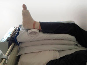
Of course, most people don't have a functional bed at home, as shown in this picture. However, the same effect can be achieved on a regular bed or sofa by placing a pillow under the foot. Do not put your foot up when sitting on a chair. Here, too, we recommend staying at home for the first two weeks.
Keep the foot dry and cool to minimize the risk of infection. Avoid excessive moisture and heat. Wear a tight bag over your foot when showering.
Move your foot and ankle regularly to prevent venous thrombosis. Drink plenty of fluids. If you have risk factors for thrombosis, you should tell your doctor; If necessary, he can prescribe anticoagulants for you.
Two weeks after surgery
You will be visited by your GP and your wounds will be dressed. Once your wounds have completely healed, you will be explained how to massage the tissue at the surgical site. Measures to reduce the sensitivity of the scar begin only after the wound has completely healed. For this purpose, you can use a massage cream (e.g. E45) rubbed into and around the scar area. Only when the wounds have completely healed can you expose the surgical area to moisture and shower.
Once the swelling has largely subsided after the operation, you can stand your foot up more often, but you should keep it horizontal for as long as possible.
However, driving is only possible for a short time if you have had surgery on your left foot and your car has an automatic transmission. If you have had surgery on your right foot, you will not be able to drive a car until 6 to 8 weeks after surgery.
At this stage, you may be referred to the physiotherapy department for early rehabilitation and exercises to strengthen the posterior tibialis muscle.
Conservative treatment of calcaneal fractures
The treatment plan for a heel bone fracture depends on the severity of the fracture, the patient's health, their activity level, and the desire for a quick recovery.
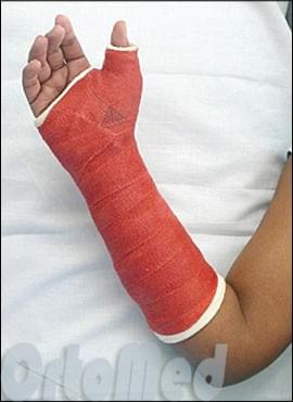
If the fracture is not displaced, treatment is non-surgical. This typically requires immobilization (casting) of the wrist and first toe with slight abduction for 2 months while the fracture heals. In some cases, patients with non-displaced fractures require surgery to shorten the time of immobilization and restore hand function as quickly as possible. This approach is still somewhat controversial among orthopedic surgeons.
Fracture of the lower third of the navicular bone (closer to the fingers)
Fractures of the lower third of the navicular bone usually heal within 4-6 weeks with appropriate immobilization (cast). This part of the navicular bone has a good blood supply, so there are usually no problems with non-healing.
Fracture of the upper third of the navicular bone (closer to the forearm)
If a fracture of the navicular bone occurs in the middle third (waist) or closer to the forearm (proximal pole), healing is more difficult. These areas of the navicular bone have poor blood supply.
The healing time depends on age, blood circulation in the hand and metabolism. Therefore, regular check-ups with the doctor and X-rays or CT scans are necessary to determine whether the fracture has healed.
Surgical treatment of calcaneal fractures
The aim of surgical treatment is to stabilize the fracture of the scaphoid bone so that the blood supply to the fracture is restored early and healing occurs.

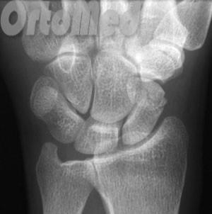
The operation is usually carried out on an outpatient basis under regional anesthesia (brachial plexus) or local anesthesia. During the operation, the bone fragments are repositioned and fixed with an implant (screw) for stabilization. During the operation, X-rays are taken to confirm the restoration of bone anatomy and the correct fixation of the fragments. In most cases, a single screw is used to stabilize the bone fragments.
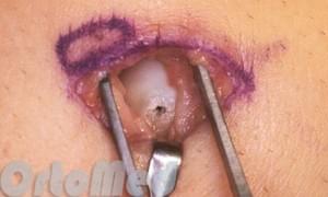
The cut can be made on the hand or the back of the wrist. Where the doctor makes the surgical incision and how large it is depends on which part of the navicular bone the fracture occurred. Most often, fresh fractures are fixed by making a 3 to 5 mm incision with a screw because the dislocation is easily repaired and does not require a large incision to access the bone.
If the fracture is old and has not healed properly, a sufficient incision is required to perform an osteotomy (artificial fracture) of the healed fragment to restore correct anatomy and fix the bone with a screw.
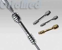
Most orthopedic surgeons repair fractures with screws:
- This method of fixation is much safer than spokes because the screw completely sinks into the bone;
- Early development of wrist movements is possible.
Periarticular necrosis
In navicular fractures, especially displaced fractures, the blood supply to the bone is impaired. If the blood supply to one of the fragments is severely impaired or completely interrupted, the bone no longer receives nutrients and its cells die. Under such conditions, a normal anastomosis is not possible. This condition is called avascular necrosis.
The most effective treatment for this condition is bone grafting with a vascularized bone graft until severe bone loss or osteoarthritis develops.
arthrosis
An incorrect joint or avascular necrosis of the navicular bone can lead to osteoarthritis of the wrist. This condition is characterized by wear and damage to the articular cartilage.
Treatment of osteoarthritis is primarily aimed at controlling the symptoms of the disease. These include nonsteroidal anti-inflammatory drugs or over-the-counter pain medications, immobilizing the wrist, and avoiding activities that increase pain in the joint. Sometimes pain relief can be achieved by injecting a corticosteroid into the joint.
If conservative treatment does not work, the doctor may recommend surgery. There are several surgical treatment options available for wrist osteoarthritis.
Rehabilitation after a foot fracture
For complicated injuries, a plaster or fixation bandage is applied for 3-4 weeks to several months. Rehabilitation begins after immobilization and lasts up to a year.
Removal of the cast should be prepared in advance. For minor injuries, it is advisable to remove the bandage and move the ankle joint 2-3 weeks after immobilization without putting any strain on the limb. Circulation exercises that do not put any strain on the injured leg but improve its blood circulation are also recommended.
Once the bandage is removed, rehabilitation begins.
- Massage;
- Physical therapy;
- PHYSICAL THERAPY;
- diet that begins during treatment;
- wearing special shoes, insoles and foot pads.
A holistic approach to recovery allows you to regain full mobility more quickly.
massage
The massages are carried out before the bandages on the free part of the limb are loosened. Once the leg is free, you will learn the basic techniques from the nursing staff and perform the foot massage alone or with the help of someone close to you. A professional massage performed by a professional is best.
The massage improves blood and lymph circulation, tones blood vessels and muscles, and helps reduce swelling.
physical therapy
Treatment with devices can begin during immobilization and continue after the cast is removed. The following treatments are indicated:
Device treatments improve microcirculation, promote the resorption of edema and hematoma, and accelerate the formation of bone callus.
For example, after a double ankle fracture with foot subluxation, rehabilitation includes the following measures:
The order of treatments is determined by the doctor. Prolonged immobilization after this fracture means the foot takes a long time to recover.
PHYSICAL THERAPY
Depending on the location and type of injury, a set of physiotherapeutic exercises is selected individually for each case. Standardized exercises help restore muscle memory, improve tissue trophics, and prepare the foot for full participation in movement with simple activities.
Stages of rehabilitation.
During cast treatment, hip and knee exercises must be performed to maintain muscle tone and improve blood circulation.
Rehabilitation measures must be carried out consistently so as not to overload the limb through excessive exertion. The main stages of correct rehabilitation:
- Phase I. Rehabilitation should begin with warming up the foot with massage. Simple kneading movements of the foot without any effort are suitable for this. The movements are light, kneading and stroking. Don't try to walk right away, especially without crutches. This mistake is often made during the rehabilitation of patients with metatarsal fractures.
- second phase. In the second phase, a specially selected set of physical exercises should be performed, periodically increasing the load. Walking is of utmost importance. It is important to walk correctly from the start and avoid a limp, which can become permanent. The arch of the foot, whose muscles have atrophied due to immobility, should be trained.
- Third step. In the final stage, you should not stop exercising, continue your diet and go to physiotherapy. During this time, exercises to develop the spring function - small jumps - are carried out.
The result of the rehabilitation period should be full joint mobility and a smooth gait without limping. Supinators should be retained to facilitate walking. This is necessary for patients with any type of injury, including a fracture of the 5th metatarsal, which is the most common.
prophylaxis
To prevent meniscal necrosis, it is important to protect the wrist and avoid hand trauma. In the event of trauma or unreasonable pain of any intensity, a doctor should be consulted. Timely treatment of a fracture of the navicular bone can restore full trophism and avoid complications.
Kinbeck disease is very rare. Doctors are more likely to detect a fracture of the navicular bone after a hand injury if it is not treated in a timely manner. It is important to know that the bones of the wrist are very small and fragile and can be easily injured when the hand is moved in a certain way or when the hand is thrown and dropped.
It is better not to hesitate and seek help immediately after an injury. An examination will show whether there is a fracture and allow treatment to begin in a timely manner. Delay is dangerous for the patient, as the function of the wrist is significantly reduced, which inevitably affects the ability to work and the overall quality of life. Ankle necrosis can be treated surgically if the pathology is detected in a timely manner.
rehabilitation
Surgical treatment requires a hospital stay of 3 days. After the operation, the wrist is immobilized with a plaster cast for 5 weeks. A control x-ray is then taken in two projections. When the bones have successfully solidified (healed), the cast is removed and the patient receives physical therapy to restore joint function.
The complaints are referred to an orthopedist/traumatologist, who orders further examinations and makes an objective diagnosis as a basis for treatment. A rheumatologist can also be consulted during the diagnosis.
In advanced cases, surgical treatment may not be advisable. The possibility of surgical intervention is examined in each individual case.
An unsatisfactory treatment result is possible if the rehabilitation recommendations are not followed. For example, the patient may re-injure the wrist or strain the joint. It is important to strictly follow the doctor's recommendations.
Korshunov VF, Vidasova EV. Features of treatment and rehabilitation of patients with aseptic necrosis of navicular and hemilateral bones // Materials of the seventh city scientific conference 'Medical rehabilitation of patients with pathology of the musculoskeletal and nervous system'. ¬ Moscow, 2006. ¬ С.203.
Korshunov VF, Vidasova EV. Surgical treatment of aseptic bone necrosis of the scaphoid and semilunar hand // Kremlin Medicine. ¬ №2. ¬ Moscow, 2007. 18¬20.
Andrushko NS, Solodko VI Results of conservative treatment of Kinbeck's disease. – In book: The conservative treatment of traumatological and orthopedic diseases of the upper limbs. – Bitter, 1971 С.251-253.
Read more:- Fracture of the calcaneus of the foot.
- Bone structure of the navicular foot.
- Completion of the scaphoid on the foot.
- Bones of the tarsal bone of the hand.
- fracture of the elbow in the foot.
- tibia and fibula.
- Displacement fracture of the heel bone.
- Fracture of the heel bone.