After surgical treatment of a rupture of the radius-clavicular joint, patients are recommended to participate in mechanical physiotherapy and hypothermia to relieve pain and swelling.
- Anatomy of a radial head fracture
- Causes and mechanism of radial head fracture
- Anatomical Features
- Causes of X-ray oclavicular joint injuries
- Types of radial bone fractures (brief classification)
- Colles fracture
- Smith's fracture
- Classification of fractures of the radial bone of the hand:
- INTRODUCTION
- classification
- complications of treatment
- forecast
- Causes of forearm bone fractures
- This article has been reviewed by
- Symptoms of a broken forearm bone
- treatment options
- conservative method
- surgical treatment
- frequently asked Questions
- Symptoms of ACS injury
- diagnosis
- Diagnosis of a ruptured ligament
- What are the Symptoms of a Shoulder Sprain?
- Rehabilitation after injury
Anatomy of a radial head fracture
The structure of the elbow joint. The radius articulates with the humerus and the proximal elbow. This joint allows for flexion and extension of the forearm, as well as pronation (downward rotation of the hand) and supination (upward rotation of the hand) of the forearm.
The head of the radial bone is covered with articular cartilage. This allows the joint surface to slide in two planes, which is extremely important for the elbow joint. Fractures of the joint associated with post-traumatic arthritis can therefore result in mechanical limitation of movement.
The head is also an important stabilizer of the elbow joint.
In addition to acute pain, a fracture of the radial head is characterized by:
- Significant limitation of mobility of the elbow joint, including passive and rotational movements,
- hemarthrosis,
- Deformations of the outer surface of the elbow joint.
If there is a suspicion of a fracture of the radial head at the elbow joint, a fracture of the radial head with a fracture of the intercondylar membrane should be ruled out. Therefore, if a fracture is suspected, the adjacent joints should also be examined.
Most radial head fractures are isolated but are sometimes accompanied by subsequent injuries
- Fracture of the coronoid process of the ulna
- Tear of the collateral ligament of the elbow joint
- Tear of the intercondylar membrane
- Fracture displacement of the Goliacci
This injury, which is also accompanied by a fracture of the head of the humerus, can be accompanied by damage to the medial collateral ligaments and a fracture of the ulna with its shortening.
Causes and mechanism of radial head fracture
Usually, the fracture is the result of indirect trauma, that is, a fall on an outstretched arm with minimal to moderate flexion of the elbow with concomitant pronation of the forearm. The main axial load in this case comes from the radius arm joint. The injury is caused by collision of the radial head with the humeral block. Direct impact to the head of the radius bone rarely results in a radius bone fracture.
This injury results in:
- acute pain localized in the elbow joint,
- swelling of the elbow joint,
- restriction of flexion and/or extension of the forearm,
- acute pain when axial pressure is applied to the arm.
Anatomical Features
The collarbone, humerus and shoulder blade form the shoulder crown. The clavicle-shoulder joint is a low-mobility joint that connects the clavicle and shoulder blade.
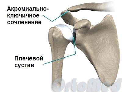
The joint is supported by bony structures that anchor it with strong ligamentous tissue. The ends of the joints are surrounded by a closed capsule filled with synovial fluid.
Causes of X-ray oclavicular joint injuries
Among the main causes that lead to a tear of the radioclavicular joint, medical professionals state:
- Injuries during sports competitions (such a tear is not uncommon in goalkeepers of football and hockey teams who forcefully lower their shoulders to protect the goal);
- injuries in contact sports (judo, boxing, taekwondo or sumo);
- Injuries caused by falling onto an outstretched arm (often from slipping on ice in winter)
- Injuries resulting from an active lifestyle (after careless roller skating, skiing or ice skating).
Types of radial bone fractures (brief classification)
Fracture of the distal part of the radius bone almost always occurs about 2-3 cm from the wrist.
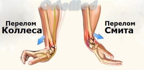
Colles fracture
One of the most common fractures of the distal radial bone is the Colles fracture, in which a fragment (fragment) of the distal radial bone migrates to the back of the forearm. This fracture was first described in 1814 by Irish surgeon and anatomist Abraham Colles.
Smith's fracture
Robert Smith described a similar fracture of the radial bone in 1847. The cause of this fracture is believed to be exposure of the back of the hand. The Smith's fracture is the opposite of the Colles' fracture, ie the distal fracture is displaced palmar.
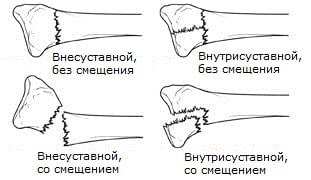
Classification of fractures of the radial bone of the hand:
Another classification of fractures of the radial bone:
- Intra-articular fracture: A fracture of the radial bone where the fracture line extends into the carpal joint.
- Extra-articular fracture: A fracture that does not reach the articular surface.
- Open fracture: When the skin is broken. Skin injuries can impact the bone from the outside (primary open fracture) or impact the bone from the inside (secondary open fracture). These types of fractures require immediate medical attention due to the risk of infection and serious problems with wound and fracture healing.
- fragmentation fracture. When a bone is broken into 3 or more fragments.
It is important to classify fractures of the radial bone because each type of fracture needs to be treated according to specific standards and tactics. Intramedullary fractures, open fractures, displaced fractures, and fractures of the radial bone should not be left untreated, whether by closed fracture reduction or surgery. Otherwise, the function of the hand cannot be fully restored.
Sometimes a fracture of the radius bone is accompanied by a fracture of the adjacent ulnar bone.
INTRODUCTION
In recent decades, interest in the diagnosis and treatment of carpal ligament injuries and their consequences in the medical literature has continued unabated worldwide. In our country and near abroad, these issues have been studied [1-6], but they remain relevant, which is explained by the prevalence of these injuries in young people with the need for increased functional load on the hand [7] and the fact that the difficulties in diagnosing and treating this pathology persist.
Diagnostic errors and treatment failures can cause scapholunate joint instability and lead to advanced scapholunate collapse (SLAC) [4, 6, 8, 9] [4, 6, 8, 9]. According to the literature, injury to this ligament can be associated with fracture of the distal radial bone [10-14], fracture of the navicular bone, and other wrist injuries [8, 15] in 16-40 % of cases.
This ligament is C-shaped and belongs to the internal interosseous ligaments of the wrist, which run parallel to and stabilize the navicular and semilunar bones [3, 7, 8, 16-21]. During the projective movement, beginning with wrist extension at radial deviation and ending with wrist flexion at ulnar deviation, minimal load is placed on the semiannular navicular ligament. This peculiarity of wrist kinematics is taken into account in rehabilitation after injuries to the navicular-semiannular ligament [7, 22, 23].
The diagnosis of ligament damage is based on physical findings such as pain in the scapholunate compartment, Watson's sign [1, 24] and indirect radiological findings such as an enlargement of the scapholunate space by more than 2 to 4 mm, an increase in the scapholunate angle by more than 60° degrees and the scapholunate 'ring' sign [3, 7, 9, 19, 24-27]. However, there is no radiological evidence of ligament damage in the early stages of so-called 'pre-dynamic' instability, although ligament rupture can be demonstrated with modern imaging techniques or during surgery. Contrast wrist arthrography, proposed more than half a century ago, has been criticized for its poor specificity and accuracy [28]. Ultrasonography [29, 30], computed tomography (CT) [3, 31] and MRI techniques of the wrist have been suggested to visualize ligament injuries. Many studies show the superiority of contrast-enhanced magnetic resonance arthrography (MRA) [3, 7, 8, 28, 32-35]. Currently, wrist arthroscopy is recognized worldwide as the gold standard for imaging ligament injuries, as it allows direct examination of the scaphoid-hip space and assesses the extent of ligament injuries [7, 9, 21, 24].
classification
A review of the literature reveals several classifications of scapholunate ligament injuries. The WB Geissler and Garsia-Elias et al. Proposed classifications take into account the arthroscopic damage to the ligament and treatment guidelines. JC Messina et al. presented the European Wrist Arthroscopy Society (EWAS) classification, which is essentially a modification of WB Geissler's classification with a more detailed description of ligament tears [7, 9, 13, 21, 24]. R. Luchetti et al. proposed a detailed clinical classification of scapholunate ligament injuries with recommended treatment methods. Depending on the degree of severity, they defined these as pre-dynamic, dynamic, static with possible reduction of the scapholunate ligament and static instability with an irreducible gap [36].
N Morrell et al. (2017) presented an algorithm for the treatment of collateral ligament injuries (Table 1) [24].
A review of the available literature and treatment algorithm by N. Morrell et al. concluded that treatment tactics for injuries to the scapholunate ligament largely depend on the extent of the injury, the severity of the degenerative changes in the wrist, and the time since the injury. J. Andersson, in a 2017 review, suggested that ligament injuries should be considered fresh or acute within four weeks of injury and subacute between four and six weeks [13]. Injuries are considered chronic after six weeks after injury [37]. Many authors note that treatment efficacy and prognosis are poorer in chronic injuries, underscoring the importance of early recovery [38,39].
In our review, we divided the treatment methods for injuries of the scapholunate ligament into conservative methods (Table 1. 2) and operative (Table 3).
Tags: wrist
234567 Start of activity (date): 03/02/2021 15:47:00
234567 Created by (ID): 989
234567 Keywords: Wrist, Wrist joint, Ligamentum scapholunatum, Injury.
12354567899
complications of treatment
Possible complications of conservative treatment are:
- Inability to return to previous loads
- Frequent recurrence of symptoms such as B. an inability to throw with full power or from full distance, pain when throwing, and loss of ball control, especially if exercise resumed soon after the injury
- damage to other structures of the elbow, including the cartilage of the outer part of the elbow; limited mobility of the arm, damage to the ulnar nerve, medial epicondylitis and sprain of the flexor tendons of the hand.
- Damage to articular cartilage leading to inflammation of the elbow joint
- Elbow stiffness (limited range of motion)
- Symptoms of ulnar nerve neuropathy
Possible complications of surgical treatment are:
Specific complications of surgical treatment of the disease:
- failure to restore normal stability
- Inability to return to previous level of activity
- Damage to the ulnar nerve
- Irritation of skin areas associated with removal of the graft from the palmar muscle tendon
forecast
Injuries to the ulnar collateral ligament usually do not heal completely with conservative treatment methods. Returning to sport often requires surgical treatment. It can take three to six months to return to sport after an injury without surgery, and nine to 18 months after surgery.
Rehabilitation (LFC) can increase muscle strength and endurance. The exercises should be selected in consultation with your LFC doctor.
- Proper warm-up exercises before training or competition
- Maintain adequate muscle strength in your hand, forearm, and wrist.
- Use appropriate protection techniques for falls and throws
- Functional orthoses can effectively prevent injuries, especially re-injuries, in contact sports.
Use of the material is permitted with an active hyperlink to the permanent article page.
Causes of forearm bone fractures
- Certain sports.
- Active movements that require physical contact and increase the risk of falling: soccer, gymnastics, skiing, skateboarding, etc.
- abnormalities in bone structure.
- Doctors associate osteoporosis and bone tumors with pathologic arm fractures, even when there is no high-energy force applied to the arm.
- gunshot wounds.
- An open fracture of the forearm bone from a gunshot wound is a serious injury with a high incidence of non-healing and infection.
This article has been reviewed by
Symptoms of a broken forearm bone
The clinical manifestations depend on the type of injury. With open forearm fractures, the skin is torn and bone fragments may protrude from the wound. With severe pain and concomitant blood loss, when blood vessels are damaged, traumatic shock occurs, manifested by pallor, rapid breathing, tachycardia, etc.
In less severe cases, the first sign of upper limb injury is an audible grinding/slapping sound and limb deformity. Typical features are:
- Severe pain that may increase with movement
- Swelling, bruising/irritation
- tension of the skin
- inability to move fingers
- Limb deformity – unnatural bending of the hand or wrist
Radial head pain or swelling may be the only physical symptom in patients with a reduced montage lesion or radial head fracture.
treatment options
conservative method
The conservative treatment – consists in dressing the injured hand with an immobilizing bandage (standard plaster or light plastic bandage). This treatment is used for fractures that do not require surgery. After putting on the cast, firstly, it is necessary to check that the arm is comfortable, and secondly, on the 5th to 7th day after the swelling has subsided, an x-ray must be taken to rule out an unintentional dislocation.
surgical treatment
Surgical treatment is performed for unstable and displaced fractures, severe intra-articular injuries and multiple fractures. The main treatment method is repositioning (squeezing the bone fragments together). Closed repositioning. With closed repositioning, the bone fragments can be reassembled by the hands of a specialist without surgery - the trauma surgeon compresses the fracture with special movements. Open repositioning. Open reduction is performed when it is not possible to repair the fracture using another method. During the operation, an incision is made over the injury site to access the broken bone, mobilize the bone fragments, remove the dislocation and immobilize them with a special structure - osteosynthesis.
* The administration of the clinic tries to update the price list published on the website in a timely manner, but in order to avoid possible misunderstandings, we advise you to pay the cost of the services at the reception or in the contact center by calling 8 (495) 268-12 -12 to ask. The published price list is not an offer. Medical services are provided on a contract basis.
- Extensive practical experience since 2004.
- Participated in advanced training courses in Austria, Germany and Russia.
- Performs arthroscopy, osteosynthesis of limb fractures, correction of deformities, joint arthroplasty and other procedures.
- He is a member of the European Association of Osteosynthesis (AO Foundation), the Union of Traumatologists and Orthopedists of Russia and the Russian Arthroscopic Society.
frequently asked Questions
The length of rehabilitation depends on the severity of the injury: for a non-dislocated fracture, full recovery takes about 3-4 weeks. In the case of a moderately severe injury with bone displacement, rehabilitation takes 1-3 months.
The fracture is characterized, first of all, by pain, which during a fall is strong and violent, sometimes even a characteristic crunch is heard. The pain from a bruise, on the other hand, varies in intensity and gradually subsides after a few hours, resulting in relief.
For treatment of a radius fracture in Moscow, please contact the Elena Malysheva Medical Center. Experienced doctors will diagnose the problem, determine the presence of complications and prescribe effective treatment. Make an appointment for a consultation.
Symptoms of ACS injury
If the acromion is dislocated, pain and restricted movement are the most characteristic symptoms. Swelling in the area of injury quickly occurs and the dislocated segment rises and moves slightly backwards. There is almost always a specific 'key' symptom: when the protruding rim is pressed in, it swings back when released. Improper extension of the arm occurs for the first few hours after the injury.
In contrast to a partial trauma, a complete rupture of the ligaments of the clavicle-shoulder joint is accompanied by pronounced instability of the joint. A massive contusion occurs on the left or right side of the collarbone. The pain is unbearable and leads to immobilization of the upper extremity.
diagnosis
Imaging methods are used to determine the type of injury to the clavicle joint:
In the past, a fracture of the shoulder joint detected on the x-ray was considered sufficient for the diagnosis. However, with incomplete dislocations, the radiological picture does not always reflect the actual condition of the patient. Therefore, MRI is considered to be the most conclusive method for diagnosing ACL injuries. With this safe and painless imaging method, the condition of all soft tissues can be assessed and the extent of damage to the intra-articular ligaments, capsule and synovial membrane of the joint can be determined with the highest accuracy.
Diagnosis of a ruptured ligament
A proper diagnosis can only be made by a qualified trauma surgeon experienced in treating this type of injury. The examination usually begins with a medical history, in which the fact and nature of the injury play an important role. A clinical examination follows, which always begins with a healthy joint. A series of clinical tests are performed that clearly indicate a specific intra-articular injury. In most cases, the tests give a clear picture of the lesion and a preliminary diagnosis is made. Additional examination methods are often used to confirm the diagnosis
- X-ray – always done to get a picture of the joint and to rule out bony or traumatic injury. An x-ray of a healthy joint is also often recommended;
- MRI – allows for a slice-by-slice visualization of soft tissues and intra-articular masses to determine the type, extent and degree of damage to ligaments, muscles, cartilage, etc.
- ultrasound examination;
- Computed Tomography;
In difficult situations, when the injury is serious or long-lasting, or when the clinical examinations do not reveal a clear clinical picture, various additional examinations can be carried out to clarify the diagnosis.
What are the Symptoms of a Shoulder Sprain?
Immediately after a shoulder injury, the following symptoms appear
- Sharp pain at the site of injury;
- Increased discomfort when trying to move shoulder;
- limitation of shoulder mobility;
- slight swelling and swelling at the site of injury.
In addition, the body temperature can rise, which can indicate both a ligament injury and an incipient inflammation. If the person doesn't take action - doesn't see a doctor, keeps actively moving the shoulder, the pain will increase and the swelling will get worse. All of these symptoms can indicate tendonitis – an inflammation of the tendon. In such a situation, there is an urgent need to visit the nearest medical institution for specialized treatment.
Rehabilitation after injury
The ligaments in the shoulder can heal quite quickly after a dislocation. A lot depends on the individual's ability to regenerate, the severity of the injury and whether the patient follows all the recommendations of the specialist. Certain diseases such as diabetes, immunodeficiency, thyroid disease, etc. can delay the process of tissue regeneration and rebuilding. During rehabilitation, the body needs a lot of vitamins, proteins and micronutrients - proper nutrition that meets all the needs of the body during this period is very helpful.
For first and second degree sprains, rehabilitation usually lasts no more than 7-10 days. In the case of a complete rupture of a ligament, recovery takes time longer – up to six months.
After the main treatment is completed, the trauma surgeon will recommend physical therapy and exercise therapy. The aim of rehabilitation is to restore the mobility of the joint, strengthen the ligaments and maintain their flexibility.
In the rehabilitation period after a shoulder injury, physical therapy is very important: during inactivity, connective tissue fibers form in place of the damaged elastic tissue. This is scar tissue that cannot stretch like the ligaments normally should. Any training should be gentle, without sudden movements and with a gradual increase in load. Under no circumstances should you train when you are in pain, as this can lead to renewed traumatization of the ligaments of the joint. The first 2-3 times physical therapy should be carried out under the guidance of a specialist who will monitor the technique in order to avoid unnecessary loads.
After the rehabilitation phase, the person concerned should take certain preventive measures to prevent further dislocations. This includes maintaining a normal weight, regular physical activity and a sensible diet.
Read more:- Pronator - what does that mean?.
- The pronator is in anatomy.
- Displacement fracture of the heel bone.
- Fracture of the lateral condyle.
- DMS rupture.
- shoulder supinators.
- Collateral ligament rupture.
- Thigh tendon rupture.