Top doctor, neurologist, physiotherapist, UWT specialist, leading specialist in the Health Plus network

- Tear of the collateral ligament
- Structure of the thumb joint
- ICD-10
- Ulnar tendon injury
- Treatment
- Information .
- sources and literature
- information
- Causes of SCA Injuries
- Diagnosis of SCA injuries
- Concurrent SCA violation
- Causes of knee ligament strains
- Symptoms of a knee sprain
- treatment of a dislocation
- Symptoms of a patellar ligament tear
- Diagnosis of patellar ligament damage
- Symptoms and diagnosis of a knee ligament strain
- Treatment options for dislocated knee ligaments
- Treatment of knee ligament sprains with UHT
- Collateral ligament injury: course and prognosis
- Lateral Collateral Ligament Injury: Prevention
- Doesn't that sound like your condition?
- Symptoms of a rupture of the collateral ligaments in the knee
- Treatment of torn collateral ligaments of the knee joint
Tear of the collateral ligament
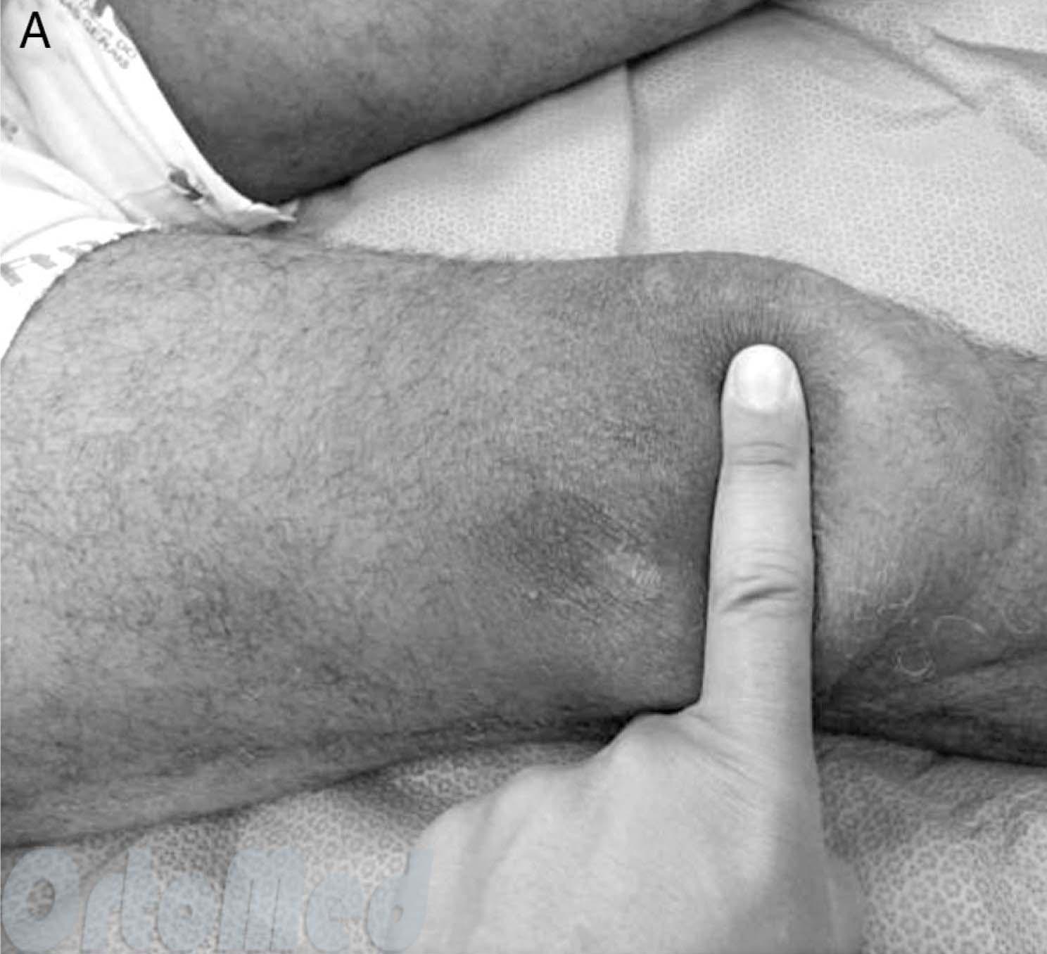
This injury, often referred to as 'hunter's finger' or 'skier's finger', involves a tear in the collateral ligament of the thumb.
The first term is typical for chronic injuries, the second for acute injuries. The 'hunter's finger' was first described in 1955. This chronic injury is typical of Scottish hunters who killed injured rabbits by pinching their necks between their index finger and thumb.
Injuries in which the collateral ligament of the big toe joint tears are typical for ski athletes. Occasionally, these injuries occur in athletes who participate in contact sports such as boxing and in sports where the athlete may fall while leaning on his hand. The injury, which is specific to skiers, was first described in 1939. It is the second most common injury in skiers (about 10 % of cases) and upper limb injuries (37 % of cases).

The ligament injury occurs when the athlete falls on the snow. At this point the thumb is maximally extended and retracted while the hand is holding the ski pole. To avoid the serious consequences of a fall, the skier instinctively pulls his hands forward, but the ski pole remains in his hand. This significantly increases the risk of injury to the thumb.
To avoid injuries, skiers were advised to use ski poles without straps. The belt prevented her from removing her hand from the stick in time if she fell. Some manufacturers developed a new type of 'handle' for the sticks, but even this did not completely solve the problem.
Structure of the thumb joint
This joint is unique in both its functional biomechanics and its anatomical structure. Its stability is essential for creating leverage and executing a strong grip. The mobility of the joint varies from person to person: some people cannot extend it beyond normal, while others manage to bend it completely. The flexion angle varies between five and one hundred and fifteen degrees, the radial deviation in some cases reaching 30 degrees in the upright position and up to 15 degrees in full compression.
There are different degrees of collateral ligament damage:
- Grade I, which occurs in skiers in almost 35 % cases. This grade is characterized by a small tear of the ligament without loss of integrity.
- Grade II is characterized by slight tearing of the fibers and stretching of the fibers, but without loss of integrity. This occurs in 47 % cases.
- Grade III, which is characterized by a complete rupture of the ligament. It arises at the distal end near the entry into the proximal phalanx. A third-degree injury occurs in 18.2 % cases.
A fracture accompanied by a torn ligament is quite common, accounting for 23.3 % of cases.
Timely and correct treatment of the injured person is only possible after a thorough examination. If the problem is ignored, it can lead to a complication in the form of chronic finger dysfunction.
ICD-10
Injury to the ligaments of the upper limbs – This is a very common group of injuries that includes damage to the rotator cuff, elbow and wrist ligaments, and damage to smaller joints of the hand.
In traumatology, the term 'shoulder rotator cuff' refers to a group of tendons located around the shoulder joint (scapular, obturator minor and supraspinatus muscle tendons). The job of this group of tendons is to ensure mobility and stability of the shoulder joint.
Rotator cuff injury is one of the most common and serious upper limb ligament injuries. It can be partial or complete, traumatic or degenerative. Traumatic rotator cuff tears are caused by a muscle strain resulting from a fall on the shoulder. Less commonly, the tendons are damaged by a direct blow to the shoulder joint.
Degenerative tendon damage can also occur with minor injuries. In contrast to traumatic rupture, the main cause in this case is a change in tendon structure due to trophic abnormalities, repeated microtraumas, or congenital connective tissue weakness.
A patient with a torn shoulder rotator cuff complains of pain, usually on the upper lateral surface of the shoulder near the insertion of the deltoid tendon. If the rotator cuff of the shoulder is partially torn, the joint's range of motion is retained and pain increases when the arm is moved laterally. If the shoulder rotator cuff is completely torn, the range of motion of the joint is limited and the patient has difficulty raising the extended arm.
If the tendon is permanently torn, a gradual subluxation of the humeral head occurs. This leads to degenerative changes in the shoulder joint. The mobility of the arm is restricted even more. A tear of the rotator cuff of the shoulder can be complicated by damage to the nearby nerves of the brachial plexus and inflammation of the tendon capsule under the scapula (subacromial bursitis).
Ulnar tendon injury
This is a rare form of upper limb ligament injury. Tears and ruptures of collateral ligaments are usually associated with joint capsule tears and burst fractures. The elbow joint is swollen and enlarged due to blood accumulation. Excessive sideways movement is observed in the upright position. Treatment includes puncture of the elbow joint, immobilization with a posterior plaster splint for 3 weeks, physiotherapy and exercise rehabilitation.
It is a common ligament injury in the upper limbs. The ulnar collateral ligament on the elbow side is most commonly injured. It is caused by a fall with an outstretched arm. There is pain when moving and swelling of the dorsal and lateral articular surfaces.
The wrist is immobilized with a plaster splint for a week. Physical therapy is then recommended.
Treatment
DIAGNOSIS AND TREATMENT METHODS, APPROACHES AND PROCEDURES
Purpose of the procedure/intervention: Restoration of lateral stability of the knee joint. Any instability of the knee leads to changes in the nutrition of the articular cartilage, which irreversibly lead to osteoarthritis of the knee.
Indications and contraindications for surgery/intervention:
Indication for surgery/intervention: Collateral ligament damage associated with lateral knee instability.
Contraindications for the operation/intervention:
Absolute contraindications:
- Severe patient condition;
- decompensation of chronic diseases;
- Inflammatory skin lesions in the area of the procedure.
- Significant vascular and neurological pathology in the affected limb;
- Noncompliance with postoperative protocol.
Additional diagnostic measures:
- blood count;
- urine test;
- blood group and Rhesus factor test;
- coagulation picture;
- biochemical blood test;
- blood test for human immunodeficiency virus;
- blood test for Wasserman's test;
- blood sugar test;
- blood for HbsAg, anti-HCV;
- ECG;
- X-ray examination of the injured knee joint in two projections;
- Ultrasound examination of the knee joint;
- MRI of the knee joint (if no MRI findings are available);
- Consultation with relevant specialists if there is a concomitant disease, indicating the necessary additional examinations and treatment measures.
Requirements for security measures, sanitary and anti-epidemic regimes
: in accordance with the sanitary regulations 'Sanitary and anti-epidemiological requirements for healthcare facilities', approved by Order No. 357 of the Minister of Health of May 31, 2017.
Information .
sources and literature
- Minutes of the meetings of the Joint Commission on the Quality of Medical Services of the Ministry of Health of the Republic of Kazakhstan, 2018.
- 1. 'Traumatology and Orthopedics', ed. by NV Kornilov, GE Gryaznukhin, SP. – Hippocrates, 2006 – VOL. 3 – PP. 284-312. 2. Zubarev AR, Nemenova NA Ultrasound diagnostics of the musculoskeletal system in adults and children. – М., 2006. 3. Friemert B., Oberländer Y., Schwarz W. Diagnosis of chondral lesions of the knee joint – can MRI replace arthroscopy? // Knee Surg. Sports Traumatol. Arthrosc. – 2003. – №8. – P. 56-75. 4. Kim Y., Ihn J., Park S. Arthroscopic analysis of lateral meniscus variants and comparison with MRI findings // Knee Surg. Sports Traumatol. Arthrosc. – 2006. – №14. – P. 20-26. 5. Ververidis A., Verettas D., Kazakos K. Meniscus spoon-pedicle tears: a retrospective study of arthroscopy and the relationship to MRI // Knee Surg. Sports Traumatol. Arthrosc. – 2006. – №14. – P. 343-349.
information
ORGANIZATIONAL ASPECTS OF PROTOCOL IMPLEMENTATION
List of protocol converters with qualification data:
- Raimagambetov Yerik Kanatovich – candidate of medical sciences, traumatologist and orthopedic surgeon of the highest category, senior researcher of the orthopedic department, head of the orthopedic department ¹5 RSE at the PCV 'Research Institute of Traumatology and Orthopedics' of the MoH.
- Korganbekova Gulzhanat Sansyzbayevna – MD, orthopedic traumatologist of the highest category, senior researcher of the orthopedic department, assistant doctor of the orthopedic department ¹5 RSE at the PCV 'Research Institute of Traumatology and Orthopedics' of the MoH.
- Rymbaev Darkhan Rymkhanovich – Assistant doctor in the Department of Adult Orthopedics of the KGP 'Prof. HJ Makazhanov regional center for traumatology and orthopedics'.
- Akhmetzhanova Gulmira Okimbekovna – clinical pharmacologist of the RSE 'Research Institute of Traumatology and Orthopedics'.
Causes of SCA Injuries
Various types of violent (mechanical) blows to the knee area that occur in the following situations can be the cause of injury or rupture of the SCA:
- Falling on a bent knee, not only during active play, but also during normal walking;
- Strain of the knee or sprain of the lower limbs (so-called hyperextension of the ACS), etc.
It should be noted that a serious injury to the knee joint is sometimes indicated by a corresponding cracking sound in the knee.
Diagnosis of SCA injuries
During the first consultation, the attending physician explains the circumstances of the injury, examines the joint and obtains information from the patient about pain in the knee area.
After discussing the clinical picture, the doctor will conduct a physical examination to determine the current condition of the damaged joint components. It is important to note that in a severe SCA injury, the joint may be rotated backwards and the tibia may be abnormally displaced (still backwards) relative to the hip.
Additional examination techniques for maximum diagnostic accuracy are MRI and X-ray. However, if the injury occurred more than 3 months ago, the presence and extent of the damage are difficult to determine using these procedures.
X-ray. Although it cannot detect damage to the vascular membrane, it is very effective in detecting the presence of a so-called burst fracture, in which the vascular membrane has been torn from its attachment along with a bone fragment.
Magnetic resonance imaging (MRI). With the help of MRI, not only the soft tissues of the knee joint can be visualized, but also the SCA itself.
Concurrent SCA violation
A rupture of the GSL is often accompanied by injury to the collateral ligaments, especially the outer ligaments. If damage to the collateral ligament is not recognized in a timely manner and/or not repaired properly, the knee is more likely to lose its normal stability. In this case, the stress is mainly distributed to the reconstructed ACL, which eventually leads to re-tear of the ACL.
In addition, completely inadequate treatment of the damaged cruciate and collateral ligaments leads to the development of severe degenerative and deforming changes in the joint.
Causes of knee ligament strains
- Impact of the knee due to falls on hard surfaces, injuries during fights, sharp blows against hard vertical surfaces (e.g. concrete wall);
- careless, sudden movements of the leg that place unusual strain on the thigh and shin muscles;
- Subluxation and dislocation of the knee joint.
Statistically, people who are overweight, have a sedentary lifestyle and lack regular physical activity are most at risk of knee injuries. People with poorly functioning vestibular systems have an unfavorable background for knee injuries because they can easily slip and fall directly onto their knees.
When the knee is subjected to extreme stress, the muscle fibers are overstretched and microscopic damage to the tendons occurs. Although the tears are not visible to the eye, the band loses its elasticity and can no longer stretch properly. The injured person feels severe pain when moving.
Stretching of the knee ligament can result in a high risk of bursitis or synovitis. This complication is caused by the leakage of fluid and blood into the knee joint cavity.
Symptoms of a knee sprain
The first clinical symptoms of a ligament injury in the knee joint are characterized by pain and limited mobility. After the injury, the person has difficulty straightening or bending the leg at the knee without assistance. The foot and toes, on the other hand, can move without restriction or pain. The characteristic feature that supports the clinical differential diagnosis between a sprain and a ligament tear is the stability of the joint. This is preserved in sprains and lost in more serious injuries. In the latter case, the knee begins to move in an abnormal path.
A few hours after the injury, swelling begins to form around the knee joint. At the end of the first day, single or multiple bruises can be noticed. These arise from small capillary breaks caused by damage to individual ligament fibers. The remaining blood can pool around the tendons and enter the knee joint cavity. In the latter case, there is a high risk of developing post-traumatic bursitis. In this case, a puncture may be necessary to remove the accumulated fluid.
treatment of a dislocation
Treatment of dislocated knee ligaments takes place in several phases:
Immediately after the injury, the patient needs absolute rest. To avoid unnecessary movement of the leg, a tight bandage is applied and the patient is placed on a firm surface - he is not allowed to stand or walk. The bandage should not be too tight - the patient should not have numbness or pain in the extremity. When applying the bandage, the pulsation in the foot should be checked - it should be maintained and the foot should not have a bluish tint. A cold compress should always be applied over the bandage - this should not be applied for longer than 48 hours.
Painkillers are recommended to relieve the symptoms. Non-steroidal anti-inflammatory drugs can be used: they are not only effective in relieving pain, but also prevent aseptic inflammation in the popliteal fossa.
If the pain and swelling increase and the body temperature rises, it is advisable to immediately consult a doctor for further diagnosis. If bursitis is confirmed, an operation is performed to remove the fluid from the bursa.
Even if the clinical symptoms of the sprain have subsided, it is advisable to wear a special orthosis for 3-4 weeks. This is necessary to prevent re-injury of the ligaments in the knee as they need time to fully recover.
Symptoms of a patellar ligament tear
Typical mechanism of damage to the patellar ligament:
- Strong contraction of the quadriceps muscle when stretching, lifting,
- crunching, snapping,
- Acute pain occurs,
- Movement of the knee joint becomes impossible.
If you notice any of these symptoms, you should see your doctor immediately.
The trauma surgeon looks at the knee joint, feels the knee joint and can detect hemarthrosis or blood in the knee joint. If the ligament is completely torn, the pull of the thigh muscles causes the kneecap to move upward, preventing the knee joint from straightening. In some cases, it is possible to feel the location where the patellar ligament should be.
Diagnosis of patellar ligament damage
X-rays of the knee joint can help diagnose torn and damaged patellar ligaments and also rule out other injuries, especially fractures. When viewed from the side, the kneecap is higher than on the healthy leg. This is a 100% diagnosis of a patellar ligament rupture. Sometimes the ligament is torn along with a piece of bone - either the lower pole of the kneecap or the tibial tuberosity. An ultrasound scan of the knee joint or an MRI scan is also helpful for diagnosis.
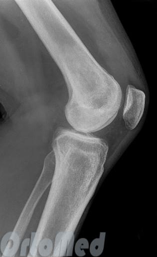
Knee x-ray, lateral view – patellar ligament intact, kneecap in typical position
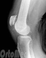
Rupture of the patellar ligament with upward displacement of the kneecap due to traction on the quadriceps.
Symptoms and diagnosis of a knee ligament strain
Before recommending tests, the doctor will examine and palpate the knee for the following symptoms of a sprain
- limitation or difficulty moving the knee;
- a grinding or clicking sound that accompanies movement of the joint
- significant pain when palpating the knee, pain when trying to step on the joint or turn it in the direction of the sprain
- Swelling of the knee joint, sometimes accompanied by bruising, redness and increased skin temperature (occurs some time after the injury and increases in intensity).
After the initial diagnosis, the doctor prescribes an ultrasound, X-ray, CT or MRI scan to confirm the diagnosis and determine the extent of the injury. These examinations also serve to detect accompanying injuries such as fractures. Once the diagnosis is established, it is time to begin treatment.
Treatment options for dislocated knee ligaments
Illness can be better prevented. That's why it's important to know how to protect yourself from a sprain. When exercising, it is best to wear appropriate clothing and shoes and not put too much strain on your body. Excessive weight creates dangerous conditions for sprains because it puts additional strain on the knee joints. Moderate exercise, a balanced diet and an active lifestyle, on the other hand, can strengthen the ligaments. However, if a sprain cannot be avoided, it is important to see a doctor immediately and take a number of measures to bring about healing as quickly as possible.
Regardless of the severity of the sprain, the limb should be protected as much as possible in the first few days. In severe cases, the knee should be secured with a special bandage or a tight cast. Every 3-4 hours, ice cubes should be placed on the injured joint with a damp towel for a maximum of 20 minutes. Painkillers such as aspirin or paracetamol can be taken. Once the swelling subsides, simple strengthening exercises and exercises should be performed regularly. This is necessary for the ligaments to function properly in the future, otherwise the new tissue may become stiff.
Treatment of knee ligament sprains with UHT
Together with other measures, the use of UWT (shock wave therapy) allows the ligaments to be repaired in the shortest possible time. The treatment uses sound waves with a specific frequency. Their effect causes activation of tissue regeneration. A pronounced anti-inflammatory and antipruritic effect also helps to quickly restore the function of the knee joint.
The main advantages of UHT are:
- disappearance of pain, inflammation and swelling;
- rapid recovery of function and all functions after a knee ligament sprain;
- lower likelihood of recurrence of joint damage;
- the opportunity to return to normal physical activity as quickly as possible (which is especially important for athletes).
Collateral ligament injury: course and prognosis
To achieve a faster and more complete recovery, it is important to diagnose the injury early and provide comprehensive treatment and rehabilitation.
- Isolated tibial ligament injuries usually heal through fibrosis.
- Grade III injuries, particularly those associated with a torn cruciate ligament, can lead to complications (e.g., osteoarthritis of the knee). In this case, the symptom of varus instability persists for a long time.
Lateral Collateral Ligament Injury: Prevention
A tear in the collateral ligament is difficult to prevent. However, there are some things you can do to help:
- Use the correct technique During sports.
Proper technique that allows for proper biomechanics of the knee joint reduces the risk of injury, especially when jumping, twisting, or lifting weights. - Stretching and strengthening the leg muscles.
- Knee ligaments.
During active sports, it is advisable to use special knee pads to prevent excessive lateral movement of the bony components of the joint.
Prevention is particularly important if you have suffered a torn ligament in the past. Once the tissue is damaged, there is an increased risk that it will be injured again.
Doesn't that sound like your condition?
A torn ACL is just one possible cause of pain on the outside of the knee. So if you need help figuring out what's going on with your knee, visit Knee Pain Diagnostics.
If your knee pain is caused by a specific event, such as: B. a fall, visit the knee injuries page.
Symptoms of a rupture of the collateral ligaments in the knee
The pain and instability of the knee joint are particularly severe and localized at the site of the tear.
The joint is swollen and its outline is flattened. On the 2nd to 3rd day after the injury, there is sometimes extensive bruising that extends down to the lower leg. Free fluid (hemarthrosis) is noted: positive patellar stiffness and balloon signs. Palpation reveals local tenderness in the projection of the injured ligament.
With a rupture of the collateral ligament, excessive deviation of the tibia to the side opposite to the damaged ligament is noted. If an internal ligament rupture is suspected, the doctor holds the outer surface of the knee joint with one hand and tilts the lower extremity outwards with the other hand. The possibility that the shin bone deviates more outward than in the healthy leg indicates a tear in the medial collateral ligament. The patient's leg must be extended at the knee joint during the examination. In acute injuries, these examinations are performed after procaine has been injected into the knee joint cavity and the knee has been anesthetized.
After the acute phase has subsided, the instability of the knee joint ('wobbling') remains, so that the patient is forced to strengthen the joint with bandages or by wearing a special knee orthosis. Gradually, muscle atrophy of the limbs develops and symptoms of deforming gonarthrosis appear.
Laboratory and instrumental studies
When deforming gonarthrosis begins to develop, the clinical diagnosis can be confirmed by an X-ray examination using a device provided by the clinic. The x-ray clearly shows an enlargement of the joint space on the side of the injury.
[1], [2], [3], [4], [5], [6], [7]
Treatment of torn collateral ligaments of the knee joint
The acute phase of the injury is treated as an inpatient.
Conservative treatment of torn collateral ligaments of the knee joint
Isolated ruptures of one of the collateral ligaments are treated conservatively. Puncture of the knee joint, removal of the hematoma, injection of 25-30 ml of 0.5%iger procaine solution into the joint cavity. For 5-7 days (until the swelling subsides), a circular plaster cast is applied from the groin fold to the fingertips in a functionally favorable position and with excessive tibial deviation (hypercorrection) to the side of the lesion. From the 3rd day onwards, UHF and static exercises are recommended. Immobilization lasts 6-8 weeks. Once cleared, rehabilitation treatment is recommended.
Surgical treatment of knee collateral ligament rupture
There are several surgical techniques to repair the collateral ligaments of the knee joint.
Tibial collateral ligament plasty. Tibial collateral ligament tears are more common than fibular collateral ligament tears. They often occur in combination with injuries to the medial meniscus and the anterior cruciate ligament (Turner's triad).
In the past, the Campbell procedure was the most commonly used procedure to restore stability to the knee joint in cases of tibial collateral ligament tears. The material used for the plication is a strip of the broad femoral fascia.
Various methods have subsequently been proposed for surgical repair of the tibial collateral ligament: entrapment, ligament plication with lavane, and cancellous tendon.
In 1985, AF Krasnov and GP Kotelnikov developed a new method of autoplastic of this tape.
A soft tissue incision is made in the projection of the lower third of the tendon and the tendon is isolated.
A bone and periosteal suture is placed in the area of the inner epicondyle of the femur and the tendon is moved underneath. They are then sutured to the periosteum at the entrance and exit. The flap is reinforced with transperiosteal sutures. The wound is stitched up.
- Rupture of the medial collateral ligament.
- Femoral collateral ligament.
- Thigh tendon rupture.
- Rupture of the ligaments of the ankle.
- shoulder supinators.
- Rupture of the radial-clavicular joint.
- DMS rupture.
- tibial ligaments.