In these cases, the nodules are examined histologically to determine the nature of the skin lesions and determine their cause.

- Follicular hyperkeratosis
- symptoms
- ICD-10
- causes
- Radiological and clinical manifestations of Schinz calcaneal disease
- Treatment options for Haglund Schinz disease in children
- complications
- Treatment
- change of lifestyle
- Clinical manifestations of Haglund's deformity
- Specificity of the course of Haglund's deformity in pregnancy
- Clubfoot in children
- Prognosis of Schinz disease
- Benign skin tumors
- Precancerous neoplasms (precancerous lesions)
- Which doctor should I see?
- Diagnosis of leg rash in children
- Treatment of foot rash in a child
- medication
- Diagnosis of a hygroma
- surgical treatment
Follicular hyperkeratosis
Follicular hyperkeratosis is a skin disease characterized by the growth of nodules of keratinized epidermis at the mouths of hair follicles. The skin in the affected areas becomes dry and rough and is covered with numerous reddish nodules that resemble goosebumps. The disease most commonly affects the elbows and knees, the buttocks and the outside of the thighs. It does not cause illness, but can cause discomfort and dissatisfaction with the appearance due to dry skin.
There are two types of cutaneous follicular hyperkeratosis.
- Type I is caused by a vitamin A deficiency. It is characterized by excessive dryness of the skin on the elbows, knees, the outer sides of the limbs and buttocks, as well as the appearance of spiny nodules at the mouths of the hair follicles.
- Type II develops with vitamin C deficiency. It can be recognized by the darker color of the nodules that block the exits of the hair follicles, as they are made of blood or pigment. The outer thighs and abdominal area are most commonly affected.
It can be congenital or acquired.
symptoms
The main symptom of follicular hyperkeratosis is goose bumps, accompanied by dryness and roughness of the skin in the affected areas. The keratinized nodules appear as a small reddish or yellowish rash that occurs on the arms and thighs, knees, buttocks and, less commonly, other parts of the body. They are located at the base of the hair follicles and form a small red border around each keratinized element. The nodules and plaques are no larger than a matchstick and can remain on the affected skin for many years. In some cases, follicular hyperkeratosis occurs on the face or scalp. The generalized form is characterized by extensive skin involvement of the trunk and extremities.
Only a doctor can accurately diagnose the disease. Don't delay your consultation - call +7 (495) 775-73-60
ICD-10

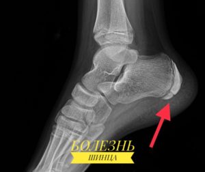
Schinz disease (osteochondropathy of the heel tip, Haglund-Schinz disease) is an osteopathy of the apophysis (tip) of the heel bone. It is caused by constant overuse of the foot (usually during sports) and repeated, sometimes mild, trauma to the heel. This osteochondropathy usually develops in girls between the ages of 10 and 16, but is less common in boys. It is not uncommon for both heels to be affected. The disease resolves spontaneously as the child gets older. Heel pain can last for a long time, sometimes until the child has finished growing. It is more common in female athletes, but can also occur in inactive children. It is one of the diseases of adolescence and childhood and is very rare in adults.
causes
Schinz disease is caused by aseptic necrosis of the calcaneus, which can arise from a genetic predisposition, metabolic disorders, neurotrophic disorders, a history of infections, and frequent trauma to the foot. The triggering factor is high mechanical stress on the heel bone, the foot tendons and the Achilles tendon.
Genetic predisposition determines the small number or reduced diameter of the vessels involved in the blood supply to the heel bone, while infections, trauma and other circumstances negatively affect the arteries. The excessive strain disrupts the vascular tone, the bony area is no longer adequately supplied with nutrients and aseptic necrosis occurs (destruction of the bone without inflammation or infectious agents).
Radiological and clinical manifestations of Schinz calcaneal disease
Schinz's calcaneal disease has a long-lasting course without obvious clinical signs. Gradual disruption of blood supply in the periosteal region of the calcaneus leads to gradual thinning of the trabeculae. The fragility of the bone increases and it tends to form isolated foci of increased pressure.
With prolonged physical stress on the calcaneal tuberosity, typical clinical symptoms can occur:
- Severe pain in the back of the heel;
- inability to take a step on the heel;
- Swelling of the heel in the back area;
- The skin is hyperemic;
- Skin feels hot and dry;
- In the initial period, there may be general disorders, weakness, a slight increase in body temperature to a subfebrile level.
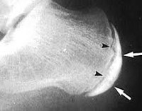
There are typical radiological signs of Schinz disease that are clearly visible when both heel bones are compared. However, it is not uncommon for both lower limbs to be affected at the same time. In this case, an MRI or CT scan may be indicated for differential diagnosis. Н
X-rays show foci of replacement tissue in the thickness of the heel bone. The edge of the heel bone may be lumpy and irregular. Foci of complete darkening in most cases indicate the second stage of the inflammatory process, when fibrous exudate accumulates in necrotic cavities. The presence of fresh calluses in areas where the bone ridges have dissolved is more typical of the final stage of the pathological process. At this stage, the patient no longer experiences severe pain when walking.
Treatment options for Haglund Schinz disease in children
Official medicine uses a number of conservative methods to treat Schinz disease. If these do not show a clear positive effect, surgery is carried out. This involves cutting certain areas of the femoral nerve and the saphenous nerve. These supply the bursa area with nerves. Once they are severed, sensitivity is completely lost. The child can then walk without pain.
However, one should be aware that such a surgical intervention interrupts the natural process of innervation of soft tissues. Under such conditions, the destruction of bone tissue will be much more intense. This leads to a complete narrowing of the small blood vessels, and the soft tissue suffers from a lack of nutrients and oxygen. Atrophy of the periosteum of the tibia occurs.
Schinz heel disease can only be treated conservatively in the acute stage. There are no medications that protect against recurrence of aseptic bone necrosis. Prescribed medication and physiotherapy can accelerate the resolution of the inflammatory reaction and initiate tissue regeneration. Drug therapy can shorten the course of Schinz's disease in children to 12-14 days. However, after the first major load on the heel, all symptoms may return, as the aseptic inflammatory process can be triggered by any traumatic load.
For successful treatment of Haglund-Schinz disease and long-term prevention of recurrence, all pathogenetic factors must be eliminated. It is important to identify the cause of this pathology and, if possible, exclude it. Rehabilitation therapy should then be carried out to effectively restore microcirculation in the heel bulb area.
Children with Schinz disease should be treated by an orthopedic surgeon. In the initial phase of treatment, special gel insoles can be used under the heel. This ensures more comfort when walking. However, with properly developed individual treatment, the patient's well-being improves quickly. Reliable protection against a recurrence of the disease is provided.
complications
Complications of Osgood-Schlatter disease are rare. Chronic pain or local swelling may occur, which can be well treated with cold compresses and NSAIDs. Often, even after the symptoms have subsided, a nodule can remain on the lower leg at the site of the swelling. This nodule may persist to some extent throughout life, but usually does not affect the function of the knee.
The medical history is important for diagnosis, and the doctor will need the following information:
- A detailed description of the child's symptoms
- Relationship between symptoms and physical activity
- Information about previous medical problems (especially previous injuries)
- Information about medical problems in the family
- Any medications or supplements the child is taking.
To diagnose Osgood-Schlatter disease, the doctor examines the child's knee joint to see if it is swollen, painful, and red. The extent of movement in the knee and hip is also assessed. The most common instrumental diagnostic method is an x-ray of the knee joint and tibia to visualize the insertion area of the patellar tendon on the tibia.
Treatment
Typically, Osgood-Schlatter disease is self-treating and symptoms resolve once bone growth is complete. If symptoms are severe, treatment includes medication, physical therapy, and physical therapy.
Drug treatment involves prescribing painkillers such as acetaminophen (Tylenol, etc.) or ibuprofen. Physiotherapy can reduce inflammation, swelling and pain.
The therapist should use exercises that stretch the quadriceps and tendon muscles, thereby reducing the stress on the insertion area of the patellar tendon on the shinbone. Hip strengthening exercises also help stabilize the knee joint.
change of lifestyle
- Relieving pressure on the joint and limiting activities that worsen symptoms (e.g., kneeling, jumping, running).
- Apply cold to the affected area.
- Use of the patellofemoral joint during sports.
- Replacing sports that involve jumping and running with activities such as cycling or swimming for as long as it takes for symptoms to subside.
Use of the material is permitted with an active hyperlink to the permanent article page.
Clinical manifestations of Haglund's deformity
Initially, the symptoms of Haglund deformity are often confused with a large cornea. The appendix is relatively soft, but over time it becomes denser and the mass becomes inflamed.
At the beginning of the inflammation, the patient complains of other symptoms of Haglund's deformity:
- redness and swelling;
- Blistering at the affected area;
- Discomfort and pain when taking off or putting on shoes;
- Pain when walking, sometimes even at rest;
- Pain when touching the growth.
X-rays show pointed 'high heel bones' with sharp spikes.
In most cases, Haglund deformity is perceived by patients as a cosmetic defect without causing severe pain. It is the pain syndrome, the symptoms of pronounced inflammation and the difficulties in adjusting shoes that prompt the patient to visit a specialist. In advanced cases, excessive and sustained compression of bone growth can lead to a rupture of the Achilles tendon, even in the absence of obvious physical pressure.
Specificity of the course of Haglund's deformity in pregnancy
The likelihood that Haglund deformity will develop or worsen during pregnancy increases as the woman's weight increases and the strain on her legs increases. The deformity itself is not dangerous for the pregnancy or the unborn child, but its painful manifestations significantly worsen the psycho-emotional state of the woman.
To avoid exacerbation of Haglund deformity during pregnancy, it is recommended to wear only comfortable shoes, avoid excessive weight gain, and use orthopedic devices to reduce the load on the feet. If symptoms are unpleasant, conservative treatment is recommended. Pharmacological treatment is indicated in severe cases and must take gestational age into account.
Clubfoot in children
Most often occurs in girls at the age of 7-8 years, in boys less often at the age of 9-11. It is very rare in adults. The disease is accompanied by gradual destruction of the heel bone, which is accompanied by pain.
Inadequate treatment can lead to lameness. Both or one limb can be affected by the pathological process. The heel bone is the largest bone in the foot and carries much of the load when running, walking and jumping. On the posterior surface of the bone affected by Schinz's disease there is a prominent tubercle of the heel bone. The cause of this disease is not yet clear. However, there are some factors that can contribute to this condition:
- overweight;
- Frequent trauma to the ankle joint;
- clubfoot;
- ill-fitting footwear;
- Poor blood circulation.
Prognosis of Schinz disease
Cheap – symptoms subside within 1.5 to 2 years. Sometimes the pain can last as long as the foot grows, but with appropriate treatment the patient will eventually recover.
It includes the elimination of excessive loads, a well-thought-out training regime during sports activities.
The Vlasov Medical Center Ltd. applies a complex of specialized procedures that have a permanent and rapid result in relieving the main complaints of this pathology.
Benign skin tumors
In benign tumors, the cells' ability to differentiate is usually not impaired. This means that they retain their original functions and are structurally similar to normal cells. They also grow slowly and can press on adjacent tissue but never penetrate it.
Types of Benign Skin Tumors:
- A myxoma is a tumor of the sebaceous gland that results from a blockage in the gland. It usually occurs where the most sebaceous glands are located: on the neck, back, head, groin.
- A hemangioma is a vascular tumor that develops from blood vessel cells. Its color ranges from red to blue-black.
- papillomas and warts. A growth in the form of a small nodule or wart. Caused by the human papillomavirus (HPV). Usually occurs against the background of stress, weakened immunity and vegetative disorders. They often occur in the armpit or groin area. Papillomas are also the most common skin tumors on the eyelids.
- Lymphangiomas are tumors in the walls of lymphatic vessels that form in the womb. They appear as small masses with a lumpy surface and are blue or reddish-brown in color.
- A lipoma is a tumor of the fat layer ('fatty tumor'). It is located in the subcutaneous tissue, most commonly in the upper back, on the edge of the shoulder and on the outside of the thighs. The tumor is painless, soft and mobile.
- Fibroadenoma – is a new formation of connective tissue. It occurs most commonly in young and middle-aged women. It looks like a new growth on the skin in the shape of a ball that protrudes above the surface of the skin.
- A neurofibroma is a tumor that arises from nerve sheath cells. It looks like a dense nodule 0.1-2.3 cm in size.
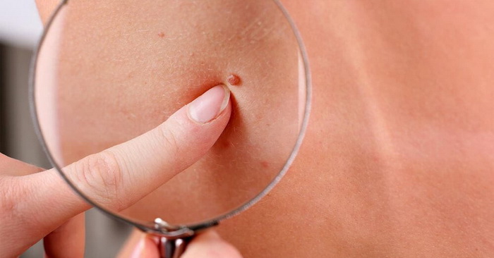
Another group of tumors are birthmarks (nevi). These can be brown, red, black, purple, etc. In most cases, a nevus is a congenital malformation of the skin. However, nevi can also appear over the course of life, usually when exposed to sunlight. Nevi are not susceptible to malignant transformation, but in some cases this can occur as a result of damage or trauma to the skin on the nevus.
Precancerous neoplasms (precancerous lesions)
Precancerous lesions are tumors that have developed a tendency to become cancerous for congenital or current reasons. These are usually chronic diseases that occur in a person over a long period of time.
Precancerous lesions are dangerous skin tumors that can lead to the development of cancer. This includes:
- Senile keratoses are keratoses in which dry crusts and scales appear on the skin of older people. If they peel off, they may bleed easily.
- Xeroderma pigmentosa is a hereditary neoplasm that develops due to increased sensitivity of the skin to ultraviolet light. It is rare and presents as pigmented patches that become papillary hyperplasia.
- Cutaneous keratosis is a cone-shaped tumor that resembles a horn. It occurs on exposed areas of the body that are regularly rubbed or squeezed. It often occurs in older people.
- Bowen's disease is an intraepithelial neoplasm. If left untreated, it can develop into invasive skin cancer. In the early stages, Bowen's disease appears as a small reddish-brown spot measuring 2-50 mm with a scaly surface and raised, jagged edges. When the scales are removed, a moist but not bleeding surface is left behind.
Which doctor should I see?
A rash on a child's feet is not the only symptom that occurs with skin diseases. The child itches, his general condition worsens, appetite decreases and sleep is disturbed. Even if your child doesn't have a fever, you should see your family doctor.

It may be necessary to consult an infectious disease pediatrician, allergist, or dermatologist to determine the cause of the rash.
Diagnosis of leg rash in children
Differential diagnosis of the rash requires history and examination.

If an allergy is suspected, special tests are carried out. For skin diseases, an examination by a dermatologist is sufficient. If necessary, the rash is scraped out. If the cause of the rash cannot be determined, in severe cases the cerebrospinal fluid is examined to rule out meningitis.
Treatment of foot rash in a child
The rule of thumb for treating skin diseases in infants and one-year-old children is: hygiene. Avoid contact between the child and the allergen. Maintain a comfortable temperature and humidity in the room. Clothing and underwear should only be made of cotton fabrics. Wash them with special powder or baby soap.
Use anti-allergenic cosmetics for skin care. The baby should be bathed frequently. When using mixed or infant formula, avoid allergy-causing foods.
medication
Treatment is prescribed by the doctor depending on the diagnosis, including antivirals, antifungals and antibiotics.

- Antiallergic drugs are indicated to relieve swelling and reduce itching - these include Suprastin, Diazolin, Zirtek.
- Antiseptics such as chlorhexidine, miramistin, betadine can be used to treat the rash.
- Topicals such as Bepanten and Fenistil gel can be used in young children to reduce inflammation and itching and to promote rapid healing of the skin.
- Older children can be treated with topical corticosteroid ointments and creams such as prednisolone and Kenalog.
- In severe cases, immunostimulants, vitamin complexes and local mud and radon baths are prescribed.
Each disease has its own prescription, which can only be issued by a doctor. Treatment begins as soon as the cause of the rash is eliminated. To get rid of the rash, it is enough to organize appropriate care for the child. Food allergies require a special diet, and scarlet fever should only be treated in an inpatient setting.
Diagnosis of a hygroma
A hygroma can be diagnosed by an orthopedist or surgeon, with diagnosis made by palpation. Examination reveals a small, round nodule 1-5 cm in diameter with a smooth surface and soft elasticity to the touch. If necessary, the doctor will recommend additional instrumental examination methods, such as an X-ray of the joints.

- As part of conservative treatment, a puncture of the hirgroma is carried out for therapeutic or diagnostic purposes. The nodule is punctured with a puncture needle and its contents are drained. Sclerosing agents are then injected into the cleaned defect and a pressure bandage is applied.
- The limb is immobilized with an orthopedic splint or cast for 7-8 days to prevent movement of the tendon (or damaged joint), which reduces synovial fluid production. A major disadvantage of this treatment method is the high relapse rate.
- If necessary, physiotherapy (UV light) or paraffin compresses are prescribed.
surgical treatment
- Excision of the hirgroma – this method is considered an effective method for the treatment of the hirgroma. It essentially consists in the complete removal of the thickened capsule, which is not carried out in the altered tissue, but is closely sutured to the subcutaneous fat of the stump. The method has the advantage that the recurrence rate is significantly lower compared to other procedures.
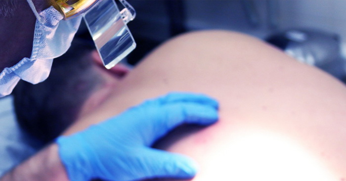
- Laser removal of a hematoma on the foot involves heating the tumor with a laser until it is completely destroyed, leaving neighboring tissues unaffected. This shortens the healing time and has the great advantage that no scars are left behind.
Indications for removal are rapid growth, significant tumor size, cosmetic defect, development of complications (abscess, inflammation, swelling, redness or severe pain). When surgically removing a nodule, all procedures are performed under local anesthesia, which eliminates any pain sensations.
Read more:- Heel bone human anatomy photo and description.
- Shoes for valgus deformities in children.
- Toe movement in children.
- What is the name of the ankle bone?.
- Footwear for children's clubfoot.
- Shallow valgus deformity in children.
- Do orthopedic shoes help children?.
- Orthoses for valgus in children.