Of course, it will take longer for the bones to heal if you don't support them with a special diet. So what should you eat to help injured limbs heal faster? One of the most important substances is protein. This is found in cheese, meat, eggs and fish. These foods also contain phosphorus, a trace mineral that is equally important for healing injured bones. Another useful element is calcium. All dairy products are rich in it. It is possible to prepare such a cocktail according to a recipe: to do this, add a few sesame seeds and flax seeds to a glass of kefir. They have an anti-inflammatory effect on the body, promote wound healing and allow a broken ankle to heal faster. To refine the smoothie and give it a varied taste, you can add fruit and walnuts before chopping the ingredients in the blender.
- broken ankle
- ICD-10
- Foods that affect healing
- Effective methods for healing an ankle fracture in children
- ankle
- Video anatomy of the ankle joint and tibial head
- Causes of ankle fractures
- classification
- Causes of ankle sprain
- Types of ankle sprains
- Causes of injury
- The diagnosis.
- How long does a broken ankle take to heal?
- Factors affecting the speed of rehabilitation after an ankle fracture
- surgical treatment
- Possible complications
broken ankle
broken ankle – is a disruption of the integrity of the ankle joint as a result of a traumatic impact. It usually occurs when the foot is turned outwards or inwards. It manifests itself in pain, swelling, bruising and a restriction of support and movement. In some cases, grinding, deformation, and abnormal mobility occur. An x-ray of the ankle joint is recommended to clarify the diagnosis. Treatment is conservative in most cases, with repositioning and casting of the ankle if indicated. If closed reduction of the ankle joint is unsuccessful, surgery is required.
ICD-10

Ankle fractures are one of the most common injuries to the skeletal system. They can affect people of all ages and genders, but are more common in middle-aged and older people due to reduced movement coordination and general physical fitness. The frequency of ankle fractures increases significantly in winter, especially when there is black ice. The injury may be associated with or without ligament tear, subluxation, and sprain. It can be a single, double or triple ankle fracture. The prognosis, as well as the tactics and timing of treatment, depend on the characteristics of the fracture.
The pathology may be combined with fractures of other limbs, thoracic trauma, CMT, pelvic fracture, blunt abdominal trauma, kidney injury, etc. Isolated ankle fractures are usually closed. Open injuries and contusions are not uncommon in combined injuries. The treatment is carried out by trauma surgeons.

Since an ankle fracture is an intra-articular injury, an intra-articular injury must be treated not only by a traumatologist, but also always by a rehabilitation specialist.
This is much more common in the winter time. Therefore, ankle fractures are the most common winter injuries. The foot can be twisted outwards and inwards. In general, an ankle fracture is a combination of ligament and bone injuries, with ligament injuries playing just as big a role as bone injuries and fractures. The possibility of conservative or surgical treatment depends on this combination of soft tissue injuries and bone fractures.
If you have an ankle fracture, it is important to see a doctor as soon as possible. A correct diagnosis is important here as ankle fractures can vary greatly. It is important to get a specialist's opinion on whether and how surgery is necessary.
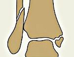
As far as the anatomy of the ankle is concerned, there is the lateral malleolus, which is the natural extension of the fibula, and the medial malleolus, where the tibia ends. The outer malleolus and the inner malleolus, together with the tibia, form what is known as the foramen of the ankle joint. In this foramen is the talus, whose main movements are flexion and extension. The human foot can rotate outwards and inwards, it can supinate or pronate, that is, it can bend inwards or outwards, but only within certain limits. When these boundaries are violently and unintentionally violated, a rupture occurs. For example, a person slips and all the weight falls on one foot - the bone cannot support the load. A triquetral fracture and a 'posterior ankle' are sometimes referred to. In reality, there is no such thing as a back ankle. However, such a term exists because there are powerful posterior intercondylar syndesmotic ligaments attached to the back of the tibia. This is sometimes referred to as Folkman's triangle. In some injuries, the ligaments are stronger than the bone and the Folkman triangle is torn. This is called a fracture of the triangle.
Foods that affect healing
Of course, it takes longer for the bones to heal unless a special diet stimulates healing. So what should you eat to help injured limbs heal faster? One of the most important substances is protein. This is found in cheese, meat, eggs and fish. These foods also contain phosphorus, a trace mineral that is equally important for healing injured bones. Another useful element is calcium. All dairy products are rich in it. Special shakes can help the fracture heal quickly.
To do this, add a few sesame seeds and flax seeds to a glass of kefir. They have an anti-inflammatory effect on the body, promote wound healing and the ankle fracture heals faster. To fill the smoothie with vitamins and add variety, you can add fruit and walnuts to the smoothie and grind the ingredients in a blender.
Effective methods for healing an ankle fracture in children
The healing time for a broken ankle in children depends on several factors:
The larger the broken bone and the older the child, the longer it will take to heal. For example, it takes an average of two to three weeks for the phalanges to heal and two to three months for the shinbone to heal. For injuries that involve a dislocated bone, such as: B. the shinbone, healing can take up to four or five months.
Rehabilitation of a child after a fracture is not much different from that of an adult. It is important that the child exercises regularly to increase strength, taking care not to put too much strain on the injured limb and eating a healthy diet.
ankle
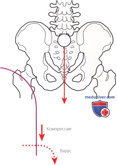
Figure 1: The ankle joint lies to the side of the center of gravity and is therefore susceptible to varus and compression.
a) ankle. The ankle joint is a synovial joint and is formed by three bones: tibia, fibula and talus. Despite their close relationship, the ankle and foot have different, independent functions. The ankle joint is an extremely stable connection between the lower limb and its supporting base, the foot. Because the ankle joint lies to the side of the body's center of gravity, it is exposed to varus and compression loads (Fig. 1). The distal ends of the tibia form the foramen or acetabulum of the ankle joint. There is an medial malleolus formed by the distal end of the tibia and the distal articular surface, and an medial malleolus formed by the distal end of the fibula. The lesser lateral malleolus is located distal and posterior to the medial malleolus.
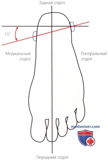
Figure 2: The intercondylar axis is rotated outwards by 15°.
As a result, the axis passing through both ankles and the frontal plane of the tibia form an angle of approximately 15° (Fig. 2). From the front, the ankle joint is reinforced by the anterior intercondylar ligament. Posteriorly, the joint is supported by the convexity of the posterior edge of the lower articular surface of the bone (posterior hock) and the posterior intercondylar ligament. The shaft of the talus bone is covered by the fork of the ankle joint. Its convex upper articular surface (block) articulates with the lower articular surface of the tibia. The talus also has articular surfaces of the ankle joint. The talar block is wider anteriorly than posteriorly. At maximum extension, the talar bone is firmly locked in the ankle joint by the wedge. This causes internal-external tension on the distal syndesmosis and tibiofibular ligaments. In an intact ankle, the talus moves primarily in one plane (flexion-extension) with only a small amplitude of displacement in the anteroposterior direction. The increased stability of the ankle during posterior flexion allows assessment of the integrity of the medial and lateral ligaments of the ankle as well as the inversion-extension mobility of the subtalar bone.
Video anatomy of the ankle joint and tibial head
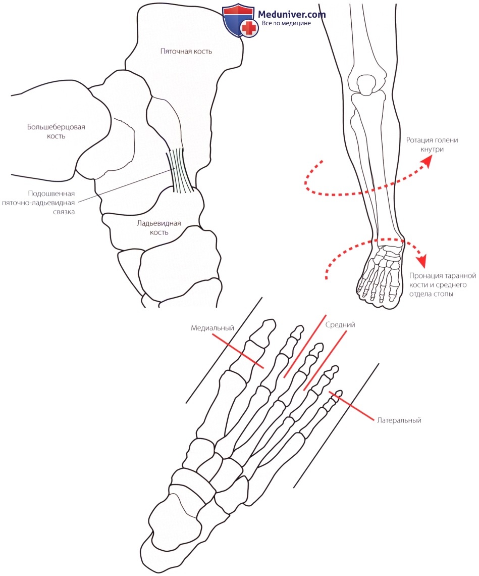
b) Foot. The main functions of the foot are to provide stable support to mitigate shock loading on the limbs during movement and to allow efficient forward movement of the trunk. Three parts of the foot - the hindfoot, the midfoot and the forefoot - are involved in carrying out these tasks, which in turn include a series of mobile and semi-mobile joints that allow the foot to adapt to different surfaces. The bony elements of the foot form the longitudinal and transverse arches. The sole parts of these vaults are reinforced by bands that act as shock absorbers (Fig. 3a).
The foot consists of 26 bones (Fig. 3b). The rear foot takes up a third of the length of the foot. It consists of the two largest bones of the foot: the larger metacarpal and the smaller talus. The heel bone is inferior and supports the body of the ankle bone. The talus is the only bony connection between the shinbone and the foot and connects to the shinbone in the middle of the hindfoot.
The metatarsal consists of the small ankle joint, the three sphenoid bones (inner, middle and outer) and the cuboid bone. This part of the foot takes up just over a sixth of the foot. Only small movements take place at the metatarsal joints.
The forefoot takes up half the length of the foot. It consists of small, long bones, including five metatarsals and 14 phalanges.
The structural integrity of the foot is ensured by the formation of the articular surfaces of the bones and the support of the soft tissues. All joints of the foot are synovial in nature. Soft tissue support is provided by static (ligamentous) and dynamic (tendinous-muscular) stabilizers. Instability of the joints or soft tissues leads to ankle joint dysfunction, foot dysfunction, reduced movement efficiency, the development of osteoarthritis and even bone damage (stress fractures).
Causes of ankle fractures
The most common cause of ankle instability (approximately 80 %) is a fracture of the lateral malleolus. Fractures of the medial malleolus are less common.
Immediately after the injury, severe pain occurs at the fracture site, making it impossible to stand on the injured leg. With a simple ankle injury, the injured person can sometimes walk unaided using the heel.
Local swelling, hematomas and deformities in the ankle area occur very quickly. When palpated, the most painful area is 3-4 cm above the ankle. Comminuted fractures with displaced bone fragments result in extensive bruising of the hindfoot and sole of the foot. There is a characteristic angle between the tibia and the distal limb and there is pathological mobility and a pronounced cracking of the bone (crunching).
A medial ankle fracture is characterized by increased pain in the area of the medial malleolus, which is formed by the tip of the tibia. The severity of the clinical symptoms of the injury largely depends on the degree of displacement of the bone fragment and damage to the ligamentous apparatus.
classification
All fractures of the ankle joint are classified according to the mechanism of injury:
- Pronation and abduction (excessive outward rotation of the foot)
- Supination-adduction (inward rotation of the sole of the foot)
- Rotation (excessive flexion of the sole of the foot)
There are open, closed, isolated, fused and united, oblique, transverse, single, double and triple ankle fractures. In the latter case, it is a single-stage fracture of the medial malleolus, medial malleolus and tibial plateau.
Causes of ankle sprain
- Increased mobility of the joint
- Congenital hypoplasia (underdevelopment) of the ankle
- History of repeated sprains of the ankle, habitual sprains
- high arch
- Age-related changes and destructive degenerative diseases of the musculoskeletal system.
The clinical picture is acute pain in the ankle at the time of injury. This then leads to swelling, hematomas and impairment of movement and support of the limb. Liveliness and tension in the area of injury, cold foot, lack of pulsation and sensitivity indicate a neurovascular disorder. Subluxations result in a moderate disruption of the relationship between the anatomical structures, and dislocations result in pronounced deformity. Crepitations indicate a simultaneous fracture.
Types of ankle sprains
A posterior sprain is the most common type of ankle injury. The talus is displaced posteriorly relative to the distal tibia as the force pushes the foot backwards. With a lateral ankle injury, a tear of the intercondylar syndesmosis or a fracture is detected.
An anterior dislocation is caused by immobilization of the foot, force on the tibia, and forced dorsiflexion of the ankle.
A lateral sprain occurs due to forced inversion (twisting), eversion, or external/internal rotation of the ankle joint. The injuries are associated with fractures of the ankle and/or distal fibula.
The upper ankle joint is the result of upward displacement of the talus as a result of a jump from a height. The patient is also examined for a heel bone fracture and spinal cord injury.
In 50 % cases it is an open dislocation of the ankle joint in which the integrity of the skin is disturbed by a fragment fracture.
Causes of injury
This type of injury is often caused by the following causes
- Rolling the foot inwards.
- Sudden twisting of the foot
- Direct impact on the foot or toes
- An unfortunate fall or leap onto your foot from a great height
It is extremely important to consult a qualified doctor in a timely manner, otherwise the consequences of a sprained foot can be catastrophic: there is a risk of developing diseases such as arthritis or arthrosis; mobility of the injured joint may be impaired after healing; Partial or complete muscle loss and circulatory problems in the limb may occur.
The diagnosis.
After the accident, the injured person must be taken immediately to the accident center and must not be able to move their leg (actively or passively). After admitting the patient, the doctor conducts a detailed examination of the injured limb and takes a complete anamnesis. Only with the help of an x-ray can the trauma surgeon make an accurate diagnosis (determine the type of dislocation) and initiate appropriate treatment.
Treatment should only be performed by a qualified traumatologist.
- Reduction of a dislocated foot under local or general anesthesia
- Immobilization of the dislocated joint with a plaster cast
- Immobilization for 8 to 12 weeks (depending on the type of sprain and the severity of the injury)
- Administration of pain medication (if necessary)
- For the first few days, you should take an elevated and cold position.
- It is forbidden to lean or stand on the injured limb while it is healing.
How long does a broken ankle take to heal?
Of course, the time it takes to heal the injured limb depends primarily on the severity of the fracture. According to medical practice, healing a fracture without dislocation takes one to two months if the process is not hindered by negative factors (age, chronic diseases).
In order for the ankle joint of the lower limbs to heal properly and quickly after a serious injury, significantly more time is required. Proper positioning and immobilization of the leg after applying the cast is very important. If this is the case, the rehabilitation course is automatically extended by at least one month.
There are situations in which both ankles are broken at the same time without any displacement of the bones. In such cases, the cast can be removed after six to eight weeks. If the injury to both legs was aggravated by displacement of the bones, healing will take about two and a half months.
For an open ankle fracture with muscle damage and bone displacement, the healing time is three to four months. Rehabilitation after removing the cast depends on the measures the patient takes.
To make the treatment as effective as possible, the patient should visit the doctor regularly while wearing the cast. X-rays make it easier to monitor the bone healing process. It is strictly forbidden to remove the plaster cast yourself. Doctors also note that people with diabetes, osteoporosis and other chronic diseases that make healing difficult should wear a cast twice as long.
How quickly a limb recovers after a fracture depends on the accuracy of the cast. It is important to make sure there are no folds or kinks in the cast to avoid putting additional pressure on the broken bone. The cast can be applied without pads (for short-term use) or with a cotton swab (for long-term use).
Factors affecting the speed of rehabilitation after an ankle fracture
The goal of treating an injured limb is, on the one hand, to restore its integrity and structure and, on the other hand, to avoid complications.
The treatment methods can be divided into three types:
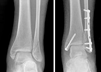
The surgical method involves an operation: bone fragments are fixed with metal plates or spokes. The damaged ligaments are stitched up. Healing occurs relatively quickly if the necessary treatment procedures are followed.
Conservative treatment consists of a cast and administration of medication to relieve pain and speed healing.
In general, this method guarantees callus formation within two to three weeks; After this period, an x-ray is usually taken and the cast is removed or treatment is extended based on the x-ray.
Important indicators of how quickly the leg will heal after an ankle fracture are:
- The age of the injured person.
- The type of fracture and its severity.
- The extent of the surgical procedure.
- Various diseases associated with bone and joint pathology, such as arthrosis, osteopathy and others.
For better healing of the limbs, traumatologists recommend including calcium and phosphorus in the diet and taking vitamin preparations.
Physical exercises for rehabilitation promote the restoration of bone integrity. These include the following methods:
Wearing a cast correctly ensures that the pain that many people experience during the healing process of an injury is avoided. Pain in the foot area can be due to impaired bone integrity, the effects of bone calluses and weakening of the muscular system due to prolonged lack of exercise.
Although there are differences in the treatment of a fracture with or without dislocation, rehabilitation treatment is equally effective in both cases. General recommendations for pain relief include bed rest, using assistive devices, and elevating the leg.
surgical treatment
If non-surgical treatment does not work or the ankle is too unstable, the doctor may recommend surgery on the broken ankle and subsequent rehabilitation. After surgery, it is important to put weight on the leg and follow all recommended measures to shorten the rehabilitation time.
Special features of the operation: If the fractures are out of place, the bone fragments must be surgically repositioned. The fragments must be returned to their normal position and fixed with special screws and metal plates that are attached to the outer surface of the bone. Surgery may include a bone graft to allow the new bone to actively grow together. This is fixed with screws and a metal plate. This can reduce the risk of osteoarthritis and speed recovery of mobility.
Possible complications
After treatment, it is important that you follow your surgeon's instructions. Failure to follow them can lead to infections, deformities, arthritis and chronic pain Source:
Errors and complications in the treatment of complex fractures of the ankle region. Salikhov RZ, Pankov IO, Plakseychuk YA, Solovyov VV Practical Medicine, 2014. pp. 128-131.
- Errors and complications in the treatment of complex fractures of the ankle region. Salikhov RZ, Pankov IO, Plakeichuk Yu.A., Solovyov VV. Practical Medicine, 2014. pp. 128-131
- Analysis of ankle joint injuries. Tailashev MM, Salatin PP, Sobolev VV, Pozikov VV, Kolesnikov AS Acta Biomedica Scientifica, 2008. pp. 144-145
- Comprehensive diagnosis of ankle injuries. Kim LI, Diachkova GV. Genius of Orthopedics, 2013. pp.20-24
- Fractures in the ankle joint area and methods of their treatment. Cherednik AA, More Gautam Sacherbao, Al-Fakih Abdulaziz. Bulletin of Online Medical Conferences, 2014. P.432
- Therapeutic tactics for intra-articular fractures of the ankle (literature review). Zedgenidze IV, Tishkov NV Acta Biomedica Scientifica, 2013. pp.178-182
- Broken ankle.
- Photo: Outer malleolus fracture.
- tibia and fibula.
- The lateral ankle is.
- Bones of the human ankle.
- The hock in which the person is located.
- The ankle bruise is where the picture is taken.
- fracture of the lateral malleolus.