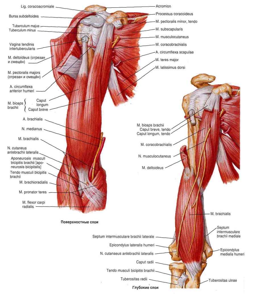
Effective supination of the forearm is an important factor in success in hook fighting. This movement is performed by the supinator muscle of the forearm (Fig. 1.6), the biceps brachioradialis muscle, and partially by the brachioradialis muscle. To further improve this movement, it is advisable to develop the strength of the driving muscles of the arm.
- forearm muscles
- deep layer
- Clinical Significance
- etymology
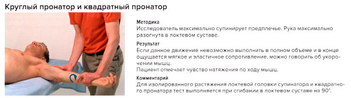
- circular pronator
- Initiation [ edit edit source ]
- Attachment[ edit ]
- Denervation [ edit edit source ]
- pronation of the forearm. Muscle function tests [edit| edit code ]
- How often should I train my forearms?
- Exercises for the forearm muscles
- forearm muscles. Front group, deep layer
- FOREARM MUSCLES, FRONT GROUP:
- forearms
- superficial layer
- How do you build a forearm?
- Training program for the forearm
- What about shoulder pronation and shoulder supination?
- What is HBA?
- Why is this necessary?
- muscles of the shoulder girdle
- Peripheral muscles of the upper limbs
forearm muscles
pronator muscle of the arm The pronator teres muscle of the forearm (Figs. 111, 115, 116, 117, 125) provides for the pronation of the forearm (rotating it forward and inward so that the hand points backwards (down) and the thumb inwards rotated toward the center of the body) and participates in its flexion. A thick and short muscle composed of two heads. The large humeral head (Caput humerale) begins at the medial epicondyle of the humerus and the intermuscular septum of the medial humeral fascia, while the small humeral head (Caput ulnare) attaches to the spinous process of the tuberculum ulnare. The two heads join to form a flattened abdomen. The middle third of the spoke is the starting point.
Brachioradialis muscle (M. brachioradialis) The brachioradialis muscle (Figs. 90, 111, 113, 114, 115, 116, 118, 121, 125) flexes the forearm and is involved in both pronation and supination of the forearm (rotating it so that the hand rotates forward (upwards) and the thumb rotates outwards in the medial plane of the body) of the radial bone. The muscle is fusiform, inserting at the humerus above the lateral epicondyle and at the intermuscular septum of the lateral fascia of the humerus, and inserting at the base of the body of the radius.
The radial flexor of the hand (M. flexor carpi radialis) (Figs. 90, 115, 121, 125) partially flexes and curves the hand. It is a long, flat, bipartite muscle whose proximal part is covered by the aponeurosis of the biceps brachii. It arises from the medial epicondyle of the humerus and the fascia of the forearm, and its insertion point is at the base of the palmar surface of the 2nd
Palmaris longus (m. palmaris longus) (Fig. 115, 125) lengthens the palmar aponeurosis and is involved in the flexion of the hand.
This muscle is characterized by a short spindle-shaped abdomen and a long tendon. It inserts on the medial epicondyle of the humerus and on the fascia of the forearm, inside the radial flexor of the wrist, and inserts on the palmar aponeurosis (aponeurosis palmaris).
deep layer
Long flexor of the thumb (m. flexor pollicis longus) (Figs. 115, 116, 120) flexes the distal first phalanx (thumb). The long, flat, single-fingered muscle originates in the upper two-thirds of the anterior surface of the radius bone, in the interosseous membrane (Figs. 117, 125) between the radius and ulna, and partially in the medial epicondyle of the humerus. It attaches to the base of the distal phalanx of the thumb.
Deep flexor tendons of the fingers The deep flexor tendon muscle (flexor digitorum profundus) (Figs. 116, 119, 120, 125) flexes the entire hand and the distal phalanges of fingers II-V. It is characterized by a pronounced flat and broad abdomen, and its insertion point is on the upper two-thirds of the anterior surface of the ulna and the intercondylar membrane. The point of attachment is at the base of the distal phalanges of fingers II-V.
Pronator quadrate (M. pronator quadratus) (Figs. 116, 117, 120, 121) internally rotates the forearm (pronation). This muscle is a thin sheet of quadriceps that lies at the distal ends of the forearm bones. It begins at the medial edge of the ulnar shaft and inserts at the lateral edge and anterior surface of the radius bone.
Clinical Significance
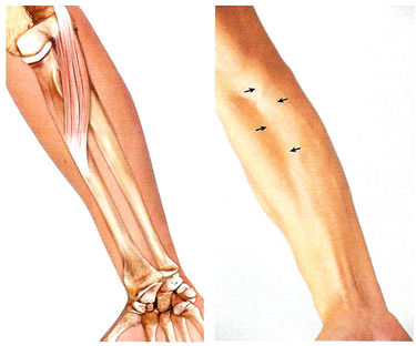
Pronator circular syndrome is one of the causes of wrist pain. It is a form of neurogenic pain.
- Patients with round pronator syndrome have median nerve numbness with repeated pronation/supination of the forearm and not with elbow flexion and extension.
- Premature fatigue of the forearm muscles is evident during repetitive weight-bearing movements, especially during pronation.
- The EMG may show only a slight reduction in conduction velocity.
- Despite the anatomical proximity, patients with round pronator syndrome do not have a higher incidence of AIN
- Other places with compressive changes:
- Strothers' ligament
- Lacertus fibrosis
- FDS of the proximal arch
- Rare causes such as consecutive tendon transfers in radial paralysis
- Positive Tinel's sign in forearm, not wrist
- Negative result of the Phalen maneuver
- Dysesthesia of the triangle of the hand
- Pain with resistance on pronation
- Forearm pain with resistance during isolated flexion of the PIP joint of the long and ring fingers
In C5 te patients with traplegia or radial nerve palsy, the circular pronator tendon, known as tendon transfer, can be redirected to the short wrist extensor tendon to restore wrist extension.
etymology
The word pronator comes from the Latin pronus, meaning 'leaning forward or lying face down', and refers to the muscular action of pronation of the forearm. The Latin term teres, meaning 'round or cylindrical' or 'long and round', refers to the shape of the muscle. So, an indirect translation of the word pronator teres from English means: a cylindrical muscle that rotates the forearm (and thus the hand) downwards.
 Cross section through the medial forearm.
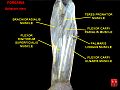 Circumferential pronator muscle.
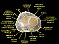 muscles of the upper limbs. transverse incision.
- Simplified representation of the circular pronator's onset
circular pronator
circular pronator (m.propatotor teres) performs the pronation of the forearm and is slightly involved in the flexion of the forearm.
Initiation [ edit edit source ]
- head of the humerus.: medial epicondyle of the humerus, medial intermetatarsal septum
- head of the humerusHumeral head : Proximal process of the ulna
Attachment[ edit ]
Denervation [ edit edit source ]
Pronation of the forearm (when the forearm is flexed).
*M brachioradialis (from supination to mid-position).
*M flexor carpi radialis (when the forearm is not bent).
*M extensor carpi radialis longus (when the forearm is flexed).
*M Brachioradialis (from pronation to middle position)
*M Extensor carpi radialis longus (in the straight forearm)
pronation of the forearm. Muscle function tests [edit| edit code ]
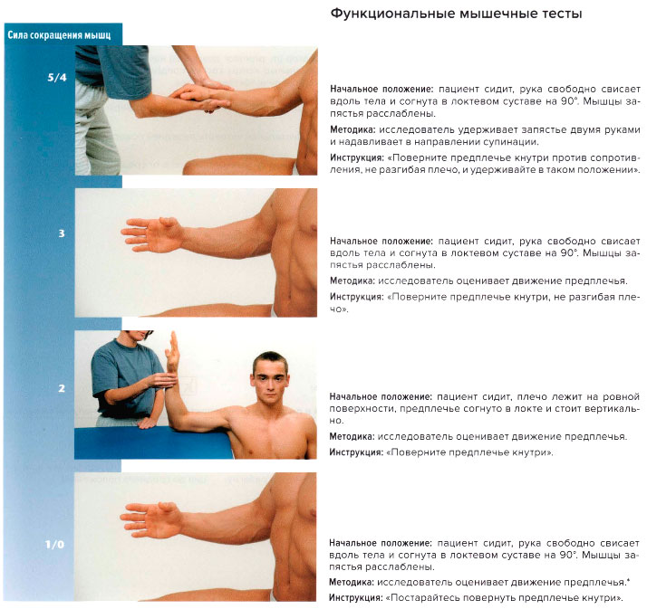
This muscle lies deep, is obscured by other muscles of the forearm, and can only be evaluated when those muscles are paralyzed or atrophied.
How often should I train my forearms?
It is often said that the forearm muscles are the most difficult of all muscles and are therefore difficult to train. One can disagree with this opinion, since the forearms are not subjected to intense loads during training. A simple weight load is necessary for the development of this group. It has already been mentioned above that the muscles of this group are inferior to the others in terms of size. This has a positive effect on their recovery speed.
That's the theory, but in practice no professional athlete pays more attention to the forearm than other muscle groups. So it doesn't make sense for all other bodybuilders to do the same. However, the number of repetitions should be increased. That's because the range of motion of the forearm is quite small, and loading time is important for the muscles. Usually 10 to 20 repetitions are enough for a full workout.
Exercises for the forearm muscles
We are now talking about an exercise program to increase muscle mass of the forearm. For increasing the strength parameters, which are of great importance in arm wrestling, we need a different program. As already mentioned, forearm training should be started after the biceps muscles, that is, in the last phase of training.
- Lifting weights with a reverse grip primarily targets the biceps, but it also engages some of the forearm muscles.
forearm muscles. Front group, deep layer
In this article, we will go further into the numerous muscles of the forearm. Myology is the branch of anatomy that deals with the structure and location of muscles in the human body. And in all myology there is no area more difficult than the forearm.
That's why I've divided the muscles of the forearm into several articles - in the last article we looked at the superficial layer of the forearm muscles. In the last article we looked at the superficial layer of the forearm muscles, now we're going to look at the deep layer.
Now recall the physiological position of the forearm so we know where the front and back of the hand are. In this illustration, the hand is in its natural anatomical position, with the front of the hand in front of us. On this side are the muscles we are looking at.
FOREARM MUSCLES, FRONT GROUP:
1.long thumb flexor (Musculus flexor pollicis longus). This is the most lateral muscle of the deep anterior group. Note the tendon - it is much shorter than the tendon of the similarly located superficial muscle.
This muscle is also characterized by the fact that its tendon runs close to the thumb.
Location: It inserts on the medial epicondyle of the humerus and on the 2/3 anterior surface of the radius bone; it attaches to the distal phalanx of the thumb;
Function: Flexion of the distal phalanx of the thumb. Here it is enough to know the starting point and the name to get an immediate idea of the function.
2. deep flexor of the thumb (Musculus flexor digitorum profundus). It can be seen that the entire deep muscle layer of the front surface of the forearm consists of two large muscles and one small muscle (see below). One of these two large muscles is the long flexor muscle of the thumb, which we discussed above. It occupies the lateral half of the forearm. The thumb flexor, which we are now considering, occupies the medial portion of the forearm.
Location: It begins on the front surface of the ulna and inserts into the distal phalanges of all the fingers of the hand except the thumb (it is flexed by the preceding muscle).
Function: Flexion of the distal phalanges of all fingers of the hand except the thumb.
3. the pronator quadratus (Musculus pronator quadratus). We've dissected the two large muscles that occupy nearly the entire anterior deep surface of the forearm. Now only the most unusual muscle of the whole layer remains, the pronator quadratus. Unlike the round pronator, which isn't particularly round, the square pronator is quite square.
forearms
In order to develop the muscles of the forearm, it is best to first familiarize yourself with the anatomy of this part of the arm. The basis is the knowledge of the superficial and deep muscles, which are strengthened with special exercises.
Functionally, the muscles of the forearm are divided into extensors and flexors. Some muscles are only responsible for the fingers, others for the whole arm. The radius is moved by the supinators and pronators, which are divided into two groups:
- The anterior groupThe front group includes the pronators and the flexors.
- The posterior group of forearm muscles (posterior group)consists of the supinators and extensors.
Both groups have both a deep and a superficial layer.
superficial layer
- Round pronator Performs flexion and pronation of the forearm.
- wrist flexors Performs the flexion of the hand and its extension towards the radial side.
- Palmaris longus muscle (Palmus longus)Flexes the hand and stretches the aponeurosis.
- Ulnar flexor of the wrist Leads and bends the hand.
- Superficial flexors of the fingers flexes the middle and proximal phalanges and activates the entire hand.
To understand exactly how the muscles of the forearm should be engaged during exercise, you have to look at the deep layer.
- Long wrist flexor of the thumbThe thumb activates the fingernail phalanx and thus the entire hand. This is important in understanding which grip will be more effective when performing forearm exercises on machines.
- The deep flexor helps flex the phalanges, both the middle and distal phalanxes of the fingers from the second to the fifth finger, thus contributing to the movement of the hand.
- The square pronator Performs forearm pronation.
- Circular pronator Performs assisted pronation.
Forearm exercises with dumbbells are a good option for men to develop strong arms and prevent shoulder disproportion that is common
How do you build a forearm?
The forearm is the part of the arm that is between the elbow and the wrist. Distinguishing feature of the forearm: it consists of a large number of small and medium-sized muscles that facilitate the movement of objects. The muscles of the forearm perform the following tasks:
- GIBBEN (anterior muscle group);
- SPREAD the forearm (posterior muscle group);
- CLICK (clench fingers);
- supination of the forearm;
- PRONATION of the forearm.
The bones of the forearm are designed to move in different directions, allowing for a variety of exercises. There are two bones in the wrist, the radial and the ulnar, which are connected by ligaments and muscles. This physiology allows the radial bone to move around the ulnar bone in a circular motion, the supination and pronation. The development of the muscles that perform this function will respond with added size. Also, the muscles of the forearm are close to the surface of the skin, and then directly to the bone.

Big muscles are important to us. Why waste your time on something that's hard to train? In the case of the forearm, it's even more difficult because there are a lot of these muscles. However, a large proportion will copy the function of the leading muscle or develop something that does not add volume. For that reason, let's break down the development of the muscles responsible for enlargement.
The brachioradialis muscle – is the main flexor muscle of the forearm. In addition, it is involved in both pronation and supination of the forearm. It is the second most important muscle in reverse-grip arm flexion.
Main Exercises: Hammer, reverse grip bicep curlFlexors (radial and ulnar) of the wrist They are located on the inside of the forearm and are responsible for the lateral movement of the hand. An additional task is the pronation of the hand (outward rotation).
Most important exercises: Forearm flexion with a straight grip or reverse grip with a barbell (dumbbells, used by trainers).Training program for the forearm
Perform the above exercises at the end of your workout or after the 'biceps'. For the latter, perform a reverse grip as this exercise works both the biceps and forearms well. Next, flex your forearm and pronate your wrist.
- Reverse Grip Bicep Curl 3-4 X 6-12
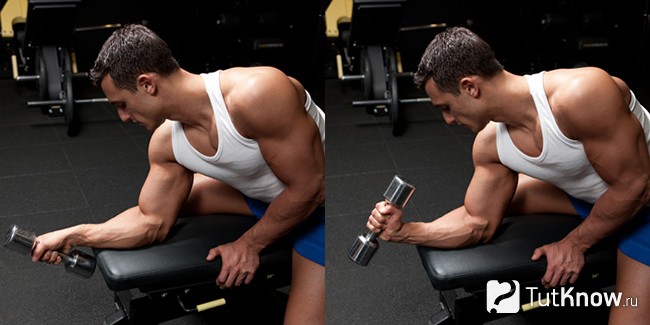
- Forearm flexion with barbell seated on a bench 3-4 X 10-20
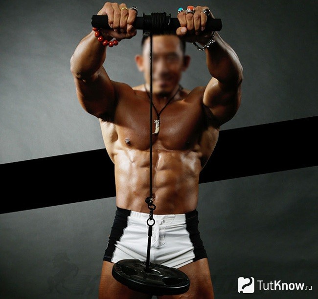
- Wrist pronation with a barbell or single-sided dumbbell 3-4 X 10-20
What about shoulder pronation and shoulder supination?
You hear that a lot less often. And rightly so.
In this photo, the lifter performs supination of the shoulder joint.What is HBA?
HBA stands for 'Outward Turn of the Shoulder'. That is, the supination of the shoulder joint.
IPA exercises are exercises that develop the muscles that externally rotate the shoulder (shoulder supinators).Why is this necessary?
At the beginning of this article, I drew your attention to getting to know the muscles and their functions. Read about the muscles that control the shoulder joints:
Pronation of the shoulder: 1) scapularis muscle, 2) pectoralis major muscle, 3) anterior deltoid muscle, 4) dorsal wideus muscle, 5) circumcus major muscle
Supination of arms: 1) biceps brachii, 2) biceps brachii, 3) biceps posterior.If we look closely at the drawing, a lot becomes clear. A - front view, B - rear view.
note!!! the pronators of the humerus are the two largest muscles in the body - the pectoralis major muscle and the dorsal widest muscle! And also the anterior deltoid. Everything is done in training to develop these muscles in every way. What is the result of this?
Well, it looks like this. Strength training causes muscles to shorten, become significantly stronger, and increase in size. Have you ever noticed the absurd way solid bodybuilders stretch their arms? One of the reasons for this is the penetrating action of the pectorals and broad muscles.
The shoulder joint gradually moves more and more into a slight but still noticeable pronation. This is not a natural position, and the shoulder joint must bear the brunt of the loads in a neutral position, not in a supinated or pronated position.
When we inflate only the shoulder pronators, we set the stage for a distortion of the normal geometry of the shoulder joints. This is a shortcut to injuries. Especially when doing bench presses, deadlifts and pull-ups. Sudden pain in the shoulder!
muscles of the shoulder girdle
Consider the muscles that flex the elbow joint. In a fight, regardless of technique, you must, among other things: maintain an optimal angle in the elbow.In addition to exercising lateral pressure during the attack. The effectiveness of this pressure is directly related to the strength of the arm muscles and the athlete's ability to use them.
When wrestling 'overhead', the muscles of the shoulder girdle and the brachioradialis are particularly stressed. The biceps brachii muscle is also involved to a small extent. During pronation, the sphincter muscle is actively involved in flexing the shoulder joint.
When doing a side push, the force of the side push is primarily determined by the stiffness and length of the collateral ligament (common flexor tendon - attachment point for all forearm flexors) and the strength of the brachialis and brachioradialis muscles.
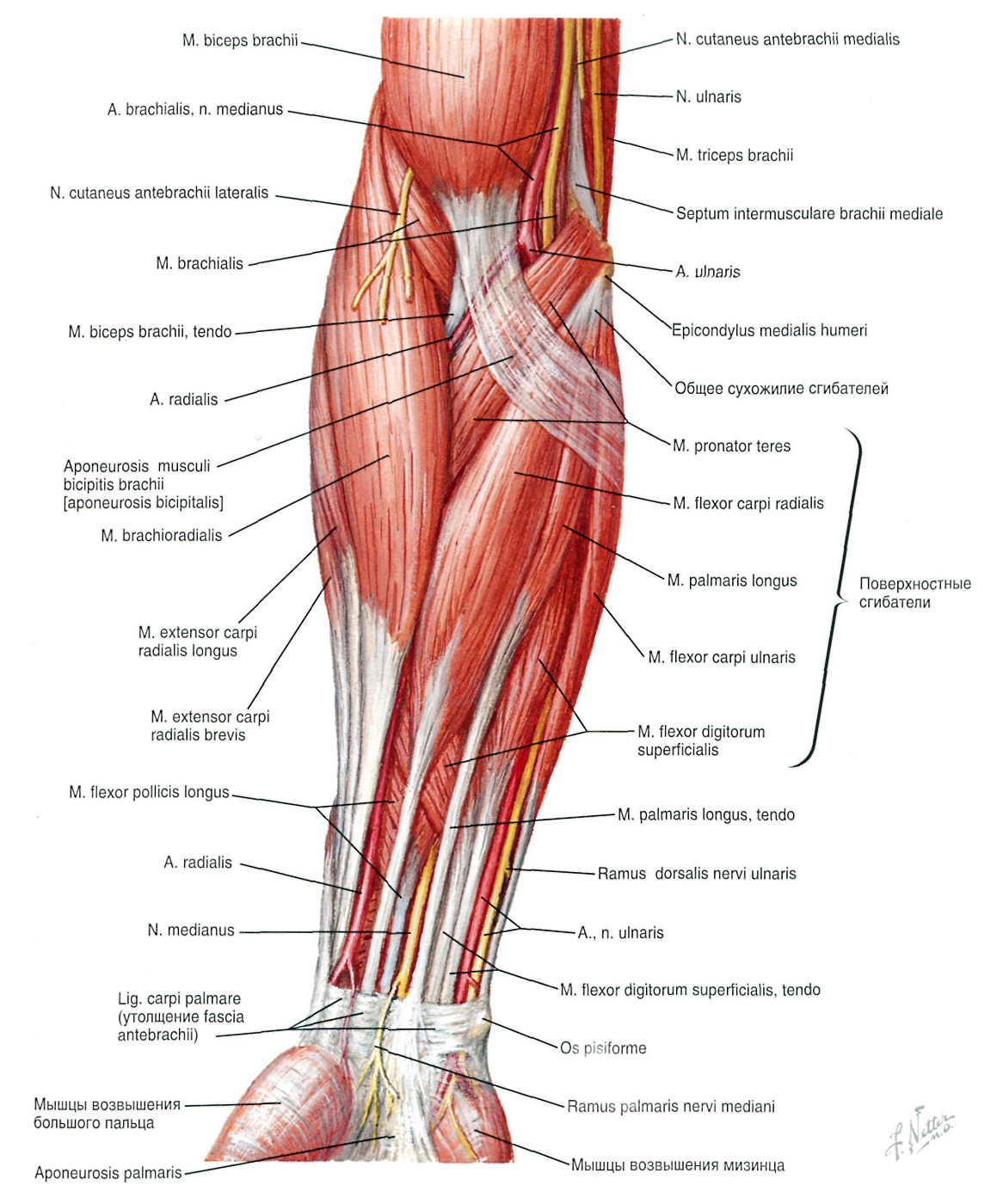
Figure 2.2 Muscles of the forearm (superficial layer): anterior view
Similar muscles are involved in hook roll, regardless of the type of hook (lower, middle, upper): the deltoids and biceps. The collateral ligament is also heavily loaded. However, it depends on the type of hook which muscle is responsible for the success of the movement. For example, if a 'bottom hook' is used, the load is almost entirely on the sideband..
Peripheral muscles of the upper limbs
With correct technique, the following muscles are also actively involved in addition to the forearm and arm muscles Upper limb girdle muscles. The involvement of these muscles is due to the athlete fixing the arm level with the trunk during wrestling. When wrestling, the deltoids, deltoids, pectorals, latissimus dorsi, and other muscles of the upper limb girdle are all actively involved.

In wrestling, regardless of technique, the athlete can perform the following movements:
1. withdrawing upon oneself;
2. Lateral pressure at a constant angle on the elbow.Figure 3.1 Muscles of the upper border of the limb
Of course, these movements cannot be separated from each other. Typically, the wrestler performs a combination of these moves.
Pulling puts a strain on you the broadest back muscles.. It also includes the posterior deltoid bundle, trapezius muscles, obturator major and minor, quadriceps, posterior obturator major and minor, and triceps.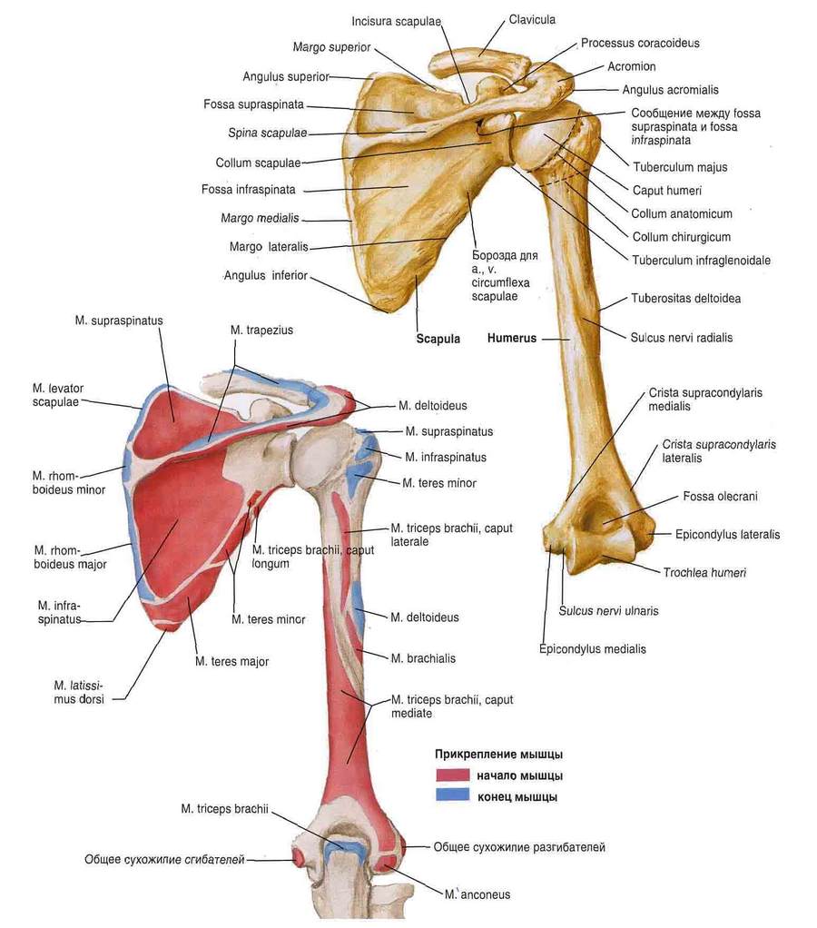
Figure 3.2 Origin and muscle insertions of the scapula and humerus: posterior view.
The side thrust engages the scapular, upper, and to a lesser extent the middle portion of the pectoralis major. In addition, the pectoralis minor, anterior deltoid, supraspinatus, scapular, and piriformis muscles are also involved.
In the video (1:34; 5:14) it is clear to see how an athlete with a delayed pectoral muscle failed to capitalize on his advantage in arm strength.
- The pronator is in anatomy.
- pronator and supinator muscles.
- These are the pronator muscles.
- pronator to.
- The pronator muscle - what it means.
- pronation and supination.
- The long section of the big toe.
- The flexor muscles of the foot.