Different types of anesthesia can be used during the operation. Shoulder arthroscopy is usually performed under general anesthesia. Combined anesthesia may also be used. In this case, doctors administer intravenous surface anesthesia. In addition, regional anesthesia (injection of drugs to block nerve impulses into the brachial plexus) is performed.

- Rotator cuff injury
- Why is this muscle group often injured?
- Why does my shoulder hurt?
- Causes of 'Frozen Shoulder'
- Causes of dislocation of the shoulder joint
- Types of shoulder dislocations
- Symptoms of a shoulder dislocation
- Treating a dislocated shoulder
- examination and diagnosis
- Treatment and prevention of osteoarthritis
- List of Sources
- Types and severities of shoulder contractures
- Post-traumatic contracture after a fracture of the humerus
- Arthroscopy of the shoulder
- recovery after surgery
- Is treatment possible without surgery?
- Causes of shoulder ligament strains.
- What are the Symptoms of a Shoulder Sprain?
- Rehabilitation after an injury
Rotator cuff injury
The shoulder joint is one of the most mobile joints in the human body. It can move in virtually any direction, meaning the shoulder can perform many complex tasks.
However, this variety of movements requires more than just good mobility of the joint itself. You also need the right muscles. The right muscles are required. The shoulder joint is surrounded by a well-developed muscle mass that supports flexion and extension, abduction, adduction, external and internal rotation. Together, these muscles form the rotator cuff of the shoulder. Rotator cuff injuries are quite common.
In the past, most shoulder pain was described with the term periarthritis.
With advances in diagnosis and experience, particularly with the introduction of shoulder arthroscopy into medical practice, partial or complete tears of the rotator cuff tendon in the shoulder have become more common. Today they are considered one of the main causes of pain and functional limitations of the shoulder. The treatment of this disease is carried out by doctors at the CELT Clinic who specialize in orthopedics and trauma surgery.
Why is this muscle group often injured?
There are several causes of common injuries:

- Tendon injuries (complete or partial)
- Minor injuries during sporting activities.
- Degenerative changes in tendons due to age-related changes.
- Poor blood circulation: There are few blood vessels in these muscles.
- Congenital connective tissue abnormalities.
- Peculiarities of the anatomy of the scapula: In some people, its protruding processes damage the muscles surrounding the shoulder joint.
- Repetitive high amplitude movements. This is particularly important for athletes and other physically demanding workers.
- Certain occupational risks, taking medications, including certain antibiotics.
- Topical administration of glucocorticosteroids
Various factors lead to degenerative changes in muscles and tendons. As a result, they gradually become thin and weak and eventually crack.
Why does my shoulder hurt?
The humerus, shoulder blade and collarbone meet in the shoulder joint and form a ball joint. The ends of the bones are held together by a dense connective tissue 'cover' - the joint capsule.
Pain can occur as a result of various pathologies, the most common being trauma, arthrosis, various neurological diseases, hemiplegia, tumors, etc. Some of these complaints manifest themselves as acute pain, others build up gradually, and in both cases the shoulder joint can become dysfunctional.
It is one of the most mobile joints in the body and has a very important 'coordination' function for all elements. In frozen shoulder syndrome (FTS), the joint capsule becomes thicker and stiffer, loses elasticity, and the amount of synovial fluid, which serves as a lubricant, is reduced in the joint.
The disease is most commonly diagnosed in people aged 40-60, of whom about 70 % are women. The joint is more commonly affected in the non-dominant hand: left in right-handed people and right in left-handed people.
Causes of 'Frozen Shoulder'

The cause of the disease can be an inflammatory disease of the ligamentous joint, trauma or surgery to the shoulder joint, circulatory disorders or the formation of a local tumor, but often the etiology of the disease is unknown. In such cases, the disease is called idiopathic or primary CJD. The prevalence of idiopathic PPH is estimated at 2 % to 5 % of the general population.
Factors that increase the risk of developing PPH include:
- high age;
- joint trauma;
- cardiovascular and cerebrovascular diseases;
- Diabetes and obesity: SPP is diagnosed in 10 to 20 % of diabetics;
- Thyroid diseases – hyperthyroidism and hypothyroidism;
- Stroke;
- history of mastectomy;
- Parkinson's disease;
- Tuberculosis.
Causes of dislocation of the shoulder joint

We have already mentioned that the shoulder joint is very mobile. It is the part of the skeleton that is most frequently 'moved' by a person. It is very sensitive and is the most commonly dislocated. It is often caused by general force and the movement itself is continuous or rotary. To cause an injury, it must occur with a simultaneous interruption of all possible joint movements.
- Excessive movement directed at this part of the skeleton is diagnosed in 12 % cases and is referred to as joint hypermobility.
- Posterior or anterior deviation can occur for a variety of reasons, but is most commonly caused by significant deviation of the socket.
- If the scaphoid socket has a low capacity, the cause changes significantly and the risk factor for dislocation increases.
- A common cause is acetabular hypoplasia, which means that the lower area of the socket is mainly changed, but many other physiological changes also occur.
- The ligaments and joint capsule are often subjected to repetitive movements that place repeated stress on the ligaments and joint capsule. Athletes (swimmers, handball players, etc.) are most often affected in this case.
There is a particular risk if the amplitude of the movements is excessive. In medicine, this type of movement is called 'generalized hypermobility'. There are several reasons for this, which are based on the anatomy of the joint. It is therefore necessary to carry out tests in advance and avoid traumatic situations.
Types of shoulder dislocations
Shoulder dislocations can be congenital or acquired. Acquired dislocations can be traumatic or nontraumatic. Non-traumatic shoulder dislocations can be either spontaneous or pathological (chronic). Traumatic dislocations can be straightforward or complicated. Shoulder dislocations can be complicated by compromised skin integrity, tendon rupture, fracture, nerve and vascular damage. Shoulder dislocations can also be chronic or pathologically recurrent.
Depending on the position of the articular surfaces of the humerus and scapula, a distinction is made between anterior, posterior and inferior dislocations. Anterior dislocations of the shoulder can be subacromial and subclavian, inferior dislocations can be axillary, and posterior dislocations can be subacromial and subscapular. Anterior dislocations account for the vast majority of shoulder dislocations (approximately 75 %), followed by axillary dislocations (24 %). Other dislocations account for only 1 % of cases.
Shoulder dislocations are also differentiated based on the time that has passed since the injury. They can be non-recent (the injury occurred more than three weeks ago), recent (three days to three weeks) and recent (no more than three days).
Any movement, even when brushing or combing, is often the cause of the discomfort. If the original dislocation is inadequately treated or left untreated, complex pathologies of recurrent traumatic dislocations can develop.
Symptoms of a shoulder dislocation

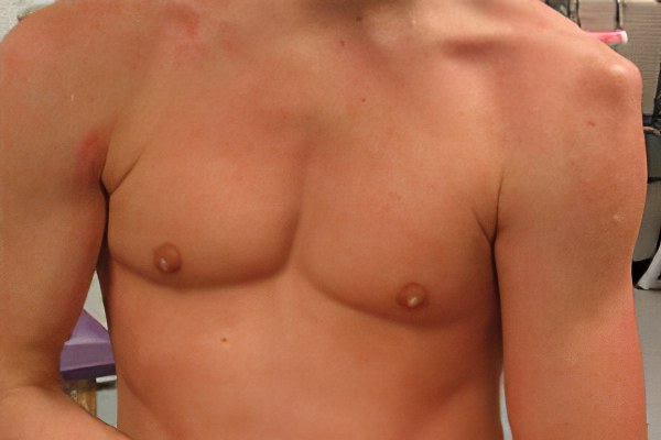
Traumatic dislocations of the humerus are accompanied by acute pain at the site of injury and deformation of the shoulder joint (it becomes angular, concave or hollow). Movement in the joint is impossible. During passive movement attempts, a characteristic spring-like resistance occurs.
In an anterior shoulder dislocation, the head is displaced forward and downward. The shoulder is in a forced position (sideways or flexed, extended and stretched). During palpation, the humeral head is not in its normal position and can be felt in the anterior region of the armpit (in anterolateral dislocations) or beneath the pectoralis major muscle of the scapula. Anterior and anterolateral shoulder dislocations are sometimes accompanied by a tear of the humerus and a fracture of the shoulder process of the scapula.
If the lower shoulder joint is dislocated, the head is displaced into the armpit. Vessels and nerves run through the armpit. When the head compresses the neurovascular bundle, numbness of the skin and muscle paralysis occur in the area innervated by the compressed nerve. In a posterior shoulder dislocation, the head shifts toward the shoulder blade.
Treating a dislocated shoulder
The initial treatment consists of immobilizing the injured joint with a Deseau bandage or a step splint. A traumatic shoulder dislocation is associated with acute pain, which is relieved by the administration of non-narcotic (Analgin) or narcotic (Promedol) painkillers. The longer the injury occurred, the more difficult it is to get the shoulder back into place. Therefore, the patient should be examined as soon as possible by an orthopedist/traumatologist in the department of traumatology or traumatology.
Upon admission, local anesthesia is administered to the shoulder joint area. A closed dislocation of the shoulder joint is performed under local anesthesia. The Janelidze, Kocher, Hippocrates and Mukhin-Kota methods are used. Sometimes a shoulder joint dislocation cannot be corrected under local anesthesia. The inability to reduce may be due to entrapment of the soft tissues or a relatively long-term dislocation. In these cases, the dislocation is reduced under anesthesia. If the joint cannot be reduced without surgery, an open reduction is performed, followed by fixation with needle or Lavsan sutures.
After the shoulder dislocation, a Desaut bandage is applied for 3-4 weeks. Once the humeral head is in place, the pain will decrease significantly and may subside after a few days, but the bandage is left in place to allow the soft tissues to heal. Once the shoulder joint capsule has healed, the bandage is removed and physical therapy and strengthening exercises are recommended.
examination and diagnosis
Your surgeon or orthopedist can help you determine the nature of your problem. The most important part of the examination is to compare active (independent) and passive movements. If you have tendonitis, you will not be able to actively move your arm, and the doctor will not try to move the joint effectively. This is the main sign of the disease when viewed visually.
You will be asked about the type and frequency of pain, as well as possible causes: lifestyle or injuries. Then it's time for a thorough diagnosis, which includes an ultrasound scan and an MRI of the shoulder. MRI is the most accurate and detailed method to diagnose joint disease.
If there is calcifying inflammation, a disease that has disrupted the metabolism must also be looked for. Arthroscopy – a minimally invasive procedure used for both treatment and examination and diagnosis – may be recommended for a thorough examination.
Treatment and prevention of osteoarthritis
There are various treatment options for treating osteoarthritis. You will be prescribed anti-inflammatory painkillers in the form of tablets or ointments and physiotherapy. Physical therapy, ultrasound and magnetic field therapy as well as massage therapy are ideal for treating shoulder osteoarthritis. All patients should be advised to take it easy and protect the shoulder from excessive strain.
Surgical intervention is only performed if conservative treatment is ineffective. However, with physiotherapy and rest, tendinitis can be easily cured in the early stages.
The most important measure to prevent shoulder problems is to avoid repetitive heavy work. If that doesn't work, you can simply warm up your body to avoid injury. If you experience pain in your shoulder during the activity, stop immediately and take a short break. For best results, eat right and do light exercise daily. Their job is to reduce the risk of sudden damage to the joint. Once he gets used to constant, easy work, it will be hard to hurt him.
Visit your doctor regularly if you have an infectious disease. Also, don't forget to treat back problems that lead to restricted mobility and inflammation of the spine.
List of Sources
- Vasiliev A.Yu. Magnetic resonance imaging of the wrist: normal anatomy and errors in image interpretation 2006 (Vestnik Traumatologii i ortopedii im. NN Priorov. 2006.
- Caravan P, Lauffer RB. Contrast media: basic principles. In Edelman H, Zlatkin MB, Crues JV, editors. Clinical magnetic resonance imaging. 3rd ed., vol. 1. Philadelphia: Saunders; 2006. p 358-376.
- KARPENKO AK, DUMANOVSKAYA O. Ultrasound contrast agents in the assessment of juvenile arthritis // 12th European Congress of Radiology, March 5-10; 2000, Vienna, Austria: Final Program. Berlin: Springer, 2000.-P.245.
- Steinbach LS MRI in the detection of malignant bone marrow infiltrates // Am. J. Roentgenol. – 2007 – Vol. 188, № 6. – pp. 1443-1445.
- Filimonova AM, Znamensky IA, Goncharov EN DIAGNOSTICS of lower limb joint injuries using magnetic resonance imaging, Current Issues of Science and Education, No. 5, 2014. ref.
- Gunicheva NV, Ahadov TA, Shubkin VN. Features of magnetic resonance imaging of the joints of the lower limbs in patients of different age groups // Siberian Medical Review, 2010. ref.
Types and severities of shoulder contractures
There are different types of shoulder contractures. They are classified according to various parameters:
- level of movement restriction (adduction or adduction, flexion or extension, rotation or full rotation);
- tissue involvement (dermatogenic – cutaneous, myogenic – muscular, neurogenic – neurological, tendogenic – tendonous, osteogenic – bony, arthrogenic – articular);
- Cause of development (post-traumatic, ischemic, inflammatory, cicatricial, degenerative, neuropathic, immobilization).
In the majority of cases, a compound contracture of the shoulder joint develops, in which all movements of the upper limbs are restricted. This often occurs in post-traumatic conditions and in patients who have suffered acute cerebral circulatory disorders. In other cases, one or two types of contractures develop at the same time.
During the initial diagnosis, it is important to determine the exact degree of contracture of the shoulder joint, as this is crucial for choosing further treatment tactics. Orthopedic surgeons distinguish three stages of pathology development:
- A true contracture is only the first stage, in which limited mobility of the joint and small skin changes with the appearance of pallor and tightness are noted;
- In the second stage, stiffness is already diagnosed, there is significant limitation of mobility, the skin is deformed and deep epidermal scarring may be present;
- the third stage of contracture development is ankylosis; permanent pathological connections of the structural parts of the joint, complete muscle atrophy and lack of mobility are observed.
Only in the first stage of contracture development can conservative treatment lead to a positive result. In cases of stiffness, an endoscopic procedure with arthroscopy may be necessary. During the procedure, the surgeon restores the integrity of the damaged tissue and removes the connections that have already formed between the individual structures of the joint parts. In the third stage, only endoprosthetic surgery (replacement) can restore physiological joint mobility.
Post-traumatic contracture after a fracture of the humerus
The most commonly diagnosed type of injury is post-traumatic contracture of the shoulder joint, which can develop after subsequent injuries
- fracture of the radius bone;
- Strain or stretching of the ligaments and tendons of the shoulder joint;
- a tear in the joint capsule;
- habitual dislocation of the shoulder.
Post-traumatic contracture of the left shoulder joint can occur with injuries to the scapula, clavicle, and spine. But the right side can also be affected to the same extent.
The contracture after a humeral fracture is often immobilized. A plaster cast is used to bring the broken bone back together and fix the joint in a specific position. The cast is removed after 25 to 35 days when the bone marrow is visible on the x-ray. At this point the mobility of the shoulder joint is no longer present.
If you develop symptoms of contracture formation after removing the cast, comprehensive rehabilitation should begin immediately. Make an appointment with your orthopedist if the following clinical signs occur:
- Feeling of stiffness when trying to bend or straighten, raise or lower, retract or pull the arm;
- Inability to perform normal injured movements;
- Limited range of motion (possibly compared to the other upper extremity);
- All movements cause mild muscle soreness or a feeling of tension in the muscles, ligaments or skin;
- External deformations of the articular bones are visible;
- The muscles of the upper limbs and shoulder girdle are deformed and have reduced volume.
Other noises (creaking, crunching, clicking, crepitus, etc.) that occur when the shoulder moves can also be cause for concern.
Arthroscopy of the shoulder

Arthroscopy of the shoulder
After shoulder arthroscopy, patients usually stay in the hospital for 1 or 2 days, rarely up to 3-4 days.
There is no significant pain after the operation. Most patients surveyed report a pain level of 2 to 3 out of 10. Painkillers are not even necessary.
The stitches are removed after an average of 2 weeks. Water should be avoided during this time. You can only shower if you cover the threads with a waterproof plaster. Once the stitches are removed, you will need to protect your scars from water for 24 hours.
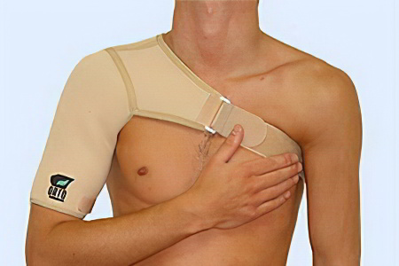
During the first month the patient must wear a bandage (orthosis). This is needed to keep the arm in a comfortable position so that the shoulder joint can regenerate. This prevents excessive muscle tension and protects the shoulder ligaments. Better conditions are created for tissue regeneration. The bandage should be worn permanently. It should not be removed overnight either. Sleeping is uncontrolled and can damage the joint, worsening the outcome of arthroscopic surgery.
Work on the operated shoulder joint begins on the second day after the arthroscopy. The exercises are carried out daily and in several stages.
- Day 2 – You are allowed to work without putting any strain on your shoulder;
- 3 days – You can drive a car if you have an automatic transmission;
- 14 days – you can sleep on the operated shoulder, go to the sauna;
- 4 weeks – even the slightest pain when carrying weights stops;
- 1 month – you can go to the gym and swimming pool, carry bags weighing 1 kg or more on the operated shoulder (gradual increase);
- 3 months – shoulder range of motion is restored to 90 %.
The time until complete recovery is individual. It depends on the reason for the shoulder arthroscopy and the patient's body (age, state of health, previous illnesses). Most people are able to make full use of the joint in daily life 3 months after arthroscopic surgery. After 4-5 months, sports training can take place and after six months the maximum load on the shoulder is back to what it was before the injury or illness.
recovery after surgery
After shoulder reconstruction surgery, the patient needs rehabilitation. The aim of this treatment is:

recovery after surgery
- Complete restoration of shoulder joint function
- shortening the duration of incapacity for work;
- elimination of pain syndrome;
- elimination of muscle contractures;
- increase in muscle strength;
- Elimination of stiffness.
After the operation, external immobilization of the shoulder is indicated. The duration of immobilization depends on the type of operation. It can range from 2 days to 1 month. During this period it is indicated:
The exercises are necessary primarily to normalize blood circulation in the limbs, which ensures complete regeneration and helps eliminate swelling. The exercises are performed on average 10 times per day with 10 repetitions.
In almost all patients, active abduction of the shoulder is impaired after removal of the immobilization bandage. This is due to a number of factors:
Further rehabilitation measures are aimed at strengthening the muscles of the shoulder girdle and normalizing the function of the joint. Physical therapy and physiotherapy are used for this purpose.
Is treatment possible without surgery?
It is known that injuries to the supraspinatus tendon do not regenerate properly. For this reason, most patients require surgery. This is successful in 85 % cases. However, recurrences often occur, limiting the patient's ability to work and requiring repeat operations.
If there is an injury to the supraspinatus muscle in the shoulder joint at stage 1 of the disease process, treatment can be conservative. Functional rest, physical therapy and physiotherapy are indicated.
Platelet-rich plasma is used to reconstruct the tendon. Platelets secrete various anabolic and trophic factors that promote healing of the lesions. These cells contain growth factors:
Causes of shoulder ligament strains.
- fall on shoulder ;
- Mechanical injury to the shoulder caused by an impact
- Acute jerking of the shoulder;
- Careless fall with an outstretched arm.
What are the Symptoms of a Shoulder Sprain?
Immediately after a shoulder injury, the following symptoms appear
- Acute pain at the site of injury;
- Increased discomfort when trying to move shoulder;
- limitation of shoulder mobility;
- Mild swelling and swelling at the injury site.
In addition, the body temperature may rise - this can indicate both damage to the ligament and the onset of inflammation. If the person does not take action - does not seek medical attention, continues to actively move the shoulder, the pain increases and the swelling becomes worse. All of these symptoms can indicate tendinitis - an inflammation of the tendon. In this case, it is urgent to go to the nearest health facility and seek treatment from a specialist.
Even if the clinical picture of a shoulder sprain is not so obvious that you rush to the doctor, it can take a long time for complications to appear. Inflammation may develop at the site of injury (tendonitis, bursitis), and sometimes shoulder arthrosis may develop. Each of these complications results in the affected person no longer being able to move freely, exercise, and carry out normal daily activities.
Further investigation is required to confirm the diagnosis. Most often, the patient undergoes magnetic resonance imaging (MRI), x-rays, and sometimes arthroscopy of the joint. During arthroscopy, the doctor can simultaneously perform a microsurgical procedure in which partially or completely torn tendon fibers are joined together. X-rays of the shoulder joint should be taken to rule out fractures and fractures, which require completely different treatment than dislocations. It is important to carry out a comprehensive diagnosis as early as possible - then the treatment measures will be as effective as possible.
Rehabilitation after an injury
The ligaments in the shoulder heal quite quickly after a sprain. A lot depends on the body's ability to regenerate, the extent of the injury and whether all the specialist's recommendations are followed. Certain diseases such as diabetes, immune deficiency, thyroid disease, etc. can delay the process of tissue regeneration and repair. During the rehabilitation phase, the body needs a lot of vitamins, proteins and micronutrients - a proper diet that covers all of the body's needs during this time is very helpful.
For first and second degree sprains, rehabilitation usually lasts no longer than 7-10 days. If the ligament is completely torn, recovery can take place up to 6 months..
After the basic treatment, the therapist prescribes physiotherapy and physical therapy. The aim of rehabilitation is to restore the joint to its former mobility, strengthen the ligaments and maintain their flexibility.
Exercise in the rehabilitation period after a shoulder injury is very important: inactivity leads to the fact that connective tissue fibers take the place of the damaged elastic tissue. This is scar tissue that cannot stretch as the ligaments normally should. All training should be gentle, without sudden movements and with a gradual increase in load. Under no circumstances should you train when you are in pain, as this can re-traumatize the joint's ligaments. The first 2-3 times you should carry out physiotherapy treatments under the guidance of a specialist who will check your technique in order to avoid unnecessary stress.
After the rehabilitation period, you need to take certain preventive measures to avoid further sprains. This includes maintaining a normal weight, regular physical activity and a sensible diet.
Read more:- Diagram of a joint with and without a dislocation.
- tearing of the joint capsule.
- How much does shoulder dislocation surgery cost?.
- Pronation and supination of the shoulder.
- There is abduction and reduction of the joints.
- shoulder supinators.
- Pronation and supination in anatomy.
- How much does shoulder surgery cost?.
