Also read: Transport Immobilization for Shin Fractures
- The main benefits
- Preparing for surgery
- What can be painful in the knee joint?
- Causes of knee pain
- Why do knees hurt?
- Posttraumatic pain.
- contraindications
- Knee replacement: types of surgeries
- Partial unicompartmental replacement of the knee
- Total (English) arthroplasty
- symptoms and severity
- Clinical signs of joint dysplasia
- diagnosis
- course of the operation.
- rehabilitation time
- Ankle Splint with Plates T-8608-1
- Option 1: self-delivery
- Generally
- Personal Information and Security
The main benefits
In contrast to classic surgery, no large tissue incisions are made during arthroscopy. The procedure involves making 2-3 small punctures, up to 5mm long, inserting special instruments into the joint and attaching a miniature camera (arthroscope). Thanks to the imaging equipment, all information about the changes in the joint is displayed on an external monitor. This enables an objective assessment of the existing damage and the implementation of the necessary treatment measures.
With minimal trauma to the joint tissue, bleeding can be avoided, early loading can be initiated, severe postoperative scarring can be avoided, and full function of the knee joint can be restored early.
- minimal traumatization of the joint tissues;
- no risk of bleeding;
- maximum precision in diagnosis and surgical manipulation;
- minimal hospital stay (up to 1 day);
- rapid postoperative wound healing time (6-8 days);
- no severe postoperative scarring;
- Possibility of early loading and walking (from the 1st postoperative day);
- early rehabilitation and full recovery of knee function.
Because of its undeniable advantages, arthroscopy has become the mainstay in the diagnosis and treatment of intra-articular knee injuries, replacing open surgery.
Preparing for surgery
An initial consultation with an orthopedic trauma surgeon is essential to determine the indications and treatment options for arthroscopic surgery. You should bring all medical documents related to the reason for your visit to the doctor: test results, reports from specialists, etc.
Before the operation, all patients must undergo a medical examination:
- General blood test, urine test
- Biochemical blood tests for total protein, bilirubin, ALT, AST, urea and other parameters;
- coagulation picture;
Before the operation, the patient is examined by an anesthetist. He assesses the examination results, the anamnesis, the general state of health and creates an anesthetic plan based on the results.
What can be painful in the knee joint?
This complex joint has a dense network of nerve fibers that coordinate the functioning of the entire complex, called the knee:
- The Nervus tuberosus major – it is responsible for perception of the posterior surface of the knee.
- The malleolus nerve conducts innervation to the anterior surface of the knee joint.
- Nerves involved in transmitting impulses to and from the meniscus, ligaments, and blood vessels.
If even a small nerve branch fails, the entire joint suffers.
Causes of knee pain
Knee pain is always a signal from the body in response to an injury. Knee pain is mostly caused by injuries that originate from the outside or from the inside of the joint itself. For example, a dislocation or tear in the ligaments of the knee joint is caused by an external injury (e.g. a blow), while inflammation caused by chronic poisoning of the body (sepsis) damages the knee from the inside.
Why do knees hurt?
The answer to this question is not as simple as one might think. The most common triggers are:
- Damage to the ligaments or menisci.
- Inflammation within the joint.
- Various forms of osteoarthritis and osteoarthritis.
- Synovitis of the knee joint, synovitis of the bursa.
- Osteomyelitic bone damage.
- Podagra.
- tuberculosis of the bone.
Posttraumatic pain.
When we consider how much strain our knees take on a daily basis, it's easy to see why more than half of all musculoskeletal injuries involve the knee joints.
An accidental bump or awkward subluxation may go unnoticed, but the resulting swelling, reddening of the skin, and symptoms of pain are clear indicators of a knee injury. In the worst case, a bone fracture or a rupture of the ligaments (menisci) occurs. Recovery from this type of injury will take a lot of time and effort.
It is more successful when the integrity of the tissue is not compromised and the damage is limited to a sprain, bruise, or dislocation. When the injury affects the meniscus, it is called meniscopathy. The first symptom of this pathology is a cracking sound in the joint when you try to move it.
A common consequence of the injury is dislocation of the kneecap. As a result of excessive stretching of the ligaments, the kneecap pops out of the thigh groove. In most cases, the kneecap returns to its place, but doctors recommend physical therapy of the knee joint after such an incident to prevent recurrence.
contraindications

The operation can be performed only if there are no contraindications:
- Severe cardiovascular disease or respiratory disease;
- Acute exacerbation of somatic diseases; – Deep vein thrombosis; – cataract and pneumothorax;
- deep vein thrombosis
- decompensated diabetes mellitus;
- paralysis of the limbs on the side of the operation;
- malignant tumors;
- recent heart attack, stroke;
- local inflammatory processes in the surgical area;
- Unresolved nidus infections in the body;
- anemia grade II or III, low blood coagulation index;
- immunodeficiencies;
- severe mental disorders;
- tuberculosis of the bone.
If the body mass index is high, prior weight correction is required. The patient must inform the doctor about all existing diseases in order to avoid negative consequences. The possibility of an endoprosthesis is decided on a case-by-case basis. If there are relative contraindications, surgical intervention can be postponed until the patient's condition returns to normal.
Knee replacement: types of surgeries
Knee cartilage replacement surgery can involve full or partial replacement of the knee joint. The choice of technique depends on the extent of the damage. The primary goals of arthroplasty are to relieve pain, restore mobility in the lower limbs, and increase activity.
Partial unicompartmental replacement of the knee
Partial unicondylar knee replacements are primarily used in older patients with limited mobility. This knee replacement surgery is uncomplicated because the damage to the knee joint is unilateral. In partial endoprosthetics, only the affected areas are removed and replaced with artificial implants. Once the dead tissue is removed, the living bone structures are preserved as much as possible.
Because the knee joint is replaced with a prosthesis on only one side, the wound is minimal and less tissue is cut. Advantages of the unicompartmental endoprosthesis
- Complete preservation of the tapes is possible;
- Minimally invasive, which translates into faster recovery;
- Partial replacement can be performed without general anesthesia;
- The effects on the surrounding soft tissues are gentle;
- Lower cost than a total knee replacement.
After a partial knee replacement, pain is relieved within a week, while it takes longer to regain walking ability. With a sedentary lifestyle, the knee joint can last up to eight years, after which a second operation is required.
Total (English) arthroplasty
With this technique, the knee joint is completely replaced with a new artificial knee joint made of high-quality materials. The service life of such prostheses is up to 20 years. A total knee arthroplasty is primarily used in people who lead an active lifestyle and are constantly on the move. During the operation, both sides of the knee joint are replaced.
symptoms and severity
Regardless of the type of injury, the disease is accompanied by severe pain in the hip area. The main symptoms of a hip dislocation in adults include:
- A change in the shape of the limb;
- shortening of the injured limb;
- restriction of mobility of the joint;
- postural restriction;
- Increased pain when performing movements.
Other symptoms of a subluxation depend on the type of joint splitting.

- bulging in the groin;
- Outward displacement of the knee
- lengthening of the injured limb;
- Extension of the limb for anteroposterior subluxation and flexion for anteroposterior subluxation.
- protrusion in the area of the buttocks;
- limb shortening;
- The injured limb is bent and bent at the knee;
- Significant deformation of the hip joint.
If the abnormality is due to trauma, bruising and swelling may occur at the site of injury.
Clinical signs of joint dysplasia
The main signs of hip dysplasia in adults are.
- asymmetry of the glutes;
- cracking of the knee when straightening the leg;
- incomplete abduction of the limbs;
- pain when walking for a long time;
- Crunching in the hip joint.
Talking about pathology in adults, it is important to consider the consequences of inadequate treatment in children in early childhood. Inadequate treatment and late diagnosis of the disease lead not only to impaired motor function, but also to other orthopedic diseases.
- The first is underdevelopment of the joint without displacement from the acetabulum at the hip bone.
- The second is a partial displacement of the condyle relative to the acetabulum.
- The third form is a complete dislocation of the joint.
diagnosis
The external appearance of the pathology and the characteristic symptoms of hip subluxation in adults facilitate the diagnosis. The patient's posture, pain syndrome and pronounced deformity of the limbs indicate joint dislocation.
X-rays of the leg can be used to determine the type of dislocation, both lateral and direct. The pathology is classified based on the position of the femoral head in relation to the acetabulum. In controversial cases, an MRI scan may be needed to determine not only the extent of bone separation but also the condition of the musculoskeletal structures supporting the joint.

When examining patients with a long-standing hip dislocation, it is not easy to make a diagnosis. Over time, the pain syndrome disappears, and the shortening of the limb is compensated by the deformation of the pelvic bones and spine. This leads to a change in gait and limping.
course of the operation.
The procedure can be performed under either general or epidural anesthesia. In epidural anesthesia, a drug is injected into the paravertebral space (puncture in the lower back) that blocks the transmission of nerve impulses from the peripheral nerve endings to the spinal cord. This leads to a sensory disturbance of the lower body. At the same time, sedatives are administered intravenously.
- Approach from the front of the knee, incision around the kneecap 12-14 cm.
- Release of the joint from the anatomical space.
- Preparation of the articular surfaces of the femur and tibia for the prosthesis. Removal of the damaged cartilage and underlying bone tissue.
- inserting the implants. The removed cartilage and bone is replaced with special pieces that reconstruct the articular surface. These parts can be cemented or pressed into the bone.
- Inserting a plastic insert between the metal/ceramic parts to create a sliding surface.
- Rinse the surgical field with an antiseptic.
- Insertion of a temporary drain to drain the wound, suturing, aseptic dressing.
Information!!! Endoprostheses are made of biocompatible materials: ceramics, innovative metal alloys and high molecular weight polyethylene. Modern artificial constructions are non-toxic, non-carcinogenic and do not cause allergic reactions.
rehabilitation time
After the operation, the knee heals for 6-8 weeks. During this time it is important to strengthen the muscles to ensure the stability of the joint. On the second day after the endoprosthesis, simple physical exercises are prescribed to restore muscle strength and a good range of motion. The recovery period lasts about six months.
After reconstructive surgery it is recommended:
- Begin walking with the help of crutches, canes, or a walker.
- Avoid movements that put stress on the joint (jumping, running, playing ball games, skiing, tennis, jogging, martial arts).
- Moderate physical activity (swimming, hiking, skiing, cycling, Nordic walking).
- Wear comfortable shoes.
Danger!!! It is important that you do not gain weight as this can displace the implant. The longevity of the prosthesis depends on compliance with the orthopedist's recommendations.
Ankle Splint with Plates T-8608-1
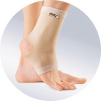
No absolute contraindications were identified.
Relative contraindications require medical advice:
- Need for an individual joint orthosis
- malignant tumors in the application area of the dressing system
- Contact dermatitis, ulcers and bedsores in the area of the dressing
- Allergic reaction to the product materials
The ankle brace should be used as directed and under medical supervision.
Option 1: self-delivery
Delivery costs: 0 rubles
Generally
Some of the items on this website are the intellectual property of 'Orthopedic Goods, Orthopedic Shoes and Medical Equipment 'ORTHOPEDIA'. The use of these items is determined by the current legislation of the Russian Federation.
The 'ORTHOPEDIA' orthopedic articles, orthopedic shoes and medical devices page contains links that will take you to other pages. Orthopedic articles, orthopedic shoes and medical devices 'ORTHOPEDIA' is not responsible for the information published on these sites and provides links to them only as a convenience to visitors to its website.
Personal Information and Security
The company 'Orthopedic goods, orthopedic shoes and medical devices 'ORTHOPEDIA' guarantees that under no circumstances will the information received from you be disclosed to third parties, except as required by the applicable law of the Russian Federation.
Under certain circumstances, 'ORTHOPEDIA' orthopedic articles, orthopedic footwear and medical equipment may ask you to register and provide personal information. The data you provide will only be used for business purposes and to access certain information.
You can change, update or delete your personal information at any time under 'Account' > 'Profile'.
In order to send you certain information, ORTOPEDIA orthopedic articles, orthopedic shoes and medical technology can, with your express consent, send newsletters to the e-mail address you provided during registration. You can change this at any time or unsubscribe from the newsletter.
Like many other websites, 'ORTHOPEDIA' uses cookie technology, which can be used to promote our product and measure the effectiveness of advertising. In addition, 'ORTHOPEDIA' uses this technology to configure itself to work with you personally. In particular, without this technology it is not possible to work with authorization in the control center.
Read more:- Axis of rotation of the knee joint.
- Orthopedic postoperative footwear.
- Pronation and supination in anatomy.
- The lateral dislocation is.
- pronation and supination.
- What is supination and pronation of the hands?.
- This is what a dislocated leg looks like.
- Pronation and supination of the shoulder.