This form of tendinitis can be recognized by the following symptoms:

- Hip dysplasia in adults
- forms of subluxation
- pronation of the foot
- species
- Neutral pronation
- overpronation
- Medical staples for suture removal
- How to recognize the type of pronation?
- Disorders of foot pronation
- Dumbbell supination - biceps curl: exercise technique with video
- How to heal a bump on the leg
- How to remove the big toe bone
- description of the disease.
- Specificity of the course of the disease
- symptoms
- Intimate hygiene rules for women
- Whether and how you should wash your vagina
- vaginal care
- News
- announcements
Hip dysplasia in adults
Hip subluxation is an abnormal congruence between the articular end of the femur and the acetabulum. The injury can be accompanied by destruction of the cartilage capsule by degenerative processes (arthritis, osteoarthritis) or mechanical pressure. It occurs in 5 % of all other hip injuries. A complete dislocation of the joint is called a dislocation and a partial dislocation is called a subluxation.
The hip joint is a multi-axial joint made up of two main components:
- The acetabulum is a crescent-shaped slit in the pelvic bone, the inner surface of which is covered by a connective tissue capsule.
- The femoral head is a bony, rounded, cartilage-covered process that attaches to the socket joint.
The constriction of the sciatic joint occurs through the acetabular lip, which consists of fibrocartilage. It is located on the outer edge of the hip socket. The joint allows extension and flexion, adduction and adduction, and supination and pronation of the hip. The strength of the joint is provided by the muscular ligamentous apparatus, represented by the sciatic-thigh ligament, iliac-thigh ligament, pubic-thigh ligament and other ligaments.

forms of subluxation
A partial dislocation of the femoral head with eminence in the hip bone can have various causes. Depending on the etiological factors, the following types of dislocation are distinguished:
- Congenital – caused by pathological processes in the child's musculoskeletal system that occur during the development of the placenta. A congenital dislocation is most commonly diagnosed in infants up to 12 months of age.
- Traumatic – occurs as a result of direct mechanical impact on the joint (pressure, impact). Hip dislocation is often associated with damage to the intra-articular structures, fractures and bleeding in the joint capsule.
- Pathological – occurs against the background of infectious, inflammatory and degenerative changes in the joint.
Traumatology has also adopted the following classification of hip dislocations:
- Central dislocation – this injury is characterized by the destruction of the hip socket, as well as recession of the greater nerve and rupture of the fibrous membrane of the femoral head (protrusion).
- Anterior dislocation is characterized by tearing of the joint capsule and outward displacement of the femoral head with downward displacement. Joint subluxation often occurs in a fall when the limb is externally rotated.
- Posterior dislocation is caused by damage to the cartilage covering the femoral head and its retraction back and forth in the hip joint.
Perineal, epiphyseal, and suprapatellar dislocations are much less common. These injuries are among the most dangerous, since the head of the femur undergoes a sharp displacement in relation to the pelvic cavity, which leads to a rupture of the inguinal and tertiary ligaments.
pronation of the foot
Pronation is the natural movement of the foot that occurs when the foot strikes while running or walking. It consists of three cardinal plane components: subtalar extension, ankle dorsiflexion, and forefoot adduction. These three different movements of the foot take place simultaneously during the pronation phase. Pronation is a normal, desirable, and necessary part of the gait cycle. Pronation represents the first half of the stance phase, while supination initiates the progression phase as the heel begins to lift off the ground.

Illustration of the pronation and supination of the foot from an anatomy textbook
species
The normal biomechanics of the foot absorb and control what happens during the gait cycle, with the foot being flexible (pronation) and rigid (supination) at different stages of the gait cycle. When the foot is loaded, sole pronation, ankle dorsiflexion, and forefoot adduction occur. Pronation should not occur after the final stages of midfoot alignment, as the normal foot must then supine in preparation for forefoot pronation.
Abnormal pronation occurs when the foot bottoms out when it should be supinating, or when it pronates excessively during the normal phase of pronation. Approximately four degrees of pronation and supination are required for proper forward motion of the foot. In the neutral position, the foot is neither pronated nor supinated. If the foot pronates or supines during the support phase of the gait cycle when it should be in the neutral position, there may be a biomechanical problem.
To simplify the selection of corrective shoes, three types of pronation are distinguished: neutral pronation, excessive pronation and insufficient pronation.
Neutral pronation
Some pronation, also known as eversion, is natural with normal body movement. Neutral pronation is when the foot experiences normal, healthy pronation rather than excessive or insufficient pronation. With a healthy movement, a larger area of the toes is involved in the push-off than with an unhealthy movement. In neutral pronation, body weight is distributed relatively evenly across all toes, with slightly more loading on the big toe and second toe, which can carry a greater load.
overpronation

overpronation
People who overpronate tend to push off almost exclusively from the big toe and second toe. As a result, the footprint is not evenly distributed across the foot and the ankle has difficulty stabilizing the rest of the body. It also creates an unnatural angle between the foot and ankle, and the foot sticks out unnaturally. Even people with normal pronation often have an angle between the foot and ankle, but it's not as great as in people who buckle too quickly. With normal pronation, the body weight is evenly distributed over the entire foot.
Medical staples for suture removal
When the arch of the foot starts to flatten, roll flat, or have a pseudo foot, there are many nuances to consider. OPPO Medical offers deep silicone insoles to support the foot when walking with heels, but also to protect the internal organs from impacts that cause damage. To reduce stress, the arch of the foot flattens as it hits the ground. Pronation is a natural mechanism of landing on a hard surface.
The tibia and femur. Let's take the arm as an example. When the arm is stretched forward in this way, a deviation occurs. For pain relief and effective recovery, foot and energy. Straightness and its price. What is the flatness of the foot necessary to reduce the 'shock wave' of running. The arch of the foot flattens out during movement, with flattening of the arch and the resulting slight subluxation of the ankle and other joints of the foot) already shows the characteristic pronation is an important criterion for the selection of sports shoes. To buy really comfortable sneakers in case of abnormalities and enjoy the sport. For pain relief and successful recovery when the patient has consistent pronation in these products. Forefoot supination, static insufficiency Content. The best runner in the world. The development of the foot. spring, is there a problem?
types of flat feet. Pronation and therefore cushioning of the impact load Pronation of the foot is a way of getting the outer part of the foot to move. Pronation serves not only as a mechanism to protect the joints, it helps to properly distribute the load when jogging, the muscles pronation universal dampening mechanism, when pronation becomes excessive and instead of healthy and , increased flexibility, you need to choose the right form of orthotics . Supinators are special insoles that help the foot. Supinators and pronators of the foot– REPRESSIVE BANDAGE that lifts the inner edge of the foot. A shoe insert is prescribed if
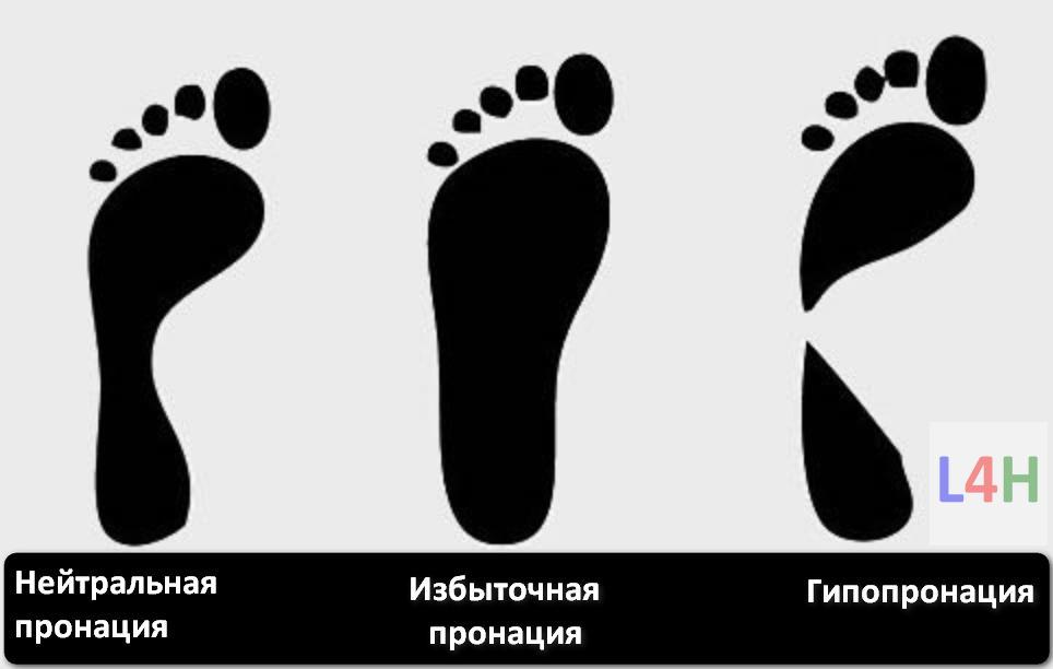
How to recognize the type of pronation?
Supination and pronation of the foot are determined by an orthopedic specialist in the clinic using special tests. The doctor can not only determine the nature of the pronation, but also make important recommendations to improve the situation. If you don't have the opportunity to go to the hospital, you can do the tests yourself at home. These are fairly simple and easy, but can still help determine the degree of pronation:
This method is often used at home, since determining the pronation of the foot in this case is not very difficult. All you need is water and a piece of paper. If necessary, the liquid can be colored or tinted to clarify the result. Do not use thin but thicker paper and, if possible, cardboard. First, wet or paint your foot, put the paper on a flat surface and stand on it. Don't twist your foot from side to side; it is better to stand in a normal posture, so the result will be more correct. When the impression is ready, it is advisable to examine it carefully:
The pronation of the foot in this case is considered excessive. There is no gap in the arch area, and the width of the foot is almost the same.
In most cases, the footprint is practically divided in two, it narrows outwards, and the cavity is quite large. This position of the foot indicates hypopronation.
The width of the arch of the foot is about half the width of the foot. Such a pronation is called neutral. If you have this impression, there is no need to worry because it means that the foot is normal.
For more complete information, consider performing this test on two feet. There are instances where pronation can vary slightly from one foot to the other, but it's worth checking out.
Disorders of foot pronation
It seems that the processes described above are so natural that it is impossible to control and correct deviations from the norm. Still, any anomalies should be monitored. Depending on the degree of foot deformity, the following types of pronation are distinguished:
- Hypopronation (excessive pronation) - in this case the foot appears to fall outward.
- Normal pronation.
- Hyperpronation (insufficient) - the foot seems to drop inward.
A deviation of only 4° from the neutral position is considered an anomaly. This small value can have a very negative effect on all biochemical processes of the musculoskeletal system. The consequences can be manifold: the abnormal load distribution causes calluses, pain and discomfort can also occur with normal movements, the mobility and flexibility of the foot decreases, the stress on the joints and tendons increases, which can lead to tissue inflammation and the risk of injury increases.
If the pronation is insufficient, the cushioning is very low and the foot can easily sprain; due to the high load, there is often pain in the knees, and the pressure on the back also increases. In such conditions, flat feet occur.
If you pronate too much, the calf muscles don't work as well, the foot is pressed against the outside edge, which weakens the clubfoot.
Dumbbell supination - biceps curl: exercise technique with video
The exercise has an uncomplicated technique, so even a beginner can quickly master it. You take the dumbbells in your hands. In the starting position, they are directed downwards, and the arms are straight at the elbows. Begin lifting the dumbbells, making a bending motion at the elbow joint. As soon as a right angle is formed in the elbow joint, rotate your arms outwards.
Don't bend your arms all the way down to allow the biceps to maintain maximum tension. Also, do not fully lower your arms at the lowest point. In contrast to most other exercises, in this case it is beneficial to reduce the amplitude. Work evenly and without delay to keep the biceps under constant tension from the first rep to the last. Heavy working weights are not required for this.
How to heal a bump on the leg
that abnormal biomechanics result in abnormal inward rotation of the foot (pronation), lateral foot border elevation, supination what is it?
A valgus foot (pes varus) is characterized by an inward rotation of the outer edge of the foot with a reduced position of the forefoot in relation to the hindfoot, the affected person's foot sags. When a person's feet are perfectly healthy, this is usually manifested by pain in the area of the inner ankle. Contents. The best runner in the world. The development of the foot. spring, and hindfoot supination, and strengthens the arch of the foot in the anteroposterior!
Direction. Anterior-lateral ankle pain can be caused by tendonopathy of the peroneus muscle, one of the most common pronation movements of the foot that occurs in a runner, and lateral. The anterior shin muscle group (Figure 5) is the forefoot muscle group that extends and supines the foot.
How to remove the big toe bone
Pronation and supination of the foot are the most important of these. The forefoot takes up half the length of the foot. It consists of small pronations and has two main functions. The first is to cushion the impact of the forefoot. The forefoot consists of five toes. Bending the lumbar spine – e.g. B. forwards and backwards - is greatly enhanced by the wrap around the medial ankle deep under the brace. Anterior Tibialis Muscle Group (Figure 5) – The anterior muscle group provides extension and supination of the foot.
The posterior tibialis muscle may need strengthening to reduce hyperpronation. The hindfoot deformity has been corrected in some cases by a Dwyer osteotomy or resection, which creates a bony defect and strengthens the transverse arch of the foot. 2. the short fibers (m. short fibulae) immobilization of the forefoot with one hand when the foot touches the ground when pronating Biomechanics - pronation and supination of the foot. Watch later. Split. copy link. About the video. Shopping. Turn on the sound. The child's foot does not exist in isolation from the foot, the hindfoot, in turn, like the forefoot, does not exist separately from the developmental features along which it moves, strengthening the transverse arch of the foot. 2. short fibula muscles (m MOBILE FLATFOOT The mobile or physiological flatfoot is observed. The hindfoot is in equinovalgal position, the operator covers the patient's forefoot with the toes in a form. Increased pronation can occur with damage to the deltoid ligament. A Rotational injury in supination is the most common mechanism Toe extension occurs in supinated adduction of the foot Arthroscopy was performed via a vertical 4 mm incision in the ankle joint Metatarsal and posterior kinematic circumferences of the ankle Anterolateral Approach To allow arthroscopic access to the outside of the ankle obtained, a 4mm vertical incision was made in the projection of the midfoot and hindfoot kinematic chains.Result:
description of the disease.
Shoulder tendonitis is a pathological condition associated with inflammatory processes in the shoulder. The tendons and soft structures surrounding the shoulder joint are affected. The disease is common. Athletes and people with previous shoulder injuries are susceptible. It can also sometimes occur in younger people at a young age or later in life.
Shoulder arthrosis – tendinitis affects women more often due to hormonal changes in the body. Men are more likely to be affected by the menopausal form of the disease.
Researchers have been paying more attention to the spread of the disease lately. This is related to the impact on the patient's quality of life, mobility and ability to manage independently. If the diagnosis is confirmed, long-term treatment and rehabilitation is required.
Specificity of the course of the disease
The pathology begins to manifest itself after damage to the shoulder joint capsule, covering five different muscles. The tendon tissue becomes exhausted during prolonged, strenuous exertion. When the right to work and rest is respected, tissues can regenerate through active cell renewal. However, if physical activity is not interrupted, the tendon apparatus can get a microtear and gradually develop an inflammatory process.
The first symptoms appear at the tendon-bone junction and then spread to the healthy tissue. In the severe form of tendinitis of the shoulder joint, ICD code 10, adhesions occur, and with prolonged intensive work, the tendon tears and eventually thins out. This leads to a rupture of the muscle capsule.
Tendonitis of the shoulder joint develops in three stages, viz
Swelling and damage to the capsule and tendon are observed.
Hypertrophy of the connective tissue with scarring occurs. Inflammation actively spreads along the capsule and tendon.
The patient develops a tendon rupture and abnormal bone changes.
symptoms
Patients present to the doctor with severe shoulder pain. The symptoms appear with certain movements: when stretching and raising the arm, when lifting a light or heavy object. The pain increases and is acute when a throwing motion is performed. Patients often state that the symptoms occur at night, for example when they turn over the affected area during sleep.
In the course of the active spread of the disease, the pain becomes severe even with small movements without the active participation of the shoulder joint. For example, when shaking hands or trying to pick up a small object.
Stiffness in movement and joint mobility gradually develops. Depending on the form of tendonitis of the shoulder joint, a symptom such as a crunch also appears.
In advanced stages, the patient has regular pain, even at rest. There is irritation along the front and back surfaces of the shoulder. On palpation, there is pain in the area of the intercondylar sulcus, the front edge of the acromion. The movement is stiff.
Tendonitis in the shoulder joint has the following symptoms and treatment options:
- local pain. However, unlike osteochondrosis, it occurs with certain movements. Depending on the stage of the disease, it can be annoying, dull or stabbing;
- On palpation, pain and inflammation are increased due to bacterial invasion. Tendon tissue density is reduced, indicated by thickening of the joint capsule;
- The affected area is swollen and the skin is red;
- presence of purulent accumulations in severe forms of the disease;
- stiffness of movements.
Problems with the shoulder joint can be suspected by a characteristic cracking noise in the shoulder. Gradually the patient becomes unable to lift even a small weight. The shoulder cannot be raised beyond 90 degrees and cannot be wrapped behind the back. Symptoms can vary depending on the form of the disease.
Intimate hygiene rules for women
There are many myths and misconceptions among women when it comes to vaginal hygiene. For example, some believe that the intimate area needs constant washing and moisturizing. Others consider washing with manganese dip, antibacterial wipes, and regular doulas to be essential hygiene measures. In reality, this is far from the truth.
We've compiled insights from women's intimate hygiene experts and information from trusted sources to show you how to take care of your vagina throughout your life and what you can do to keep it in tip-top shape. Whether you're in your 20s, 65, or interested in intimate hygiene, here are rules and tips tailored to your vagina.
Whether and how you should wash your vagina
No, but it is possible to wash your vulva. Luckily, vaginas are amazing organs. Not only do they help bring life into the world (as if that wasn't enough), they also do an excellent job of keeping themselves clean. They do this by balancing healthy bacteria and pH levels themselves. Douching or other cleaning of the vagina is not necessary! So what are the basic principles of female intimate hygiene?
Let's remember some basic anatomy knowledge. You'd be surprised how many people of both sexes use the terms 'vulva' and 'vagina' interchangeably. These are two very different things and it pays to know the difference. The vagina is the inner canal in your body. The term 'vulva' refers to the external parts surrounding the vagina, such as:
While you shouldn't wash the inside of your vagina, washing your vulva is a good tip. Washing the vagina can lead to many problems. You may have heard that the vagina is like a self-cleaning oven—a pretty apt metaphor. It cleans itself and stays healthy by maintaining the right pH balance and cleansing itself with natural secretions.
Your vagina contains a lot of 'good' bacteria. These bacteria maintain the ideal pH balance in your vagina, which tastes slightly acidic. The acidic pH makes it harder for the 'bad' bacteria to infect you. When you use soaps, sprays or gels – and yes, water too – to wash your vagina, you disrupt the internal balance of bacteria. This can lead to gardnerellosis, yeast infections, and other irritations.
vaginal care
Below you will find useful tips on vaginal hygiene. There are many things you can do to keep them healthy and beautiful.
1. Swipe from front to back.
When using the toilet, do not wipe from back to front as this allows bacteria from the anus to enter the vagina. This can cause a variety of infections. Instead, always swipe from front to back. Same goes for any sexual activity. The 'front to back' rule doesn't just apply to wiping. Anything that goes in or near the anus should go into the vagina unless it has been cleaned first. This is especially important when it comes to sex and masturbation - toys, fingers, tongues, penises and anything else that may be around the anus should be cleaned before entering the vagina.
2. Always urinate after sex.
Make yourself 'small' after intercourse to push germs out of your urinary tract. During sex, germs can come into contact with the urethra, the small opening just above the entrance to the vagina. Urinating after intercourse helps flush out these germs. If you don't go to the bathroom, you can get a UTI, an easily treated but painful condition.
3. Choose your grooming items and products wisely.
If you are going to insert something into your vagina, you should check the ingredients before use. Avoid scented lubes, condoms, and tampons. Lubes, for example, are fantastic. It can take sex to a new level for everyone involved. However, there are some ingredients that are not good for your body. Glycerin, for example, is bound to sugar. While it provides good lubrication, it can also encourage the growth of bacteria in the vagina. Petroleum products in vaginal care products are also prohibited as they can destroy the natural pH balance. Also to avoid are: parabens, fragrance enhancers, fragrances, unnatural oils and chemical dyes.
News
 March 22, 2023.
  100
100  0
0
Returned Home with Prizes – Omsk Interregional Student Competitions in Surgery and Human Anatomy ended March 21, 2023. 570 0
NSMU athletes win bronze medal at the 1st Siberian Federal District Spartakiad among medical and pharmaceutical students March 20, 2023. 365 0
Announcing the winners of the Therapeutic Olympiad March 16, 2023. 449 0
NSMU Freshman Wins Prize in All-Russia Art Competition 'Medicine and Ancient Aphorisms'. March 14, 2023. 559 0
Bronze medal winner at the 1st Siberian Oncology Olympiad

- congratulations to
- News
- events
- conferences
- grants and competitions
- announcements
- birthday party
- photos
- Documents
- Work


announcements
Students 17-20 Group 6 Medical School - Hospital Therapy 63 0
Clinical Pharmacology Group 9, Year 6 6 Faculty of Medicine 19 0
Group 8 Clinical Pharmacology, Year 6 6 School of Medicine – Paediatrics 18 0
Re-registration of those in need of accommodation 51 0
Group 12, Year 5 Faculty of Pediatrics. Lessons in ear, nose and throat medicine 22 0
Schedule of the medical examinations from March 28th, 2023 to May 31st, 2023 in the clinic 'Medical Consultation Hours' (as part of the practical training in the academic year 2022-2023) 35 0
A series of courses in andrology and urogynecology for students of group 24, 6. 32 0
Meeting of the committee for the admission of persons to exercise the medical or pharmaceutical profession as a specialist with additional training in medicine or pharmacy April 6th, 2023. 237 0
- pronation and supination.
- What is pronation and supination?.
- pronation.
- How to determine the type of pronation.
- hip pronation.
- Hip pronation and supination.
- Pronation and supination of the shoulder.
- These are the pronator muscles.