As the arthrosis progresses, the pain increases and the ankle becomes painful. Joint space decreases in size, stiffness develops, ankle mobility decreases, and leg deformities develop. Eventually, if left unchecked, patients are unable to stand on their feet and the supportive function of the joint is compromised.

- The ankle and its anomalies
- Diseases
- arthrosis
- Advantages of X-ray diagnostics in the MS clinic
- Indications for an X-ray examination of the ankle
- species
- plastic devices
- Individual characteristics
- Why swollen ankles in women
- Symmetrical swellings
- pharmaceutical treatment.
- signs
- to form
- diagnostic techniques
- Indications for the examination
- X-ray diagnosis of neoplasms of the foot
- Diagnosis of fractures of the foot
- Preparing for the examination and carrying out the procedure
- Anatomy of the ankle
- Biomechanical features of the ankle and ankle
- Preparation
- positioning of the ankle
The ankle and its anomalies
The part of the fibula that connects to the ankle is called the hock. This part acts as a stabilizer and is located on the side of the leg, slightly above the foot and protruding outwards. The other, more common name for the hock is ankle joint. The lateral ankle is formed by part of the tibia and the median ankle is formed by the fibula.
Anatomically, there are two ankles in humans: the medial malleolus and the medial malleolus. The inner malleolus is formed by part of the fibula. The hock is directed inwards and is called the medial hock. The lateral ankle joint is on the opposite side. It is formed by the protruding end of the tibia.
The inner and outer malleolus and the talus together form one of the most important joints - the ankle. It is characterized by great mobility, but at the same time it is very stable. The joint of the lower limbs performs the following functions:
- It supports the weight of the body and distributes it properly on the foot;
- It supports the foot;
- it allows an upright posture and active movements such as jumping, running and walking;
- the ankle components allow the user to rotate the body about an axis while the foot is resting on a surface;
- Ankles and ankle braces provide cushioning for movement.
Both the lateral and medial ankles are easily palpated and visually protrude like a bump on either side of the ankle in asthenic individuals.
The surface of the joint is densely covered with hyaline cartilage, which allows for smooth movement in the joint. The joint cavity is filled with synovial fluid. The structure of the ankle allows for a cushioning function and protects the cartilage tissue from wear and tear caused by mechanical friction.
Diseases
Like any other joint, the human ankle is prone to injury and joint disease. The most common diseases include arthrosis, osteoarthritis, dislocations, fractures and other injuries.
arthrosis
Arthritis involving the ankle is an inflammatory process provoked by certain pathologies. Arthritis is caused by ankylosing spondylitis, osteoporosis, rheumatoid arthritis and gout. The arthritis is caused by an infection of the ankle by hematogenous or lymphatic routes.
Arthritis can be acute or chronic. The acute stage is characterized by the development of swelling and redness of the ankle. The ankle joint is also swollen, its shape is blurred, and even in people with well-developed bones in the joint, the ankle can hardly be seen due to the swelling. The ankle itself and the foot feel hot.
If the joint inflammation is accompanied by an active discharge of purulent contents, they accumulate in the joint cavity and the patient's condition deteriorates significantly - the redness around the ankle worsens, it hurts to touch, and the patient's body temperature rises to high values . It is painful to move the leg, and acute pain occurs when trying to twist the ankle.
The symptoms of the acute form subside as the arthritis progresses to the chronic stage. At the same time, the redness decreases, the swelling can decrease significantly and an external feeling of well-being sets in. Only at night do the patients still have pain and a 'twisting' of the legs. Nevertheless, the inflammatory process is present and can lead to aggravation.

A variant of arthritis is rheumatoid arthritis. It affects chronically symmetrical joints, affecting the ankles of both the right and left legs. Doctors believe that rheumatoid arthritis is caused by genetic predisposition and the influence of negative external factors on the body.
Advantages of X-ray diagnostics in the MS clinic
X-ray examination of the ankle joints allows to detect pathological processes of different nature. X-ray examination helps in the initial diagnosis of the following diseases:
- Disorders of the integrity of the bones that form the joint – talus, distal part of the fibula and tibia (fractures, breaks are diagnosed);
- Disorders of joint congruence due to dislocations and subluxations;
- Primary tumors and secondary tumors involving bone tissue;
- calcium salt deposits in the soft tissue components of the joint;
- Necrotic processes in bony structures;
- flat feet (longitudinal and lateral), clubfoot;
- degenerative-dystrophic joint damage (arthrosis or osteoarthritis);
- decreased bone mineral density characterized by osteoporosis;
- Arthritis - an inflammatory lesion in a joint;
- Arthrosis – a purulent inflammatory lesion of the bone marrow and adjacent bone tissue.
Indications for an X-ray examination of the ankle
An x-ray examination of the ankle is recommended if indicated. These are situations where a visual assessment of the musculoskeletal structures is required to clarify the clinical situation. Indications are:
- trauma (it is advisable to take an X-ray as soon as possible after the traumatic event);
- swelling of the foot and/or ankle;
- pain and associated claudication;
- restriction of movement in the ankle;
- Pallor of the ankle and foot, especially in connection with low local temperature;
- irregular wearing of shoes;
- visible deformations of the foot.
In addition to the initial diagnosis, the X-ray examination of the ankle also serves to dynamically assess anomalies that have already been identified. A follow-up visit is usually scheduled some time after treatment or surgery. Modern digital scanners generate a minimal radiation dose, so that repeated examinations are possible if indicated.
species
Different types of splints are used for ankle injuries. When choosing the right splint, the trauma surgeon considers the extent of the injury and the area of the injury. What types of dressings are used?
- The most common are the so-called Johnson pads. They immobilize the foot, including the heel area. A plaster cast compresses the injury site so that the formation of an inflammatory swelling can be contained. Soft padding rolled in several layers is also used for immobilization. A soft flannel fabric is placed under the bandage to increase comfort and reduce pain;
A bandage is used to immobilize the foot.
- A plaster splint 10 to 15 cm thick is also called a bandage. The bandage is made to cover the back of the injured leg when it is applied. The bandage is fixed with special bandages. When wrapping, the doctor controls the tension to avoid pinching the leg and causing pain. This bandage is needed to immobilize the foot or ankle to promote tissue regeneration;
Posterior Bandage.
- Instep plaster bandages are intended for the correction of plantar fasciitis (inflammatory and degenerative changes in the plantar fascia) with their drug therapy. This type of bandage is specifically designed to treat plantar fasciitis, relieving the painful symptoms that are exacerbated by swelling. It immobilizes the toes in the up position, maximally stretching the plantar tendon and fixing the metatarsal. The splint is fixed in the area of the ankle with ligature bands using a special bandage technique.
plastic devices
A prefabricated orthopedic device that is simply attached to the leg is also known as a ligature. It is used to treat malpositions and injuries as well as to prevent tissue destruction. Hip bands are used, for example, in young children who are diagnosed with an immature hip joint. Wearing the bands helps the joint form properly. They are used for fractures of the fingers, ankles and knee joints. They are more comfortable than the plaster casts and are easier to remove and clean. The term 'bandage' for an external device is rarely used in medicine, but is very common. What it can mean:
- bandage. The easiest way to keep a leg in place and slightly limit its mobility. It is a tight elastic bandage that is attached with either straps or Velcro. The bandage is made of hypoallergenic materials, is breathable, warm and has a massaging effect;
Semi-rigid ankle brace.
- orthosis. A complex rigid structure that protects the injured parts of the leg (toe, foot, ankle or knee joint). The design of the orthosis has metal or plastic inserts for better fixation. The orthopedic product prevents overloading of the injured or affected parts of the lower limbs;
Dynamic ankle orthosis.
- orthosis. Produced in the form of a capsule into which the injured leg is inserted. This orthosis has no joints and its frame is made of plastic, less often metal. The surface of the orthosis is made of cotton or synthetic material with good air permeability. A complex attachment system is used to attach the splint to the leg and provide the required degree of immobilization.
Individual characteristics
Everyone's ankles are different. Depending on the anatomy of the skeleton, they can be high or low (tapered or widened).
An ankle injury causes severe pain, soft tissue swelling, localized fever, and restricted movement.
There are different degrees of severity of ankle ligament injuries:
- light – characterized by a slight stretch and a small amount of swelling;
- moderate – there is a partial detachment, accompanied by moderate pain;
- severe - severe trauma occurs, complete detachment and the bone is thrown to one side. At the time of injury, a cracking sound is heard, which is caused by the formation of bone fragments.
First aid to the patient consists of:
The victim's ankle hurts badly on impact. Pain relievers (ibuprofen, aspirin) or topicals (Indovazin, Voltaren) may help relieve the pain.
If the injured person's condition does not improve within 24 hours, you should see a trauma surgeon. He or she will determine whether or not there is a fracture and recommend further treatment.
Why swollen ankles in women

Any swelling is a buildup of excess fluid in a specific area. Swelling in the feet is caused by a disorder of the lymphatic drainage. Slowed lymphatic drainage can be caused by infections, injuries or chronic diseases.
The risk of slowed lymph drainage is that toxins and breakdown products from dying cells accumulate at the site of the edema. The harmful substances slow down the cell renewal process.

An initial swelling caused by external factors can lead to a serious condition. Conversely, swollen ankles can also be caused by an underlying condition. Lymphatic congestion is caused by:
- defective venous valves;
- blockage of blood vessels in the abdomen or pelvis;
- the presence of an inflammatory process in the veins of the lower limbs;
- Kidney, liver, vascular or cardiac abnormalities.
The edema can be symmetrical or unilateral. They have different origins and formation mechanisms. Symmetrical edema arises in both legs and has the same shape and volume. Unilateral edema occurs in one leg.

Symmetrical swellings
Many people confuse swollen feet with fatigue. This is a big mistake and can lead to serious illnesses. Causes of swelling that can be treated:

pharmaceutical treatment.
Topical medications—cooling ointments, sprays, and gels—are used to soothe and reduce swelling. Their action is to strengthen vein walls and improve blood circulation, including capillary circulation.
If the swelling is caused by varicose veins, one of the best remedies is Essaven gel.

It has a comprehensive therapeutic and preventive effect:
- escin Its composition tightens vessels and improves blood flow;
- heparin has an anticoagulant effect;
- phospholipids improve blood circulation.
The gel has cooling properties and reduces swelling and pain. Venitan and other horse chestnut ointments also strengthen blood vessels and regulate microcirculation.

Swollen ankles can be treated with diuretics, but these should only be taken with caution and on the advice of a doctor. When swollen ankles are due to poor heart function or high blood pressure, potassium-sparing medications such as veroshpirone are prescribed. Mild herbal diuretics are used during pregnancy - such as B. eufillin, Kanefron, phytolysin.
Even a single case of swelling should not go unnoticed. Any external factors causing them are indicative of a pre-existing condition.
signs
Symptoms of an old broken ankle with foot subluxation include pain when walking and ankle swelling. The pain in the ankle can vary in severity. They appear at the site of injury when walking and moving. The ability to support the limb is impaired (in some cases, one can no longer move independently), and the foot is numb (when blood vessels and nerves are damaged). The foot may be visually dislocated.
In the case of insufficient treatment of an ankle fracture with subluxation of the foot, residual external subluxation and unresolved distal intertibial syndesmosis (connection of the bones by connective tissue) can be detected a few months after the reduction of the fracture and the performance of transosseous osteosynthesis of the ankle fracture with the Ilizarov apparatus become.
The objective examination shows a deformation of the foot bones and an unnatural position of the foot. The ankle is swollen (extremely severe in some cases) and painful on palpation of the distal interdigital joint.
to form
The form of a protracted ankle fracture with foot subluxation depends on the fracture line, its location relative to the distal intertarsal syndesmosis, and the biomechanics of the injury.
There is a classification of fractures of the ankle according to BG Weber-Danis R. divided into three types – A, B, C, depending on the level of the fracture of the fibula in relation to the intercondylar syndesmosis.
Depending on the mechanism of injury, a distinction is made between pronation fractures (outward rotation of the foot), supination fractures (inward rotation of the spur) and rotation fractures (rotation of the tibia around its axis when the foot is stationary).
There are also fractures of the lateral and medial ankles, the anterior and posterior edges of the tibia, double ankle fractures (Malgene's supination fracture, Dupuytren's pronation fracture), triple ankle fracture (Pott-Desto), open ankle fracture, ankle fracture with damage to the syndesmosis of the intermediate bone.
diagnostic techniques
The diagnosis of a protracted fracture of the ankle with subluxation of the foot is made by a traumatologist and is based on recording the complaints, anamnesis, clinical examination and the obligatory use of instrumental diagnostic methods.
Based on the anamnesis, the fact of the injury and its mechanism can be established. The mechanism of injury can be direct (severe blow to the shin, dropping heavy objects on the foot) or indirect (acute twisting of the shin with an immobile foot). In the first case, there are transverse fractures, in the second - oblique and spiral fractures.
Bilateral x-rays are sometimes sufficient to diagnose a protracted fracture, but in some cases magnetic resonance imaging (MRI) may be needed to evaluate the muscles and surrounding soft tissues, and computed tomography (CT) to confirm the x-rays in difficult cases.
Some orthopedists find it necessary to take radiographs of the ankle in three x-ray projections: anteroposterior, anteroposterior with internal rotation of the foot 20 degrees and with the axis of the ankle parallel to the film (bifurcated view), and lateral. Ultrasonography of the lower leg is recommended in all patients with suspected damage to the neurovascular bundles of the lower leg, soft tissues, muscles, and ligaments.
General clinical examinations (general blood and urine tests) and biochemical blood tests are recommended to assess the patient's general condition, underlying disease or co-existing abnormalities.
Differential diagnoses include contusions, sprains and sprains/dislocations of the ankle ligaments.
Key instrumental studies include:
- X-ray examination of the ankle.
- Computed tomography of the ankle (if necessary).
- MRI of the ankle (if necessary).
- Ultrasound examination of the lower extremities.
Indications for the examination

Undoubtedly, osteoarthritis of the ankle is the most common foot disease, primarily due to mechanical wear and stress on the cartilage. However, the localization is not atypical for inflammatory rheumatic diseases (rheumatoid arthritis, psoriatic arthritis, ankylosing spondylitis and Reiter's syndrome), which often first appear or are diagnosed in the lower limbs. In addition, the foot is commonly affected by cystic fibrosis and diabetic neuropathic osteoarthropathy.
X-ray diagnosis of neoplasms of the foot
Occasionally, isolated tumors can occur in the bone of the distal foot. Fortunately, most of these tumors are benign. An example is a solitary bone cyst, the enchondroma.
Some lesions have distinctive radiographic features. However, some are similar to others and may not be visible on x-ray alone.
In order to assess these lesions, it is necessary to recognize radiological features. These can serve not only as diagnostic clues, but also to determine the growth rate or aggressiveness of the lesion.
A list of possible differential diagnoses can then be drawn up based on this data.
Diagnosis of fractures of the foot
In medical dictionaries, a fracture is simply defined as the collapse of a bone. However, the physician must also know the anatomical location of the fracture, its direction, and whether it is a linear fracture or a comminuted fracture, and must be able to distinguish it from a dislocation.
The biomechanics of different fractures can be different, and accordingly the speed and type of anastomosis will also vary.
An x-ray of the foot is the best way to examine and identify a fracture. The X-ray shows changes that describe the nature of the fracture and the location of the bone fragments.
Preparing for the examination and carrying out the procedure

No special preparation is required for an X-ray examination of the foot. The patient should remove clothing (including shoes), jewelry, or metal objects that may interfere with the image.
The developing fetus is more sensitive to radiation than an adult and is therefore more exposed to the harmful effects of X-rays; if the patient is pregnant, she should inform her doctor.
Although the procedure can take around 15 minutes or more, the actual exposure to radiation is usually less than a second.
The patient comes into a special room, which probably has a table and a large X-ray machine hanging from the ceiling.
Usually parents can accompany their child so they don't have to worry. The escorts must wear a special protective apron to protect certain parts of the body.
The radiographer positions the patient both on the table and off the table to get them in the right position. Then he or she goes behind a wall or into an adjoining room to operate the device.
The three x-rays are typically taken from the front, side, and angle, so the lab technician returns to reposition the leg for each new image. Sometimes doctors ask for X-rays of the other leg for comparison.
Anatomy of the ankle
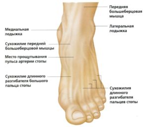
The ankle has three articular surfaces. This allows it to move in several planes:
- Anterior-posterior flexion is the movement of the joint around the talar bone, which acts as a detent in the up position of the foot.
- Plantar flexion is a supination of the foot, i.e. an inward spiral rotation of the foot.
- Dorsiflexion or pronation of the foot is an outward spiraling rotation of the foot.
In traumatology and orthopedics, a distinction is made between 2 ankles:
- Medial ankle formed by the convexity of the lower part of the tibia.
- Lateral ankle formed by the bulge of the lower part of the fibula.
- External malleolus, located behind the ankle of the foot.
- The medial malleolus, which is in front of the joint in the frontal plane (the plane parallel to the frontal bone).
The ankle has a capsule that is reinforced by a multitude of small ligaments:
- The collateral ligaments of the ankle begin at the tips of the ankle and are fan-shaped. Its end attaches to the periosteum of the lower tarsal bones (navicular bone, 3 cuneiform bones and cube bones).
- The anterior and posterior interosseous ligaments and the interossae membranes. These structures hold the distal intercondylar joint together.
Interesting!!! The interosseous ligaments are very strong, especially the posterior ligaments. They can withstand loads of up to 450 kg. Therefore, excessive loading of the foot in the sagittal plane (anterior-posterior) leads to so-called burst fractures, in which not the ligaments but the bone fragments to which they are attached are torn.
The movements in the ankle are very extensive:
They are very individual and depend on many factors (including the flexibility of the calf muscle) so the ankle has an anteroposterior range of motion of 60° to 140°.
Biomechanical features of the ankle and ankle

For a better understanding of the ankle and its biomechanics, reference can be made to illustrations and photographs in the medical literature. This subject is best described in the book 'Atlas of fractures and their treatment' by AN Shabanov and VA Sartan.
In this book, the ankle is represented as multiple axes set at specific angles to each other, forming complex triangles. Violation of the angles and planes in these figures results in dislocation and fracture of the ankle or hock.
- The A axis is a vertical line that runs down the middle of the shin bone and runs to the heel bone.
- The B axis is a line running laterally at an angle of 3-10° to the A axis.
When the ratio of the axes is altered, several types of anomalies in ankle integrity occur:
- When the angle between the A axis and the B axis decreases, the center of gravity shifts inward, resulting in fracture of the small distal tibial condyle and fracture of a fragment of the lower part of the fibula.
- As the angle between the A and B axes increases, the center of gravity shifts outward, resulting in a fracture of the tibia at the epiphysis and a fracture or fragment of the fibula.
To better understand the mechanism of these fractures, imagine a girl walking in high heels. When her foot rolls inward, the first type of fracture occurs. If it tilts outwards - the second.
This knowledge is essential for determining the cause of injury in insurance and forensic science, and for determining the cause and effect of human disease in pathology.
Read more:When the integrity of the bones, ligaments, joints and soft tissues in the lower leg is compromised, a cascade of biomechanical reactions occur, leading to swelling, poor circulation and infection in the leg.
Preparation
The procedure does not require long preparation (in terms of a special diet, rest and physical activity). Preparation can be limited to psychological and moral preparation for the upcoming investigation.
The physician should educate the patient about who will perform the test, how and for what purpose, and what is expected of the procedure. The patient should have a rough idea of the procedure, its nature and importance. He should also have an idea of the purpose of the procedure and the risks involved.
During the examination, the patient should be in a suitable position on the couch. The laboratory technician or physician conducting the examination should explain or show the patient the position to assume. Legs should be bent at the knees and feet placed on a flat surface. If an ankle injury is to be diagnosed, the x-ray is taken from the side. To do this, the patient must be in a sitting position. The injured limb must be placed on a support.
In order to determine whether a lateral or longitudinal flatfoot is present, the ankle must be flexed. When examining the arch of the foot, the patient must stand on one foot and press on the other foot.
In preparation, an initial medical history should also be taken. if e.g. B. If you have already had an X-ray 6 months ago, you should not repeat the examination as it involves a high level of radiation exposure to the body. It's also important to let your doctor know if you're pregnant or breastfeeding, as these are contraindications to the procedure. An exception is if you are seriously injured. In this case, a special lead apron is required, which can provide radiation protection.
positioning of the ankle
The ankle must be positioned correctly for the examination. First, a visual assessment of the injury is made, and then an X-ray examination is performed. The whole procedure takes no more than 10 minutes.
Direct rear projection is most commonly used. The advantage of this projection is that rotation of the foot is avoided. Examination in this position requires the patient to be in a supinated position with the legs extended horizontally on the table surface. The sagittal plane of the foot must form a 90 degree angle with the table surface.
The examination can also be performed in a simple posterior projection with foot rotation. To do this, the foot is positioned in the same way as for posterior projection (lying supine with feet flat on the table). The difference is that the foot should be turned inward, creating an angle of 15-20 degrees.
When examining the ankle in lateral projection, the patient lies on their side. The limb that is not being examined should be pressed against the abdomen, and the foot of the other side should be in contact with the lateral surface. The heel is pressed firmly against the cassette and the foot rotated inward by about 15-20 degrees.
The test can be performed with or without weight bearing on the foot.
[18]
- tibia and fibula.
- How to distinguish a fracture from an ankle sprain.
- ankle sprain.
- ankle sprain.
- The ankle bruise is where the picture is taken.
- The lateral ankle is.
- Damaged ligaments of the ankle photo.
- Structure of the human ankle.