The knee joint is formed by the fusion of three bones: the femur, the tibia, and the kneecap (patella), commonly referred to as the 'kneecap'. This joint is the most complicated - when you flex, the kneecap rests in a depression formed by the inner and outer processes of the femur.
- How the human leg is built
- bones of the lower limbs
- The hip.
- shin
- The musculoskeletal system of the foot
- Bone
- Tarsal bones (tarsal bones).
- tarsus
- distal area
- How the bones are arranged
- Vascular and nervous system of the foot
- blood
- Annoy
- Suddenly my feet start hurting badly below the knee (see below). And why?
- Lower leg muscles: anatomy, classification and function
- vessels and nerves
- Diseases of the tibia
- Peculiarities of leg muscle structure
- glutes
- Conclusion
- Anatomical structure of the human leg
- functions
- parts
- Thigh
- shin
- Foot
- areas of the legs
- ankle
- diagnosis
- How the human foot works
- function and structure
- Common diseases of the lower limbs
- bones and joints
- soft tissues.
- Treatment
- Defects in the shape of the foot
- Timely treatment of the feet
How the human leg is built
The human body evolved in response to human needs. The need to move upright greatly influenced the formation of our skeleton. The feet give the body full support and allow us to move around without the help of our hands.
In this article, you will learn about the anatomy and names of each part of the leg. We describe the composition and structure of the parts of the lower limbs and tell you which muscles, joints and ligaments help us to move.
bones of the lower limbs
The human leg skeleton includes the pelvic girdle and the skeletal structure of the free lower limbs. The leg is made up of 30 bones: 26 make up the foot, two make up the tibia, and one makes up the skeleton of the thigh. The remaining bone is the kneecap, which covers the knee joint.
The legs are divided into three areas from the hip joint to the fingertips:
To help you understand what we are going to talk about, look at the structure of the human leg and the photo with the description.

The hip.
The thigh is made up of a single bone. Its length is a quarter of human height. The structure of the femur resembles a tube with two elongated ends. The middle part of this bony tube is the diaphysis and the elongated, rounded ends are the epiphysis.
Inside the diaphysis there is a cavity called the bone canal.
The epiphyses have a spongy structure. Their shape resembles pumice stones. The upper epiphysis, the femoral head, is almost perfectly rounded. It connects to the diaphysis at an angle.
Important. The femoral neck (the part between the femur and femoral head) is a known weak point. This area is the most prone to damage, especially in the elderly.
shin
The tibia skeleton consists of the tibia and fibula. The fibula is thin and on the outside, while the tibia is strong on the inside. Both have a tubular structure.
The top of the tibia forms the underside of the knee joint. It is forked, forming two 'saucers' in which the two condyles (protrusions) of the femur rest. Below the knee is another joint - the joint between the fibula head and the tibia.
It allows a small range of motion, allowing the legs to rotate in and out more freely. The lower end of the tibia is embedded in the hock. There is a bony 'icicle' on its lower epiphysis. – ankle. This outgrowth forms the lateral surface of the ankle, the part of the leg above the foot.
The musculoskeletal system of the foot
Bone
The bones of the foot and hand are similar in structure. In anatomy, the foot is divided into the following bony sections:
Tarsal bones (tarsal bones).
Includes 7 bones. The talus and heel bones are the most massive. The talus is located between the shins and is more closely related to the ankle. It includes:
tarsus
This is a set of five bones that resemble a tubular shape. This section is medial and is responsible for the function of the fingers and the correct alignment of the arch of the foot. The bones ending in the joints lead to the beginning of the fingers.
distal area
There are 14 bones in this area. Each finger has 3 bones except for the thumb which only has 2. Between the bone formations are the joints that allow movement.
Thanks to this area of the foot, the human body maintains its balance and is able to move. Interestingly, if the hands are lost, the fingers have a backup function.
The joints are between the bones. In addition, the foot contains muscles, ligaments, nerves, and blood vessels.
How the bones are arranged
The bones need a closer look as they are the main component of the foot.
The strongest bone is the heel bone (calcaneus).
It is the strongest part of the foot. Although this part has nothing to do with the ankle, it plays a big role in distributing pressure. The shape of the heel bone resembles a triangle with a long axis in three dimensions.
The connections between the heel bone and the talus act like a link. A strong connection between these two bones is necessary for the foot to maintain its proper shape. The Achilles tendon is located at the back of the bone. You can tell by the slight protuberance. The lower part, on the other hand, provides support when walking on the ground.
Vascular and nervous system of the foot
blood
The shin arteries, located at the front and back of the foot, provide blood flow to the foot. They run along the sole of the foot itself. Smaller connections and vertebrae branch off from these main arteries.
When the foot is injured, one vertebra falls out, but the remaining vertebrae continue to supply the limb with the necessary blood.
The veins on the back are responsible for drainage. They are intertwined and supply blood to the large and small saphenous veins in the lower leg.
Annoy
They are an essential part of the normal functioning of the human foot. You are responsible for the sensation:
Nerve signals sent from the CNS via the calf, brachial, superficial, and tibial nerves reach and are processed in the spinal cord.
The nerves transmit signals to the muscles, which are essentially reflexes that are either voluntary or involuntary (independent of human will). The involuntary include the glands (sebaceous and sweat glands) and the tension of the blood vessels.
As far as the skin is concerned, several zones can be distinguished on the foot, which differ in their density, texture and elasticity. For example, the skin on the sole of the foot is dense, while the skin on the heel is thick. Initially, the skin of the hands and feet is the same, but over time and increased stress, additional layers are formed. The instep is smooth and flexible and has nerve endings.
In summary, one can say that nature has ensured that the foot can withstand enormous loads.
Suddenly my feet start hurting badly below the knee (see below). And why?
To be honest, I'm going to see the doctor on Monday. I want to know what could be the causes.
My feet started to hurt a little about 6 months ago, hardly noticeable and very rarely, I prefer not to pay attention.
Dizziness for a few weeks (if I don't eat for a long time), trembling hands and fog. Blood pressure 107-117/65-71 (am below 75, feel fine). I used to be able to go very long without eating and I was fine, my body was light - I felt good, my stomach never hurt and it doesn't hurt now, apart from dizziness and nausea. I had cramps a few times, I started taking calcium+magnesium, the cramps stopped.
Instead, my legs started hurting like crazy. After walking, after a little exertion, the area around my lower leg, that is, below the knee, in front, along the bone to the foot, hurts. Pressure on the bone is painful when I touch it from the side and apply pressure. Very strong aching and throbbing pain (to tears only, the harder the exercise/exertion, the more and longer it hurts), quite long-lasting. He tires easily, can hardly walk, is breathing heavily, is tired and has palpitations. However, a few months ago he had no problems, he could run fast and a lot. He was neither in pain nor tired.
Lower leg muscles: anatomy, classification and function
Surrounding the bony structures of the lower leg is a dense ring of muscles that keep the leg in motion from foot to knee. Depending on their location, all muscles are divided into three different groups: anterior, posterior and lateral. The anterior group of muscle fibers is responsible for supination, extension and adduction of the foot and toe extension. The posterior group acts as an antagonist and controls the flexion of the foot and toes. The lateral group muscles control the abduction, pronation, and flexion of the foot.


vessels and nerves
The blood vessels begin with the vessels in the hip. Arteries such as the tibialis anterior and posterior pull through the muscles of this part of the leg. The knee is supplied with blood by eight arterial trunks.
The veins of the lower leg are divided into deep and superficial veins. These branch into a large number of smaller vessels and are connected to the arteries by capillaries.
The nervous system is represented by the major and minor tibial nerves. These arise from the largest trunks and ensure the sensitivity of the muscle tissue. There are also subcutaneous nerves that are responsible for external sensation.
Diseases of the tibia

The human tibia, like any other part of the body, can be affected by various diseases. Pathological processes take place in the muscles, bones and blood vessels.
- myositis. This is what the inflammatory process in muscle tissue is called. It is accompanied by pain, tension and reddening of the skin. In advanced cases, muscle atrophy may occur.
- osteomyelitis. This is a purulent-necrotic disease that develops in the bones. It is caused by the negative effects of pathogenic microorganisms. Patients diagnosed with this disease have high body temperature, are prone to pain, their general condition worsens, and they experience nausea.
- Arthritis. An inflammatory process that occurs in the joints of the lower limbs. It can be acute or chronic. Symptoms include pain, stiffness, grinding during movement, changes in shape of the joint, reddening of the skin in the affected area.
- Nodules in the muscles of the lower leg. These can be benign or malignant. These abnormalities cause a variety of symptoms: pain, swelling of the limbs, aggravation. Large tumors put pressure on blood vessels, resulting in poor blood flow. The treatment of these disorders is surgical.
- varicose veins. This disease affects the veins and leads to their excessive expansion. Most often, such a pathology is diagnosed in the area of the lower legs. Blood circulation is impaired, causing leg swelling and pain.
To prevent the development of these diseases, sufferers should exercise. It is enough to exercise or run for 20 minutes a day, and the legs will become healthy.
It is also desirable to give up bad habits and follow a diet to prevent weight gain.
<
Peculiarities of leg muscle structure
Unlike other muscle groups, the legs have the most functions. It is the largest concentration of muscles that are closely intertwined. Everything is designed to stabilize and maximize the functionality of the three pairs of joints that interact with the muscles of the legs:
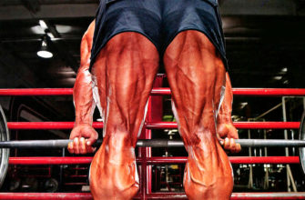
The muscles of the lower leg, the calf muscle and the neck muscle, play a special role in human life. They are giant pumps that are directly involved in blood circulation. For this reason, the calves are often referred to as the 'second heart'. For this reason Train your legs not only to build muscle mass and endurance, but also to improve your overall health.. Also, due to the number of muscle fibers and the general anatomy of the leg muscles, the lower body is designed to work hard. Not only does this allow you to work heavier weights for a strong anabolic response, but it also significantly increases your overall endurance (which depends on your legs for 50+ %).
In sports, the lower body is divided into four areas based on their main functions:
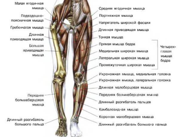
- glutes;
- the muscles of the front of the thigh;
- muscles of the back of the thigh;
- lower leg muscles.
Knowing the names of all leg muscles and looking at each muscle individually is not necessary for a productive workout. Rather, this is a medical issue. However, in order to avoid injuries and to increase the effectiveness of each exercise, it is necessary to know at least roughly the structure of the human leg muscles and the tasks of the individual areas.
glutes
As for the anatomy of the human leg muscles, the gluteal muscle group is considered to be one of the largest. It also includes the largest muscle in the body – the gluteus maximus major (gluteus maximus major). The entire region is shaped by the three gluteal muscles:
Conclusion
Remember that being able to remember the names of all the muscles on a person's legs will not give you an advantage in amateur sports; this is more important in the medical field. However, knowing the basic functions of muscles and their structure can increase the effectiveness of your training many times over. This is true for beginners who have just joined a gym, as well as athletes who have been training for years.

The easiest way to determine the ideal leg shape and joint structure is to look at the distance between the thighs, but there are many pitfalls here. First: width

What unites men and women in training is the development of the lower body. While men want to get strong muscles through their training, women need a tightening.
Like the rest of the body, the legs should be developed harmoniously. None of the large muscles should lag behind in development, including the biceps femoris.
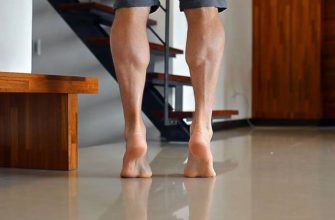
It's not easy to do calf exercises at home, especially when the shins are the slowest part of the body and develop poorly no matter how hard you train them.

Anatomical structure of the human leg
functions
parts
If you remember the anatomy, the leg consists of three parts: the thigh, the shin, and the foot.
Thigh
Has a protective function. It consists of the femur, the kneecap and is influenced by the quadriceps, biceps femoris and flexors.
shin
Has a relatively simple structure and consists of two bones of different lengths, the fibula and the tibia.
The latter connects the tibia and femur in the knee joint and is the second largest part of the human leg.
Foot
Consists of many small bones. The foot or sole is where it touches the ground. The opposite side is called the hind foot.
- The forefoot, which consists of the toes and the balls of the feet;
- the middle or arch of the foot. The arch of the foot includes the part of the foot that does not touch the ground;
- the hindfoot, the heel.
The foot is a much more complex structure, with more than 26 bones and 33 joints. The structure of the foot and hand are very similar, they differ only in the type of load. The muscles and bones of the foot are many times stronger, while the hand lacks mobility.
areas of the legs
- Front + back of the thigh;
- Front + back area of the knee;
- front + rear areas of the lower leg;
- front + rear outer + inner areas of the ankle;
- back of the foot;
- Sole.
ankle
The largest bone is the ankle bone (talus). At the top is a block with a protuberance that connects to the tibia and fibula.
diagnosis
The ankle may be damaged or defective. To diagnose the problem, a diagnostic procedure is prescribed. This can consist of:
- ULTRA SOUND. This diagnostic method is rarely used due to the small size of the ankle. However, it does allow detection of a foreign body, swelling caused by blood pooling in the joint capsule, and a view of the ligaments.
- arthroscopy. A minimally invasive method that involves inserting a video camera into the joint capsule to make a diagnosis.
- X-RAY. The most economical method. It is possible to capture images in different projections. Allows detection of tumors, fractures, dislocations and other processes.
- MRI. Best way to diagnose condition of Achilles tendon, ligaments and cartilage. Expensive but very effective.
- Computed Tomography (CT) .. Helps assess the condition of the joint. It is considered the most accurate test for osteoarthritis, cancer and broken bones.
How the human foot works

As a child grows older, the skeletal system, originally composed mostly of cartilage, hardens. This makes the bones more difficult to injure and break. Cartilage plays a very important role in joints. It ensures that the bones in the joints move easily and we can move freely. The hip, ischium, and pubic bone are the three largest bones of the pelvis. They are fused together around the hip socket and provide support for the torso. The hip joint is located in this hip socket. The head of the femur enters this joint and, through its rotation, allows the rotation of the limbs.
function and structure
The human foot is a complex structure necessary to maintain an upright posture, to absorb the force of ground contact while walking (about 70 %) and to move on different surfaces. It consists of 26 bones, differing in structure and appearance, connected by muscles and ligaments.
The function of the joints is to connect the bony structures together. They ensure the integrity and mobility of the skeleton, the consistency of the movements of each component, and the ability to perform complex gestures. A joint is a combination of bones that is able to move its parts relative to each other while maintaining its integrity.
The surfaces involved in the formation of the joint are covered by cartilaginous tissue with extremely low roughness. The space between the bones is filled with lubricating synovial fluid that facilitates gliding. All components are enclosed in a joint capsule that protects the system from malfunction and damage to its components.
The joints of the feet are often prone to injury. A fall or poor positioning of the leg can lead to a dislocation or fracture. To avoid complications, these injuries should be treated by a qualified medical professional. The bony structure of the foot is described in detail below.
The foot is divided into three functional parts:
- Distal part - the toes, which are made up of small, moving parts.
- The tarsus – the middle part made up of long bones that are similar to each other.
- The tarsal is a complex supporting part.
It is not known if the structure of the foot is limited to the skeleton. The muscular composition of the human foot region, like that of the joint region, is very different.

The table shows the muscles and their groups descending from the lower leg to the foot.
Common diseases of the lower limbs
Some of the sick are elderly people. Younger patients may suffer from a pinched sciatic nerve as a result of a herniated disc or severe hypothermia.
In older people, the limbs are more often affected by vascular diseases. Muscle and joint diseases are pushed into the background. Both the arterial and venous systems are affected. Varicose veins become noticeable earlier. These are vascular webs on the legs, followed by gross knots with signs of inflammation. Spasms and trophic changes in the skin in later stages are of concern.
With chronic arterial insufficiency as part of atherosclerosis of the lower extremities, unbearable pain in the legs appears, forcing to stop and rest.
This is called intermittent claudication syndrome. The skin on the limbs atrophies and the condition of the nails worsens. Cramps and sensory disturbances are common. The disease can lead to gangrene and require amputation.
bones and joints

There are 2 main causes of bone damage:
Joint injuries are due to the following diseases:
- arthrosis (osteoarthritis);
- Rheumatoid arthritis;
- psoriatic arthropathy;
- peripheral form of ankylosing spondylitis (Bechterew's disease);
- reactive arthritis.
Arthrosis is primarily about the limited mobility of the joint, while osteoarthritis is more about the pain. Damage to the joint with an exacerbation is accompanied by inflammation of the joint capsule and the development of synovitis. At the same time, there is swelling of the joint. The periarticular tissue can also become inflamed.
soft tissues.
Not only the joints, bone structures and blood vessels are damaged. There are a number of collections called the periarticular tissues. They are also referred to as periarticular tissues. This group includes tendons, ligaments, muscle masses, muscle tendon sheaths, fascia, aponeuroses and entheses. The latter includes the protrusions on the bones to which the tendons are attached.
Treatment
The type of treatment depends on the disease in question. Bruises and injuries must be treated by a surgeon or trauma surgeon. Surgery is required for severe wounds and open wounds.
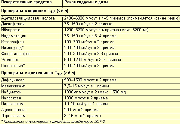
Joint disease requires appropriate anti-inflammatory treatment in its early stages. These include NSAIDs or glucocorticoid hormones. In the remission phase, chondroprotectors are prescribed to nourish the cartilage and prevent the disease from progressing. Physiotherapy can also be helpful. If conservative treatment is ineffective, an endoprosthesis is used.
Phlebologists treat venous disease at an early stage. They recommend wearing elastic stockings and phlebotherapy. In later stages with deformed veins, removal of the diseased vessels is indicated.
The same treatment tactics are used in chronic arterial disease. Instead of phlebotonics, vasodilators are indicated to improve microcirculation of the tissues of the lower limbs.
With neuropathy, thioctic acid preparations are prescribed. To date, this is the only remedy whose effect on this pathology has been proven.
The lower limbs of man are affected by diseases of the vessels, joints and nerves. Bones, muscles or tendons can be injured. Only knowledge of the topography of these structures enables a correct diagnosis and the initiation of an appropriate treatment.
Defects in the shape of the foot
If an examination of the foot reveals irregularities in shape, a doctor should definitely be consulted. Flat feet can be caused by genetic changes; this is difficult to correct. However, if one pays attention to the irregular shape of the foot in childhood, it can still be corrected. In childhood, the bones are still very soft and fragile, so you can combat the defect with exercise and special insoles.
Some parts of the foot are particularly vulnerable to damage. For example, the deformation of the first toe (especially the metatarsophalangeal joint). This can also include the heel bone and hammer toe.
Orthotics can help with this. It is enough to visit a traumatologist or orthopedist at least once a year to avoid further development of foot deformities.
[39], [40], [41], [42], [43], [44]
Timely treatment of the feet
If a doctor is consulted in good time, foot deformities can be corrected at an early stage, when the affected person has no suspicion of an abnormal development. If the abnormal development of the foot is ignored, then over time the situation will worsen under the influence of mechanical factors - walking, friction, pressure, increased loads.
Therefore, it is always important to pay attention to even the most seemingly insignificant changes in foot structure. For example, a bump on the heel, loose foot hair, a growing or painful bunion on the foot, or even calluses that weren't visible before. And ask your doctor about your foot health right away.
Read more:- The sit bones.
- Photo of the right leg.
- Heel bone human anatomy photo and description.
- What parts of the leg are called.
- The lateral dislocation is.
- The parts of the human leg are named.
- The leg of a healthy person.
- What is the connection between the femur and tibia?.